. 2022 Feb 16;14(1):212-244.
doi: 10.3390/neurolint14010018.

Hypomyelinating Leukodystrophy 8 (HLD8)-Associated Mutation of POLR3B Leads to Defective Oligodendroglial Morphological Differentiation Whose Effect Is Reversed by Ibuprofen

Affiliations

Hypomyelinating Leukodystrophy 8 (HLD8)-Associated Mutation of POLR3B Leads to Defective Oligodendroglial Morphological Differentiation Whose Effect Is Reversed by Ibuprofen

Sui Sawaguchi et al. Neurol Int. .

Abstract

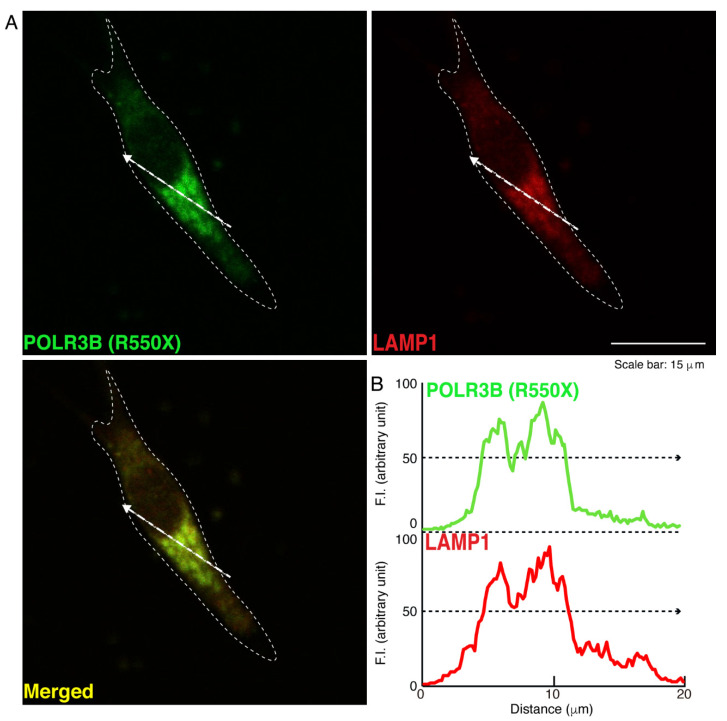
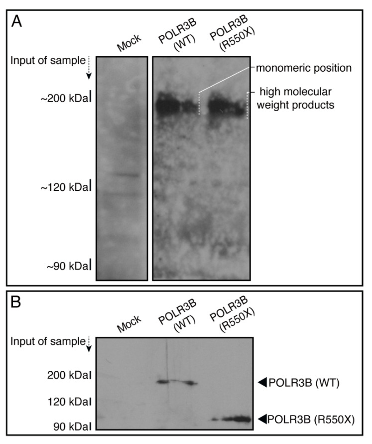
POLR3B and POLR3A are the major subunits of RNA polymerase III, which synthesizes non-coding RNAs such as tRNAs and rRNAs. Nucleotide mutations of the RNA polymerase 3 subunit b (polr3b) gene are responsible for hypomyelinating leukodystrophy 8 (HLD8), which is an autosomal recessive oligodendroglial cell disease. Despite the important association between POLR3B mutation and HLD8, it remains unclear how mutated POLR3B proteins cause oligodendroglial cell abnormalities. Herein, we show that a severe HLD8-associated nonsense mutation (Arg550-to-Ter (R550X)) primarily localizes POLR3B proteins as protein aggregates into lysosomes in the FBD-102b cell line as an oligodendroglial precursor cell model. Conversely, wild type POLR3B proteins were not localized in lysosomes. Additionally, the expression of proteins with the R550X mutation in cells decreased lysosome-related signaling through the mechanistic target of rapamycin (mTOR). Cells harboring the mutant constructs did not exhibit oligodendroglial cell differentiated phenotypes, which have widespread membranes that extend from their cell body. However, cells harboring the wild type constructs exhibited differentiated phenotypes. Ibuprofen, which is a non-steroidal anti-inflammatory drug (NSAID), improved the defects in their differentiation phenotypes and signaling through mTOR. These results indicate that the HLD8-associated POLR3B proteins with the R550X mutation are localized in lysosomes, decrease mTOR signaling, and inhibit oligodendroglial cell morphological differentiation, and ibuprofen improves these cellular pathological effects. These findings may reveal some of the molecular and cellular pathological mechanisms underlying HLD8 and their amelioration.

Keywords: POLR3B; Pelizaeus–Merzbacher disease (PMD); hypomyelinating leukodystrophy (HLD); ibuprofen; lysosome; oligodendrocyte.


Conflict of interest statement

The authors declare no conflict of interest.

Figures

Figure 1
Figure 1
R550X mutant proteins of POLR3B accumulate in punctate structures in FBD-102b cells, whereas the wild type proteins are expressed throughout the cell body. (A,B) FBD-102b cells, surrounded by dotted lines, were transfected with the plasmid encoding the wild type (WT) POLR3B or the R550X mutant constructs. Transfected cells were detected using transfected proteins (green) and nuclear DAPI (blue). Scan plots were performed along the white dotted lines in the direction of the arrows in images. Graphs showing the fluorescence intensities (arbitrary units) along the white dotted lines in the direction of the arrows can be seen in the bottom panels. (C) Percentages of cells with punctate structures were statistically assessed (** p < 0.01; n = 3 fields).
Figure 2
Figure 2
Wild type POLR3B proteins are not co-localized with the ER in cells. (A,B) Cells were transfected with the plasmid encoding the wild type (WT) POLR3B and detected with transfected proteins (green) and an antibody against the KDEL antigen (red). Scan plots were performed along the white dotted lines in the direction of the arrows in the color images (green and red as well as merged images). Graphs showing the fluorescence intensities (arbitrary units) along the white dotted lines in the direction of the arrows (black dotted lines in right bottom panels) can be seen in the right bottom panels.
Figure 3
Figure 3
POLR3B mutated proteins are not co-localized with the ER in cells. (A,B) Cells were transfected with the plasmid encoding POLR3B (R550X) (green) and stained using an antibody against the KDEL antigen (red). Scan plots were performed along the white dotted lines in the direction of the arrows in the color images (green and red as well as merged images). Graphs showing the fluorescence intensities (arbitrary units) along the white dotted lines in the direction of the arrows (black dotted lines in right bottom panels) can be seen in the right bottom panels.
Figure 4
Figure 4
Wild type proteins are not co-localized with the Golgi body in cells. (A,B) Cells were transfected with the plasmid encoding the wild type (WT) POLR3B (green) and stained using an antibody against the GM130 antigen (red). Scan plots were performed along the white dotted lines in the direction of the arrows in the color images (green and red as well as merged images). Graphs showing the fluorescence intensities (arbitrary units) along the white dotted lines in the direction of the arrows (black dotted lines in right bottom panels) can be seen in the right bottom panels.
Figure 5
Figure 5
Mutated proteins are not co-localized with the Golgi body in cells. (A,B) Cells were transfected with the plasmid encoding POLR3B (R550X) (green) and stained using an antibody against the GM130 antigen (red). Scan plots were performed along the white dotted lines in the direction of the arrows in the color images (green and red as well as merged images). Graphs showing the fluorescence intensities (arbitrary units) along the white dotted lines in the direction of the arrows (black dotted lines in right bottom panels) can be seen in the right bottom panels.
Figure 6
Figure 6
Wild type proteins are not significantly co-localized with the lysosome in cells. (A,B) Cells were transfected with the plasmid encoding the wild type (WT) POLR3B (green) and stained using an antibody against the LAMP1 antigen (red). Scan plots were performed along the white dotted lines in the direction of the arrows in the color images (green and red as well as merged images). Graphs showing the fluorescence intensities (arbitrary units) along the white dotted lines in the direction of the arrows (black dotted lines in right bottom panels) can be seen in the right bottom panels.
Figure 7
Figure 7
Mutated proteins are mostly co-localized with the lysosome in cells. (A,B) Cells were transfected with the plasmid encoding POLR3B (R550X) (green) and stained using an antibody against the LAMP1 antigen (red). Scan plots were performed along the white dotted lines in the direction of the arrows in the color images (green and red as well as merged images). Graphs showing the fluorescence intensities (arbitrary units) along the white dotted lines in the direction of the arrows (black dotted lines in right bottom panels) can be seen in the right bottom panels.
Figure 8
Figure 8
Mutated but not wild type proteins exhibit dimeric and high molecular weight structures in non-denaturing polyacrylamide gel electrophoresis. (A,B) The lysates of cells transfected with an empty vector or with a plasmid encoding the wild type (WT) POLR3B or POLR3B (R550X) were subjected to non-denaturing (upper images) and denaturing (lower images) polyacrylamide gel electrophoresis and detected using immunoblotting. The position corresponding to the molecular weight of the wild type monomer or the R550X monomeric or polymeric (including high molecular weight products) structures is shown.
Figure 9
Figure 9
Cells harboring the mutants fail to undergo morphological differentiation. (A) Cells harboring the wild type (WT) or POLR3B (R550X) were allowed to differentiate for 5 days. Some cells in the bottom images are surrounded by white dotted lines (a and b). The square fields a and b indicated by the dotted lines in the bottom panels are magnified in the upper panels a and b. Images of cells at 0 day are also shown. (B) Cells with widespread membranes were statistically assessed (** p < 0.01; n = 5 fields (50 cells in total)). (C) The lysates of the respective cells were immunoblotted with an antibody against PLP1 and CNPase, cell lineage marker Sox10, and control actin. (D) Their expression levels are shown and statistically compared to their respective controls. (* p < 0.05 of Student’s t-test; n = 3 blots).
Figure 10
Figure 10
Cells harboring the mutants decrease phosphorylation levels of ribosomal S6 and 4E-BP1 proteins at the cell and protein levels. (A,B) Cells harboring the wild type (WT) or POLR3B (R550X) (green) were stained with an anti-(pS240 and pS244)ribosomal S6 protein (pS6) or anti-(pT37)4E-BP1 (p4E-BP1) antibody (red) that shows that phosphorylation acts downstream of mTOR signaling. Their phosphorylation levels are shown statistically compared to those in their respective control cells under each image (** p < 0.01; n = 3 fields). (C,D) The lysates of cells harboring the wild type (WT) or POLR3B (R550X) were immunoblotted with an anti-(pS240 and pS244)ribosomal S6 protein (pS6), anti-(pT37)4E-BP1 (p4E-BP1), anti-S6, and anti-4E-BP1. Statistically significant differences are shown in their immunoreactive bands (* p < 0.05 of Student’s t-test ** p < 0.01; n = 3 blots).
Figure 11
Figure 11
Ibuprofen improves phenotypes in cells harboring the mutants. (A) Cells harboring POLR3B (R550X) were allowed to differentiate in the presence or absence of ibuprofen for 5 days. Some cells in the upper images are surrounded by white dotted lines. The square fields indicated by the dotted lines in the upper panels are magnified in the respective lower panels. (B) Cells with widespread membranes were statistically assessed (** p < 0.01; n = 5 fields (50 cells in total)). (C) The lysates of the respective cells were immunoblotted with an antibody against PLP1, CNPase, Sox10, and actin. (D) Their expression levels are shown statistically compared to their respective controls (** p < 0.01; n = 3 blots).
Figure 12
Figure 12
Ibuprofen improves phosphorylation levels of ribosomal S6 and 4E-BP1 proteins and aggregate-like punctate structures in cells harboring the mutants. (A,B) Cells harboring POLR3B (R550X) (green) in the presence or absence of ibuprofen were stained with an anti-(pS240 and pS244)ribosomal S6 protein (pS6) or anti-(pT37)4E-BP1 (p4E-BP1) antibody (red). Their phosphorylation levels are shown statistically compared to those in their respective control cells under each image (** p < 0.01; n = 3 fields). (C,D) Cells harboring POLR3B (R550X) (green) were treated with or without ibuprofen. Percentages of cells with punctate structures were statistically assessed (** p < 0.01; n = 3 fields).
Figure 13
Figure 13
Ibuprofen does not have significant effects on phenotypes in cells harboring the wild type POLR3B. (A) Cells harboring the wild type were allowed to differentiate in the presence or absence of ibuprofen for 5 days. Some cells in the upper images are surrounded by white dotted lines. The square fields indicated by the dotted lines in the upper panels are magnified in the respective lower panels. (B) Cells with widespread membranes were statistically assessed (n = 5 fields (50 cells in total)). (C) The lysates of the respective cells were immunoblotted with an antibody against PLP1, CNPase, Sox10, and actin. (D) Their expression levels are shown statistically compared to their respective controls (n = 3 blots).
Figure 14
Figure 14
Ibuprofen does not have significant effects on phosphorylation levels of ribosomal S6 and 4E-BP1 proteins and aggregate-like punctate structures in cells harboring the wild type POLR3B. (A,B) Cells harboring the wild type (green) in the presence or absence of ibuprofen were stained with an anti-(pS240 and pS244)ribosomal S6 protein (pS6) or anti-(pT37)4E-BP1 (p4E-BP1) antibody (red). Their phosphorylation levels are shown statistically compared to those in their respective control cells under each image (n = 3 fields). (C,D) Cells harboring the wild type (green) were treated with or without ibuprofen. The percentage of cells with punctate structures were statistically assessed (n = 3 fields).

References

    1. Bernard G., Chouery E., Putorti M.L., Tétreault M., Takanohashi A., Carosso G., Clément I., Boespflug-Tanguy O., Rodriguez D., Delague V., et al. Mutations of POLR3A encoding a catalytic subunit of RNA polymerase Pol III cause a recessive hypomyelinating leukodystrophy. Am. J. Hum. Genet. 2011;89:415–423. doi: 10.1016/j.ajhg.2011.07.014. - DOI - PMC - PubMed
    1. Saitsu H., Osaka H., Sasaki M., Takanashi J., Hamada K., Yamashita A., Shibayama H., Shiina M., Kondo Y., Nishiyama K., et al. Mutations in POLR3A and POLR3B encoding RNA polymerase III subunits cause an autosomal-recessive hypomyelinating leukoencephalopathy. Am. J. Hum. Genet. 2011;89:644–651. doi: 10.1016/j.ajhg.2011.10.003. - DOI - PMC - PubMed
    1. Tétreault M., Choquet K., Orcesi S., Tonduti D., Balottin U., Teichmann M., Fribourg S., Schiffmann R., Brais B., Vanderver A., et al. Recessive mutations in POLR3B, encoding the second largest subunit of Pol III, cause a rare hypomyelinating leukodystrophy. Am. J. Hum. Genet. 2011;89:652–655. doi: 10.1016/j.ajhg.2011.10.006. - DOI - PMC - PubMed
    1. Daoud H., Tétreault M., Gibson W., Guerrero K., Cohen A., Gburek-Augustat J., Synofzik M., Brais B., Stevens C.A., Sanchez-Carpintero R., et al. Mutations in POLR3A and POLR3B are a major cause of hypomyelinating leukodystrophies with or without dental abnormalities and/or hypogonadotropic hypogonadism. J. Med. Genet. 2013;50:194–197. doi: 10.1136/jmedgenet-2012-101357. - DOI - PubMed
    1. Garbern J., Cambi F., Shy M., Kamholz J. The molecular pathogenesis of Pelizaeus-Merzbacher disease. Arch. Neurol. 1999;56:1210–1214. doi: 10.1001/archneur.56.10.1210. - DOI - PubMed

LinkOut - more resources