. 2022 Jan 18;10(1):6.
doi: 10.3390/jdb10010006.

Differentiation of Cells Isolated from Human Femoral Heads into Functional Osteoclasts

Affiliations

Differentiation of Cells Isolated from Human Femoral Heads into Functional Osteoclasts

Daniel R Halloran et al. J Dev Biol. .

Abstract

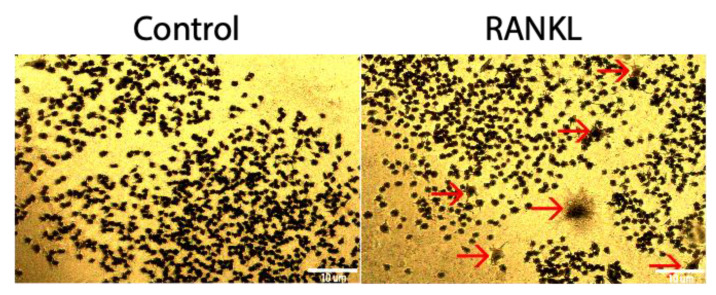
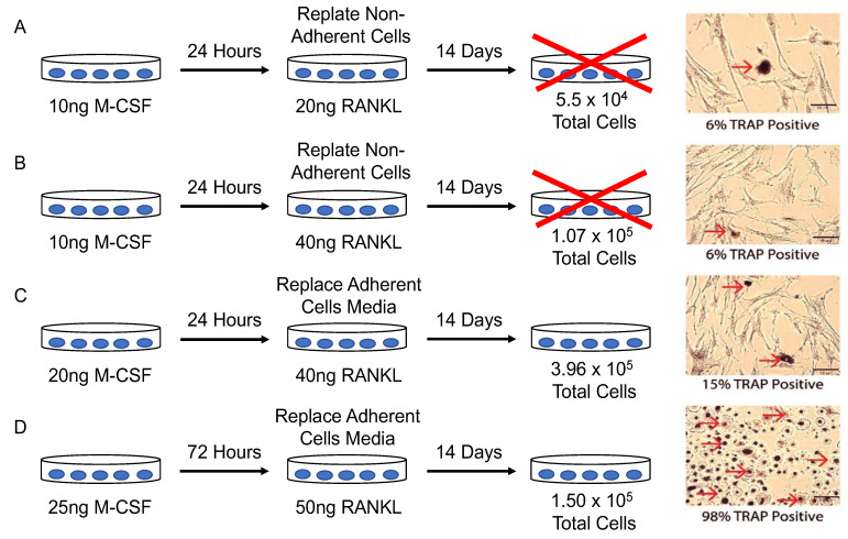
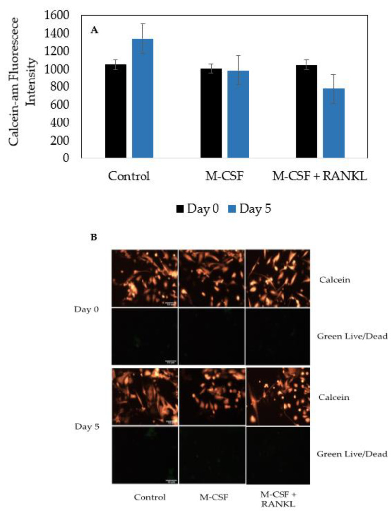
Proper formation of the skeleton during development is crucial for the mobility of humans and the maintenance of essential organs. The production of bone is regulated by osteoblasts and osteoclasts. An imbalance of these cells can lead to a decrease in bone mineral density, which leads to fractures. While many studies are emerging to understand the role of osteoblasts, less studies are present about the role of osteoclasts. This present study utilized bone marrow cells isolated directly from the bone marrow of femoral heads obtained from osteoarthritic (OA) patients after undergoing hip replacement surgery. Here, we used tartrate resistant acid phosphatase (TRAP) staining, Cathepsin K, and nuclei to identity osteoclasts and their functionality after stimulation with macrophage-colony stimulation factor (M-CSF) and receptor activator of nuclear factor kappa-β ligand (RANKL). Our data demonstrated that isolated cells can be differentiated into functional osteoclasts, as indicated by the 92% and 83% of cells that stained positive for TRAP and Cathepsin K, respectively. Furthermore, isolated cells remain viable and terminally differentiate into osteoclasts when stimulated with RANKL. These data demonstrate that cells isolated from human femoral heads can be differentiated into osteoclasts to study bone disorders during development and adulthood.

Keywords: Cathepsin K; M-CSF; RANKL; RAW 264.7 cells; TRAP; bone; osteoarthritis; osteoclasts; osteoporosis.


Conflict of interest statement

The authors declare no conflict of interest.

Figures

Figure 1
Figure 1
Development of an active osteoclast. Hematopoietic stem cells (HSCs) differentiate into GMPs, which express c-fms receptors that M-CSF ligands can bind. After binding, GMPs differentiate into monocyte/macrophage precursors, that further differentiate into pre-osteoclasts and active osteoclasts when RANKL binds to RANK receptors. Active osteoclasts are multinucleated and express TRAP and Cathepsin K.
Figure 2
Figure 2
Femoral heads from patients diagnosed with OA or OP were obtained from ChristianaCare in Newark, DE or Wilmington, DE. The trabecular bone was removed from the heads and placed in α-MEM. The interior surface of the bone was washed with HBSS. After, the α-MEM and HBSS solutions were filtered with a 70 µm filter. The solution was spun down at 1800 RPM for 9 min at 4 °C to form a cell pellet. The pellet was resuspended in 5 mL of α-MEM and cells were plated at a density of 1 × 10⁵ cells/mL supplemented with α-MEM and 25 ng/mL M-CSF. Cells were grown and subjected to experimentation.
Figure 3
Figure 3
TRAP assay of RAW264.7 cells stimulated with RANKL. After 5 days of treatment with 10 ng/mL of RANKL, 2.5% of stimulated cells stained positive for TRAP, whereas 0.09% of control cells not treated with RANKL stained positive for TRAP. Red arrows designate a TRAP positive cell. Images were acquired with a 10× objective lens and scale bars are set to 10 μm. All of the experiments were conducted in triplicate.
Figure 4
Figure 4
Cells extracted from human femoral heads were plated with different conditions and were stained for TRAP. (A) Cells were plated with 10 ng/mL M-CSF for 24 h. Non-adherent cells were replated at 5.5 × 10⁴ total cells per well and supplemented with 10 ng/mL M-CSF and 20 ng/mL RANKL. (B) Cells were plated with 10 ng/mL M-CSF for 24 h. Non-adherent cells were replated at 1.7 × 10⁵ total cells per well and supplemented with 10 ng/mL M-CSF and 40 ng/mL RANKL. (C) Cells were plated at 1.31 × 10⁵ total cells and supplemented with 20 ng/mL M-CSF for 24 h. Adherent cells were supplemented with fresh media with 20 ng/mL M-CSF and 40 ng/mL RANKL for 13 days. (D) Cells were plated at a density of 1.50 × 10⁵ cells/mL and supplemented with α-MEM with 25 ng/mL M-CSF for 3–4 days. After, the media of adherent cells was replaced with fresh α-MEM along with 25 ng/mL and 50 ng/mL RANKL. Cells were stained with tartrate resistant acid phosphatase (TRAP) and imaged to identify functional osteoclasts. Red arrows indicate TRAP-positive cells. Experiments were conducted in triplicate for at least five different patients. Images were acquired using the Nikon Eclipse TE300 epifluorescence microscope with a 10× objective. Scale bars are set at 10 μm.
Figure 5
Figure 5
TRAP assay of the cells isolated from the femoral heads of OA patients. (A) Osteoclast count of male patients (N = 5). (B) Osteoclast count of female patients (N = 5). After stimulating cells for 14 days with M-CSF and RANKL, or M-CSF only, osteoclasts were identified as cells that stained positive for TRAP and comprised of three or more nuclei. Representative images are displayed underneath bars. Red boxes designate TRAP positive cells with more than three nuclei and green boxes denote macrophages/monocytes. Images were acquired with a 20× objective lens and all experiments were conducted in triplicate. “*” denotes statistical significance, where p is set to 0.05.
Figure 6
Figure 6
Calcein-AM-red-orange and Green Live/Dead staining for proliferation and viability. (A) Graphical representation of Calcein-AM fluorescence in different conditions. (B) Visual representations of Calcein-AM and Live/Dead stains. Cells were counted for 5 days, and immunofluorescence was captured using an epifluorescent microscope. Calcein-AM is indicative of the total number of viable cells, whereas green indicates an unviable cell. Scale bars are set to 10 μm. All experiments were conducted in triplicate and images were processed using ImageJ.
Figure 7
Figure 7
Immunostaining of cells isolated from male (N = 2) and female (N = 1) OA patients. Confocal microscopy was utilized to image cells at 20× (A) and 63× (B) magnification. The population of cells stimulated with M-CSF + RANKL was ~60%, whereas the control group had ~15% osteoclasts. As displayed in the figure, M-CSF + RANKL stimulated highly expressed TRAP and Cathepsin K cells, while the control cells produced very little of these osteoclast markers (C,D). Cells from 10 representative images obtained at random from all three patients were counted. The RANKL stimulated cells expressed significantly higher TRAP and Cathepsin K than the control groups (C,D). Experiments were completed in triplicate and processed using ImageJ. All fluorescence was normalized to the secondary control. “*” denotes statistical significance, where p is set to 0.05.

References

    1. Halloran D., Durbano H.W., Nohe A. Bone Morphogenetic Protein-2 in Development and Bone Homeostasis. J. Dev. Biol. 2020;8:19. doi: 10.3390/jdb8030019. - DOI - PMC - PubMed
    1. Vortkamp A. Defining the skeletal elements. Curr. Biol. 1997;7:R104–R107. doi: 10.1016/S0960-9822(06)00049-2. - DOI - PubMed
    1. Yang Y. Skeletal morphogenesis during embryonic development. Crit. Rev. Eukaryot. Gene Expr. 2009;19:197–218. doi: 10.1615/CritRevEukarGeneExpr.v19.i3.30. - DOI - PubMed
    1. Long F., Ornitz D.M. Development of the endochondral skeleton. Cold Spring Harb. Perspect. Biol. 2013;5:a008334. doi: 10.1101/cshperspect.a008334. - DOI - PMC - PubMed
    1. Durbano H.W., Halloran D., Nguyen J., Stone V., McTague S., Eskander M., Nohe A. Aberrant BMP2 Signaling in Patients Diagnosed with Osteoporosis. Int. J. Mol. Sci. 2020;21:6909. doi: 10.3390/ijms21186909. - DOI - PMC - PubMed