. 2022 Jan 28;13(1):13.
doi: 10.3390/jfb13010013.

Biomimetic PLGA/Strontium-Zinc Nano Hydroxyapatite Composite Scaffolds for Bone Regeneration

Affiliations

Biomimetic PLGA/Strontium-Zinc Nano Hydroxyapatite Composite Scaffolds for Bone Regeneration

Mozan Hassan et al. J Funct Biomater. .

Abstract

Synthetic bone graft substitutes have attracted increasing attention in tissue engineering. This study aimed to fabricate a novel, bioactive, porous scaffold that can be used as a bone substitute. Strontium and zinc doped nano-hydroxyapatite (Sr/Zn n-HAp) were synthesized by a water-based sol-gel technique. Sr/Zn n-HAp and poly (lactide-co-glycolide) (PLGA) were used to fabricate composite scaffolds by supercritical carbon dioxide technique. FTIR, XRD, TEM, SEM, and TGA were used to characterize Sr/Zn n-HAp and the composite scaffolds. The synthesized scaffolds were adequately porous with an average pore size range between 189 to 406 µm. The scaffolds demonstrated bioactive behavior by forming crystals when immersed in the simulated body fluid. The scaffolds after immersing in Tris/HCl buffer increased the pH value of the medium, establishing their favorable biodegradable behavior. ICP-MS study for the scaffolds detected the presence of Sr, Ca, and Zn ions in the SBF within the first week, which would augment osseointegration if implanted in the body. nHAp and their composites (PLGA-nHAp) showed ultimate compressive strength ranging between 0.4-19.8 MPa. A 2.5% Sr/Zn substituted nHAp-PLGA composite showed a compressive behavior resembling that of cancellous bone indicating it as a good candidate for cancellous bone substitute.

Keywords: PLGA; bone scaffolds; nano-hydroxyapatite; strontium; supercritical CO2; zinc.


Conflict of interest statement

The authors declare no conflict of interest. The funders had no role in the design of the study; in the collection, analyses, or interpretation of data; in the writing of the manuscript, or in the decision to publish the results.

Figures
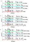
Figure 1
Figure 1
XRD pattern of Sr-nHAp (a), Zn-nHAp (b), Sr/Zn-nHAp (c).
Figure 2
Figure 2
FTIR spectra of Sr-nHAp (a), Zn-nHAp (b), Sr/Zn-nHAp (c).
Figure 3
Figure 3
TEM images of nHAp (a), Sr/Zn-1-nHAp (b), Sr/Zn-2.5-nHAp (c), Sr/Zn-4-nHAp (d).
Figure 4
Figure 4
A histogram showing the average particle size obtained from the TEM images of nHAp (a), Sr/Zn-1-nHAp (b), Sr/Zn-2.5-nHAp (c), Sr/Zn-4-nHAp (d).
Figure 5
Figure 5
XRD patterns of nHAp, PLGA, and scaffolds. Scaffold 1—PLGA-nHAp, Scaffold 2—PLGA-1% Sr/Zn-nHAp, Scaffold 3—PLGA-2.5% Sr/Zn-nHAp, Scaffold 4—PLGA-4% Sr/Zn-nHAp.
Figure 6
Figure 6
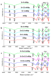
FTIR spectra of nHAp, PLGA, and PLGA-Sr/Zn-nHAp scaffolds. Scaffold 1—PLGA-nHAp, Scaffold 2—PLGA-1% Sr/Zn-nHAp, Scaffold 3—PLGA-2.5% Sr/Zn-nHAp, Scaffold 4—PLGA-4% Sr/Zn-nHAp.
Figure 7
Figure 7
Thermogravimetric analysis (TGA) of nHAp, PLGA, and PLGA-Sr/Zn-nHAp scaffolds. Scaffold 1—PLGA-nHAp, Scaffold 2—PLGA-1% Sr/Zn-nHAp, Scaffold 3—PLGA-2.5% Sr/Zn-nHAp, Scaffold 4—PLGA-4% Sr/Zn-nHAp.
Figure 8
Figure 8
SEM image demonstrating the porous structure of the bone scaffolds. PLGA-nHAp (a), PLGA-2.5% Sr/Zn nHAp (b), and PLGA-4% Sr/Zn nHAp (c).
Figure 9
Figure 9
The pore size of the scaffolds (Mean ± SEM). *** p < 0.001.
Figure 10
Figure 10
pH value (a) and weight loss (b) of composite scaffolds immersed in SBF at each time point. Scaffold 1—PLGA-nHAp, Scaffold 2—PLGA-1% Sr/Zn-nHAp, Scaffold 3—PLGA-2.5% Sr/Zn-nHAp, Scaffold 4—PLGA-4% Sr/Zn-nHAp.
Figure 11
Figure 11
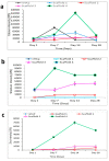
(ac) shows the release profile of calcium, strontium and zinc ions from the scaffolds after immersion in SBF. Scaffold 1—PLGA-nHAp, Scaffold 2—PLGA-1% Sr/Zn-nHAp, Scaffold 3—PLGA-2.5% Sr/Zn-nHAp, Scaffold 4—PLGA-4% Sr/Zn-nHAp. The scaffold 4 with higher zinc and strontium concentrations revealed the maximum ion release profiles for calcium (a), strontium (b) and zinc ions (c).
Figure 12
Figure 12
FTIR spectra of composite scaffolds. Scaffold 1—PLGA-nHAp (a), Scaffold 2—PLGA-1% Sr/Zn-nHAp (b), Scaffold 3—PLGA-2.5% Sr/Zn-nHAp (c), Scaffold 4—PLGA-4% Sr/Zn-nHAp (d) immersed in SBF for 2 weeks.
Figure 13
Figure 13
SEM image of crystal formation on the composite scaffold immersed in SBF (crystals shown by arrows).
Figure 14
Figure 14
TEM images of Scaffold 1—PLGA-nHAp (a), Scaffold 2—PLGA-1% Sr/Zn-nHAp (b), Scaffold 3—PLGA-2.5% Sr/Zn-nHAp (c), Scaffold 4—PLGA-4% Sr/Zn-nHAp (d) submerged in SBF for 2 weeks.
Figure 15
Figure 15
Mechanical testing of different fabricated scaffolds. (a) Stress-strain curve, (b) ultimate strength. Data is represented as the means ± SEM; n = 3. * = p < 0.05.

References

    1. Lichte P., Pape H.C., Pufe T., Kobbe P., Fischer H. Scaffolds for bone healing: Concepts, materials and evidence. Injury. 2011;42:569–573. doi: 10.1016/j.injury.2011.03.033. - DOI - PubMed
    1. Gentile P., Chiono V., Carmagnola I., Hatton P.V. An overview of poly(lactic-co-glycolic) Acid (PLGA)-based biomaterials for bone tissue engineering. Int. J. Mol. Sci. 2014;15:3640–3659. doi: 10.3390/ijms15033640. - DOI - PMC - PubMed
    1. Zhao D., Zhu T., Li J., Cui L., Zhang Z., Zhuang X., Ding J. Poly(lactic-co-glycolic acid)-based composite bone-substitute materials. Bioact. Mater. 2021;6:346–360. doi: 10.1016/j.bioactmat.2020.08.016. - DOI - PMC - PubMed
    1. Schemitsch E.H. Size Matters: Defining Critical in Bone Defect Size! J. Orthop. Trauma. 2017;31:S20–S22. doi: 10.1097/BOT.0000000000000978. - DOI - PubMed
    1. Yang L., Liu S., Fang W., Chen J., Chen Y. Poly(lactic-co-glycolic acid)-bioactive glass composites as nanoporous scaffolds for bone tissue engineering: In vitro and in vivo studies. Exp. Ther. Med. 2019;18:4874–4880. doi: 10.3892/etm.2019.8121. - DOI - PMC - PubMed