. 2022 Nov;141(11):1723-1738.
doi: 10.1007/s00439-022-02441-0. Epub 2022 Feb 28.

Usher syndrome type IV: clinically and molecularly confirmed by novel ARSG variants

Affiliations

Usher syndrome type IV: clinically and molecularly confirmed by novel ARSG variants

Hedwig M Velde et al. Hum Genet. 2022 Nov.

Abstract

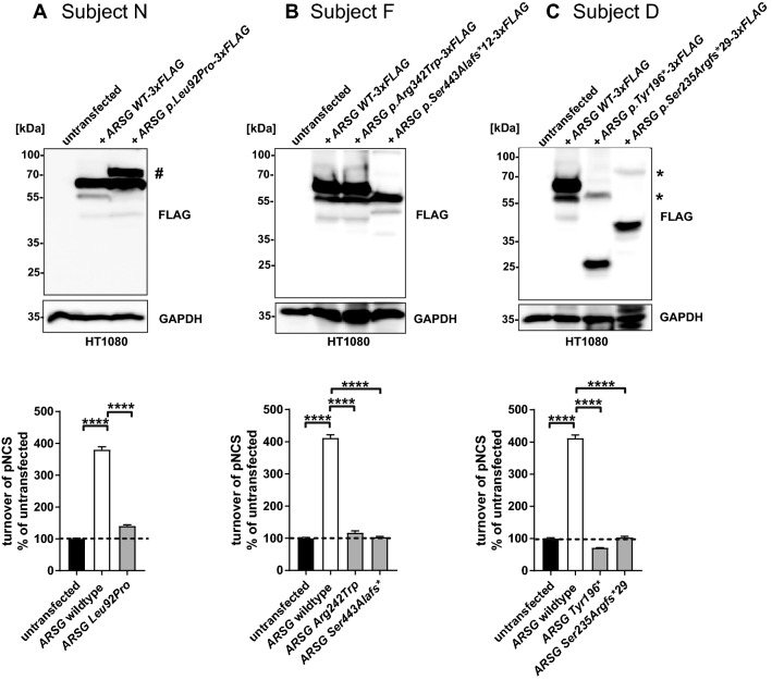
Usher syndrome (USH) is an autosomal recessively inherited disease characterized by sensorineural hearing loss (SNHL) and retinitis pigmentosa (RP) with or without vestibular dysfunction. It is highly heterogeneous both clinically and genetically. Recently, variants in the arylsulfatase G (ARSG) gene have been reported to underlie USH type IV. This distinct type of USH is characterized by late-onset RP with predominantly pericentral and macular changes, and late onset SNHL without vestibular dysfunction. In this study, we describe the USH type IV phenotype in three unrelated subjects. We identified three novel pathogenic variants, two novel likely pathogenic variants, and one previously described pathogenic variant in ARSG. Functional experiments indicated a loss of sulfatase activity of the mutant proteins. Our findings confirm that ARSG variants cause the newly defined USH type IV and support the proposed extension of the phenotypic USH classification.


Conflict of interest statement

The authors have no relevant financial or non-financial interests to disclose.

Figures

Fig. 1
Fig. 1
Audiologic features of USH IV compared to other USH types. A ARTA of USH IV compared to USH Ib (Wagenaar et al. 1999), USH IIa (Hartel et al. 2016), and USH III (Plantinga et al. 2004). Age (in years) is indicated at both ends of all lines. B Calculated age of onset of SNHL for USH IV based on individual PTA0.5–4 kHz and compared to USH IIa (Hartel et al. 2016). Black dots represent USH IV PTA0.5–4 kHz data; gray dots represent USH IIa PTA0.5–4 kHz data, connected dots represent longitudinal data when more than one audiogram was available for one subject. C Visualization of age of onset of RP (in gray) and SNHL (in black) of USH IV compared to published data for other USH types (Geng et al. ; Millan et al. ; Nisenbaum et al. ; Tsilou et al. 2002) (Table 2). dB HL decibel hearing level; RP retinitis pigmentosa; SNHL sensorineural hearing loss; y years
Fig. 2
Fig. 2
ARSG variants. A Schematic representation of the variability in location and type of novel and previously reported ARSG variants. The twelve ARSG exons are shown in dark blue squares. Protein domains are shown in light blue bars (Mistry et al. 2021). Variants that were identified in the present study are shown in bold. Truncating variants are presented above the exons, missense variants below. B Multiple sequence alignments of the sequence regions containing the two novel missense variants in 12 orthologs (Alamut Visual v.2.13). One-letter amino acid abbreviations are presented. Amino acids that do not correspond with the most conserved amino acid at their position are shown in red. The position of the variants are highlighted in blue. Both variants are located within the sulfatase protein domain
Fig. 3
Fig. 3
Results of minigene splice assays for ARSG variants c.1212 + 1G > A and c.1024C > T. In vitro splice assays were performed in HEK293T cells to validate the predicted splice defects. A A canonical splice site variant (c.1212 + 1G > A) was detected in ARSG (subject N) and was predicted by SpliceAI to cause skipping of exon 10 due to a donor splice site loss at −1nt (0.99/1) and acceptor splice site loss at −121nt (0.78/1). A minigene splice assay revealed the use of an alternative splice donor site at + 13nt in intron 10 which leads to an out of frame elongation of exon 10 (NM_014960.5:c.1212 + 1G > A r.1212_1213ins[a;1212 + 2_1212 + 13] NP_055775.2:p.(Val405Ilefs*41)). B A missense variant (c.1024C > T) was predicted by SpliceAI to cause skipping of exon 9 due to a donor splice site loss at + 67nt (0.15/1) and acceptor splice site loss at −41nt (0.25/1). A minigene splice assay did not confirm this and demonstrated no effect on splicing (NM_014960.5:c.1024C > T r.1024c > u NP_055775.2:p.(Arg342Trp)). Bp base pair; WT wildtype; MT mutant
Fig. 4
Fig. 4
Functional analysis of ARSG variants identified in subjects N, F, and D. A Subject N: Immunoblot analysis of untransfected or stably transfected HT1080 cells with plasmids coding for 3xFLAG-tagged wildtype (WT) ARSG, along with the ARSG variant p.(Leu92Pro). An additional ARSG p.(Leu92Pro)-specific band is labeled with an #. Other bands in the figure are likely to be caused by limited proteolytic cleavage of ARSG. Total sulfatase activity of cell lysates from untransfected cells, or transfected cells expressing WT ARSG or ARSG with the pathogenic variant p.(Leu92Pro) against the artificial substrate P-nitrocatechol sulfate (pNCS). N = 3 replicates. Unpaired, two-tailed t-test, Mean ± SEM of the replicates. B Subject F: Immunoblot analysis of untransfected or stably transfected HT1080 cells with plasmids coding for 3xFLAG-tagged WT ARSG, along with the pathogenic variants p.(Arg342trp) and p.(Ser443Alafs*12). Other bands in the figure are likely to be caused by limited proteolytic cleavage of ARSG. Total sulfatase activity of cell lysates from untransfected cells, or from cells expressing WT ARSG or the pathogenic variants p.(Arg342Trp) and p.(Ser443Alafs*12) against the artificial substrate pNCS. N = 3 replicates. Unpaired, two-tailed t-test, Mean ± SEM of the replicates. C Subject D: Immunoblot analysis of untransfected or stably transfected HT1080 cells with plasmids coding for 3xFLAG-tagged WT ARSG, along with the pathogenic variants p.(Tyr196*) and p.(Ser235Argfs*29). High-molecular weight bands indicating possibly SDS-resistant dimers of the truncated proteins are labeled with an *. Other bands in the figure are likely to be caused by limited proteolytic cleavage of ARSG. Total sulfatase activity of cell lysates from untransfected cells, or cells expressing WT ARSG or the pathogenic variants p.(Tyr196*) and p.(Ser235Argfs*29) against the artificial substrate pNCS. N = 3 replicates. Unpaired, two-tailed t-test, Mean ± SEM of the replicates. Antibodies in all panels are against FLAG and GAPDH, used as a loading control. pNCS, P-nitrocatechol sulfate; SEM, standard error of the mean

References

    1. Abad-Morales V, Navarro R, Bures-Jelstrup A, Pomares E. Identification of a novel homozygous ARSG mutation as the second cause of Usher syndrome type 4. Am J Ophthalmol Case Rep. 2020;19:100736. doi: 10.1016/j.ajoc.2020.100736. - DOI - PMC - PubMed
    1. Abitbol M, Thibaud JL, Olby NJ, Hitte C, Puech JP, Maurer M, et al. A canine arylsulfatase G (ARSG) mutation leading to a sulfatase deficiency is associated with neuronal ceroid lipofuscinosis. Proc Natl Acad Sci USA. 2010;107(33):14775–14780. doi: 10.1073/pnas.0914206107. - DOI - PMC - PubMed
    1. Abou Tayoun AN, Pesaran T, DiStefano MT, Oza A, Rehm HL, Biesecker LG, et al. Recommendations for interpreting the loss of function PVS1 ACMG/AMP variant criterion. Hum Mutat. 2018;39(11):1517–1524. doi: 10.1002/humu.23626. - DOI - PMC - PubMed
    1. Ahmed ZM, Riazuddin S, Bernstein SL, Ahmed Z, Khan S, Griffith AJ, et al. Mutations of the protocadherin gene PCDH15 cause Usher syndrome type 1F. Am J Hum Genet. 2001;69(1):25–34. doi: 10.1086/321277. - DOI - PMC - PubMed
    1. Ahmed ZM, Riazuddin S, Ahmad J, Bernstein SL, Guo Y, Sabar MF, et al. PCDH15 is expressed in the neurosensory epithelium of the eye and ear and mutant alleles are responsible for both USH1F and DFNB23. Hum Mol Genet. 2003;12(24):3215–3223. doi: 10.1093/hmg/ddg358. - DOI - PubMed