. 2022 May;91(5):682-696.
doi: 10.1002/ana.26338. Epub 2022 Mar 12.

Distinct Roles of Rodent Thalamus and Corpus Callosum in Seizure Generalization

Affiliations

Distinct Roles of Rodent Thalamus and Corpus Callosum in Seizure Generalization

Anastasia Brodovskaya et al. Ann Neurol. 2022 May.

Abstract

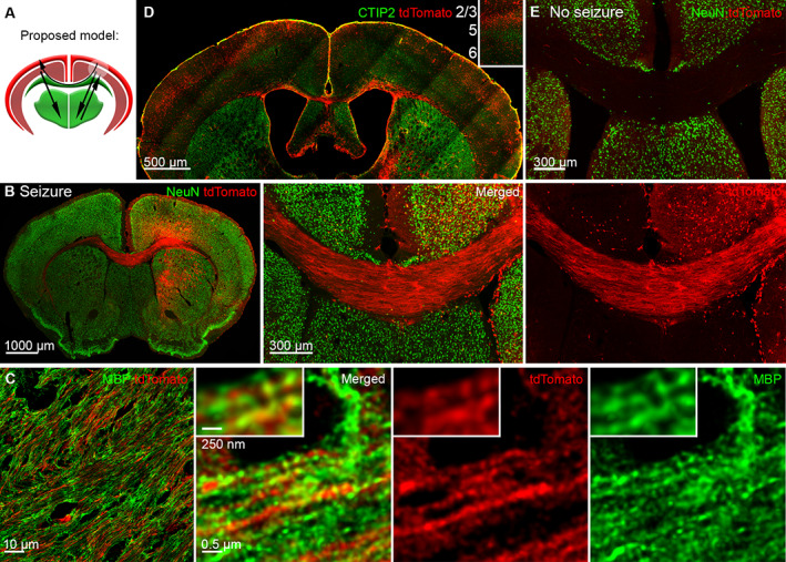
Objective: Bilateral synchronous cortical activity occurs during sleep, attention, and seizures. Canonical models place the thalamus at the center of bilateral cortical synchronization because it generates bilateral sleep spindle oscillations and primarily generalized absence seizures. However, classical studies suggest that the corpus callosum mediates bilateral cortical synchronization.

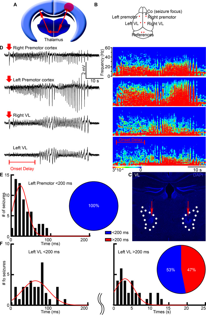
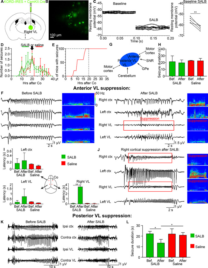
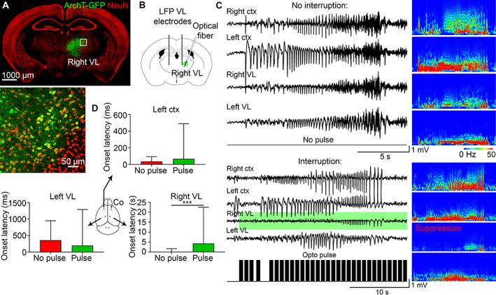
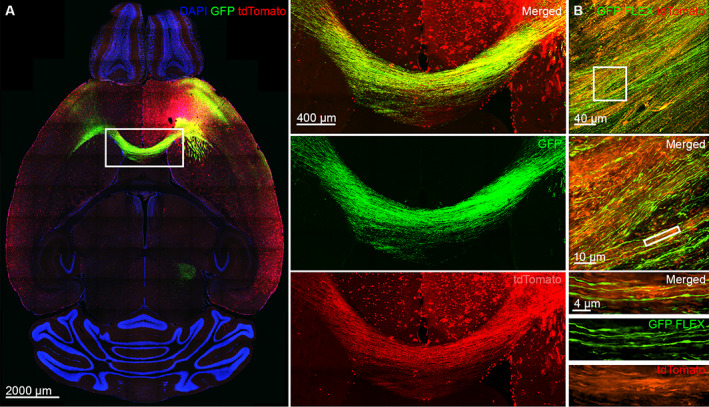
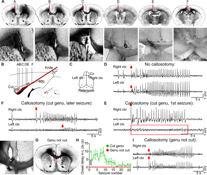
Methods: We mapped the spread of right frontal lobe-onset, focal to bilateral seizures in mice and modified it using chemo and optogenetic suppression of motor thalamic nucleus and corpus callosotomy.

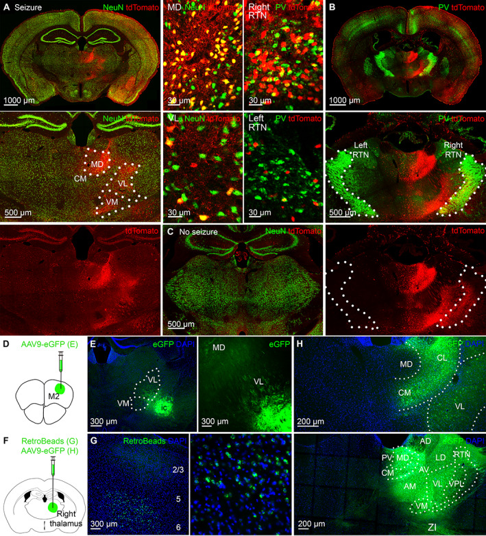
Results: Seizures from the right cortex spread faster to the left cortex than to the left thalamus. The 2 thalami have minimal monosynaptic commissural connections compared to the massive commissure corpus callosum. Chemogenetic and closed-loop optogenetic inhibition of the right ventrolateral thalamic nucleus did not alter inter-hemispheric seizure spread. However, anterior callosotomy delayed bilateral seizure oscillations.

Interpretation: Thalamocortical oscillations amplify focal onset motor seizures, and corpus callosum spreads them bilaterally. ANN NEUROL 2022;91:682-696.


Conflict of interest statement

The authors report no conflicts of interests.

Figures

FIGURE 1
FIGURE 1
Seizures spread faster to the contralateral cortex than to the contralateral thalamus. (A) A schematic illustrates the classic centrencephalic model. Red arrows represent a bilateral spread of neuronal activity. The red circle is the seizure focus. (B) A schematic shows cobalt (grey rectangle) and LFP electrode placement (red dots). (C) We marked the electrode tips' location in the VL (dotted lines) by creating an electrical lesion at the end of the recordings (red arrows). (D) LFP microelectrodes record simultaneously from the bilateral premotor cortex and bilateral thalamic nucleus VL with the corresponding power spectrums. The seizure focus was in the right premotor cortex. Red arrows indicate seizure onset (amplitude twice the baseline); the red bar indicates seizure onset delay in the left VL. (E) Frequency distribution histogram shows seizure onset times in the left premotor cortex after seizure beginning in the right premotor cortex. Pie chart: 100% of seizures required < 200 ms (blue) to arrive at the left premotor cortex. (F) Frequency distribution histogram shows seizure onset times in the left VL. Pie chart: 53% of seizures required < 200 ms (blue) to arrive at the left VL from the right premotor cortex, and 47% required > 200 ms (red). LFP = local field potential; VL = ventrolateral.
FIGURE 2
FIGURE 2
The activated ipsilateral thalamus sends minimal projections to the contralateral thalamus. (A) Only the right thalamus strongly expressed tdTomato. Dotted lines indicate nuclear boundaries. On the right are enlarged images of cellular tdTomato expression in the MD and VL immunolabeled for NeuN (green). (B) The right reticular thalamic nucleus (RTN) expressed more tdTomato than the left. RTN is immunolabeled for parvalbumin (PV, green). On the left are enlarged images of cellular tdTomato expression in the right and left RTN. (C) Minimal tdTomato expression is in the thalamus of steel wire implanted mice without seizures. (D) AAV9‐eGFP injection is in the right premotor cortex for E. (E) The right motor cortex projects to the right motor thalamic nuclei VM and VL. (F) AAV9 eGFP or RetroBeads injection is in the right VL for G and H. (G) The right motor cortex sends projections to the right thalamus from deep cortical layers 5 and 6 after retrograde RetroBeads injection in the right VL as indicated in F. (H) The right thalamic nuclei have minimal monosynaptic projections to the left thalamic nuclei. CM = centromedian; MD = mediodorsal; VL = ventrolateral; VM = ventromedial.
FIGURE 3
FIGURE 3
Chemogenetic suppression of the ipsilateral VL does not change the onset latency in the contralateral VL or contralateral cortex. (A) KORD/CamKII.Cre was injected in the right VL. (B) Patch‐clamp recordings were done on KORD‐expressing neurons in the right VL. (C) Resting membrane potential (mV) of transduced cells before and after SALB application, where the left graph is repeated recordings from a single cell, and the right graph is the average for 5 mice. (D) Mean number of seizures in C57Bl/6 mice after cobalt insertion in C57Bl/6 mice that were injected with SALB (green) or saline (red) at 20 hours after Co (black arrow) at the peak of seizures. (E) One hundred percent of all mice developed seizures by 20 hours after Co. (F) LFPs of a seizure recorded before and after SALB injection. Power was suppressed in the right VL (red box). (G) A schematic illustrates anatomical projections to the anterior and posterior VL. (H) Mean seizure duration (seconds) remained the same before (16–20 hours after Co) and after (20–24 hours after Co) SALB or saline injection. (I) Seizure onset was delayed only in the right VL after SALB injection but not in the left cortex or left VL. (J) Right cortex was suppressed after right VL suppression (in 6 out of 11 mice). (K, L) Seizure duration decreased after posterior VL suppression. Co = cobalt; VL = ventrolateral; SALB = salvinorin B.
FIGURE 4
FIGURE 4
Closed‐loop optogenetic inhibition of the ipsilateral VL does not change the onset latency in the contralateral VL or contralateral cortex. (A) ArchT‐GFP expressed in the right VL. (B) AAV9 CamKII‐ArchT‐GFP was injected in the right VL. (C) Seizures that were uninterrupted (no pulse) and interrupted (with opto pulse [black bars], green background in the right VL) in the same mouse with the corresponding power spectrums. (D) Seizure onset was delayed only in the right VL after pulse activation, whereas onset latency in the left cortex or left VL did not change. LFP = local field potential; VL = ventrolateral.
FIGURE 5
FIGURE 5
Focal to bilateral seizures activate the corpus callosum. (A) The proposed model of focal to bilateral seizures, where black arrows indicate seizure spread across the corpus callosum and to the thalamus, while a white circle in the right cortex indicates the seizure focus. (B) The corpus callosum strongly expressed tdTomato after focal to bilateral tonic‐clonic seizures. (C) SP8 super‐resolution Lightning microscopy images tdTomato positive axons expressing myelin basic protein (MBP; green) in the corpus callosum. (D) Strong activation of layers 2 and 3 in the right and left cortex, immunolabeled for CTIP2 (green) that marks layers 5 and 6, separating superficial from deep cortical layers. (E) Steel wire implanted control mice without seizures did not express tdTomato in the corpus callosum.
FIGURE 6
FIGURE 6
The seizure focus sends extensive direct projections across the corpus callosum contralaterally. (A) The tdTomato and AAV9 GFP colocalized in the axons of the corpus callosum that connects the seizure focus with the contralateral premotor cortex. The white rectangle area is magnified on the right. (B) Top: A confocal image of Cre‐driven AAV9 GFP expression in the corpus callosum of TRAP2 mice. Middle: Super‐resolution SoRa image of the callosal axons expressing Cre‐driven AAV9 GFP with a magnified view.
FIGURE 7
FIGURE 7
Anterior callosotomy prevents bilateral seizure spread. (A) Sequential light images of the coronal sections show callosotomy (red arrows, magnified views) from anterior to posterior cortex (A–F, relative brain locations in B). (B) A schematic of the callosotomy, sagittal view, illustrates knife placement (red), cut genu of the corpus callosum (gcc), and uncut splenium (scc). (C) Cobalt and EEG electrodes (red dots) were positioned in the right and left anterior and posterior cortices. (D) EEG of a seizure that was recorded in mice without callosotomy. Red arrows indicate seizure onset. (E) Representative EEG of a first seizure that was recorded after callosotomy in mice with cut genu with no seizure recorded in the left premotor cortex (red rectangle). (F) Onset oflater seizures was delayed in mice with cut genu. (G) Light image of the callosotomy where only the body of the corpus callosum was cut and the genu remained intact (red arrow). (H) Seizure onset delay (seconds) in the left premotor cortex (L ctx) after callosotomy in mice with cut genu (green) and in mice with body callosotomy and intact genu (red). (I) Mice with body callosotomy and intact genu had immediate seizure onset in the left premotor cortex just like the mice without callosotomy. EEG = electroencephalography.

Comment in

  • Focal Seizures Cruise the Callosal Highway.
    Stafstrom CE. Stafstrom CE. Epilepsy Curr. 2022 Sep 22;22(6):381-383. doi: 10.1177/15357597221125220. eCollection 2022 Nov-Dec. Epilepsy Curr. 2022. PMID: 36426194 Free PMC article. No abstract available.

References

    1. Steriade M, McCormick DA, Sejnowski TJ. Thalamocortical oscillations in the sleeping and aroused brain. Science 1993;262:679–685. - PubMed
    1. Paz JT, Davidson TJ, Frechette ES, et al. Closed‐loop optogenetic control of thalamus as a new tool to interrupt seizures after cortical injury. Nat Neurosci 2013;16:64–70. - PMC - PubMed
    1. Blumenfeld H. The thalamus and seizures. Arch Neurol 2002;59:135–137. - PubMed
    1. Huguenard JR, Prince DA. Basic mechanisms of epileptic discharges in the thalamus. In: Steriade M, Jones EG, McCormick DA, eds. Thalamus. Amsterdam, Netherlands: Elsevier, 1997:295‐330.
    1. Penfield W, Jasper HH. Epilepsy and the Functional Anatomy of the Human Brain. Boston: Little, Brown, 1954.

Publication types