

Computed Tomography Findings in Progressive Massive Fibrosis: Analyses of 90 Cases
- PMID: 35226653
- PMCID: PMC8902743
- DOI: 10.23749/mdl.v113i1.12303
Computed Tomography Findings in Progressive Massive Fibrosis: Analyses of 90 Cases
Abstract
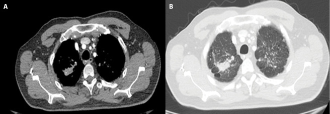
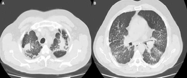
Purpose: Radiological identification of lung masses in patients with pneumoconiosis is difficult. The aim of the study is to characterize Computed Tomography (CT) findings of Progressive Massive Fibrosis (PMF).
Methods: The data of pneumoconiosis patients, who were diagnosed with PMF between 2014-2019 in a tertiary hospital, were collected. Demographic data, work-related data, Pulmonary Function Test results and radiological imaging results were gathered. Separate evaluations were made for the right and left lungs, and the CT findings and measurement results were recorded.
Results: In 90% of our cases, PMF lesions were bilaterally located. Eighty-eight point five percent of the unilateral lesions were located in the upper lobe of the right lung. Enlarged lymph nodes were found in 83.3% and calcification was found in the lymph nodes in 63% of the cases. Band structures extending between the PMF lesion and the adjacent pleura were observed in 86% of the cases, and invagination in the lung parenchyma adjacent to the PMF was observed in 80% of the cases.
Conclusion: In general, our findings were consistent with the radiologically defined PMF. In addition, pleural findings, which are not frequently studied in the literature except for asbestosis, were also described in the study.
Methods: The data of pneumoconiosis patients, who were diagnosed with PMF between 2014-2019 in a tertiary hospital, were collected. Demographic data, work-related data, PFT results and radiological imaging results were noted. Separate evaluations were made for the right and left lungs, and the CT findings and measurement results were recorded.
Results: In 90% of our cases, PMF lesions were bilaterally located. 88.8% of the unilateral lesions were located in the upper lobe of the right lung. Enlarged lymph nodes were found in 83.3% and calcification was found in the lymph nodes in 63% of the cases. Band structures extending between the PMF lesion and the adjacent pleura were observed in 86% of the cases, and invagination in the lung parenchyma adjacent to the PMF was observed in 80% of the cases.
Conclusion: In general, our findings were consistent with the radiologically defined PMF. In addition, pleural findings, which are not frequently studied in the literature except asbestosis, were also described in the study.
Figures
References
-
- Organization IL. Fourth International Pneumoconiosis Conference; Geneva, Switzerland; 1971. Report of the Working Party on the Definition of Pneumoconiosis.
-
- dos Santos Antao VC, Pinheiro GA, Terra-Filho M, Kavakama J, Müller NL. High-resolution CT in silicosis: correlation with radiographic findings and functional impairment. J Comput Assist Tomogr. 2005;29(3):350–6. - PubMed
-
- Bergin C, Muller N, Vedal S, Chan-Yeung M. CT in silicosis: correlation with plain films and pulmonary function tests. Am J Roentgenol. 1986;146(3):477–83. - PubMed
-
- Bégin R, Ostiguy G, Fillion R, Colman N. Computed Tomography Scan in the Early Detection of Silicosis1, 2. Am Rev Respir Dis. 1991;144:697–705. - PubMed
MeSH terms
LinkOut - more resources
Full Text Sources
Medical
