. 2022 Feb 28;18(2):e1009912.
doi: 10.1371/journal.pcbi.1009912. eCollection 2022 Feb.

Objective quantification of nerves in immunohistochemistry specimens of thyroid cancer utilising deep learning

Affiliations

Objective quantification of nerves in immunohistochemistry specimens of thyroid cancer utilising deep learning

Indriani P Astono et al. PLoS Comput Biol. .

Abstract

Accurate quantification of nerves in cancer specimens is important to understand cancer behaviour. Typically, nerves are manually detected and counted in digitised images of thin tissue sections from excised tumours using immunohistochemistry. However the images are of a large size with nerves having substantial variation in morphology that renders accurate and objective quantification difficult using existing manual and automated counting techniques. Manual counting is precise, but time-consuming, susceptible to inconsistency and has a high rate of false negatives. Existing automated techniques using digitised tissue sections and colour filters are sensitive, however, have a high rate of false positives. In this paper we develop a new automated nerve detection approach, based on a deep learning model with an augmented classification structure. This approach involves pre-processing to extract the image patches for the deep learning model, followed by pixel-level nerve detection utilising the proposed deep learning model. Outcomes assessed were a) sensitivity of the model in detecting manually identified nerves (expert annotations), and b) the precision of additional model-detected nerves. The proposed deep learning model based approach results in a sensitivity of 89% and a precision of 75%. The code and pre-trained model are publicly available at https://github.com/IA92/Automated_Nerves_Quantification.


Conflict of interest statement

The authors have declared that no competing interests exist.

Figures

Fig 1
Fig 1. Examples of variation in the size of nerves.
Fig 2
Fig 2. Examples of variation in the appearance of nerves.
Fig 3
Fig 3. Examples of non-specific staining.
Fig 4
Fig 4. End-to-end process flowchart.
Fig 5
Fig 5. Post-processing flowchart.
Fig 6
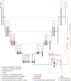
Fig 6. Proposed network architecture with augmented classification structure.
Fig 7
Fig 7. Block diagram of the proposed network with augmented classification structure.
Fig 8
Fig 8. CNN training process.
Fig 9
Fig 9. ROI extraction flowchart.
Fig 10
Fig 10. Excluded positive training data samples due to label artifacts from the colour filter.
The red box surrounds the actual nerve.
Fig 11
Fig 11. Image patches conversion flowchart.
Fig 12
Fig 12. Examples of the positive and negative training data samples.
Fig 13
Fig 13
Sensitivity of the APR-CF and APR-CNN in the detection of the expert annotated nerves (blue). Precision of APR-CF and APR-CNN in the detection of nerves (orange). The × symbol in the box indicates the mean value, while the line in the box indicates the median value. The ∘ represents an outlier, the box represents the interquartile range and the whiskers represent the upper and lower extreme, excluding the outliers.
Fig 14
Fig 14. Examples of nerves detected (i.e. true positives, TPa) by the proposed CNN based approach.
Each image patch contains one prediction instance indicated by a green box.

References

    1. Boilly B, Faulkner S, Jobling P, Hondermarck H. Nerve Dependence: From Regeneration to Cancer. Cancer Cell. 2017;31(3):342–354. doi: 10.1016/j.ccell.2017.02.005 - DOI - PubMed
    1. Magnon C, Hall SJ, Lin J, Xue X, Gerber L, Freedland SJ, et al.. Autonomic nerve development contributes to prostate cancer progression. Science. 2013;341(6142):1236361. doi: 10.1126/science.1236361 - DOI - PubMed
    1. Pundavela J, Roselli S, Faulkner S, Attia J, Scott RJ, Thorne RF, et al.. Nerve fibers infiltrate the tumor microenvironment and are associated with nerve growth factor production and lymph node invasion in breast cancer. Mol Oncol. 2015;9(8):1626–35. doi: 10.1016/j.molonc.2015.05.001 - DOI - PMC - PubMed
    1. Iwasaki T, Hiraoka N, Ino Y, Nakajima K, Kishi Y, Nara S, et al.. Reduction of intrapancreatic neural density in cancer tissue predicts poorer outcome in pancreatic ductal carcinoma. Cancer Sci. 2019;110(4):1491–1502. doi: 10.1111/cas.13975 - DOI - PMC - PubMed
    1. Rowe CW, Dill T, Griffin N, Jobling P, Faulkner S, Paul JW, et al.. Innervation of papillary thyroid cancer and its association with extra-thyroidal invasion. Scientific Reports. 2020;10(1):1539. doi: 10.1038/s41598-020-58425-5 - DOI - PMC - PubMed

Publication types