. 2022 May 5;139(18):2770-2781.
doi: 10.1182/blood.2021014267.

The SUMOylation inhibitor subasumstat potentiates rituximab activity by IFN1-dependent macrophage and NK cell stimulation

Affiliations

The SUMOylation inhibitor subasumstat potentiates rituximab activity by IFN1-dependent macrophage and NK cell stimulation

Akito Nakamura et al. Blood. .

Abstract

Small ubiquitin-like modifier (SUMO) is a member of a ubiquitin-like protein superfamily. SUMOylation is a reversible posttranslational modification that has been implicated in the regulation of various cellular processes including inflammatory responses and expression of type 1 interferons (IFN1). In this report, we have explored the activity of the selective small molecule SUMOylation inhibitor subasumstat (TAK-981) in promoting antitumor innate immune responses. We demonstrate that treatment with TAK-981 results in IFN1-dependent macrophage and natural killer (NK) cell activation, promoting macrophage phagocytosis and NK cell cytotoxicity in ex vivo assays. Furthermore, pretreatment with TAK-981 enhanced macrophage phagocytosis or NK cell cytotoxicity against CD20+ target cells in combination with the anti-CD20 antibody rituximab. In vivo studies demonstrated enhanced antitumor activity of TAK-981 and rituximab in CD20+ lymphoma xenograft models. Combination of TAK-981 with anti-CD38 antibody daratumumab also resulted in enhanced antitumor activity. TAK-981 is currently being studied in phase 1 clinical trials (#NCT03648372, #NCT04074330, #NCT04776018, and #NCT04381650; www.clinicaltrials.gov) for the treatment of patients with lymphomas and solid tumors.


Figures

None
Graphical abstract
Figure 1
Figure 1
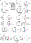
TAK-981 treatment enhances M1 polarization and NK activation in an IFN1-dependent manner. (A) Amount of IFNβ and IP-10 protein in the supernatant of 1 μM TAK-981- or 10 kU/mL IFNα2-treated hMDM at indicated time points (mean with SD; n = 3 biological replicates, 2-tailed unpaired Welch's t test). (B) Western blots of indicated proteins in TAK-981–treated hMDM at 24 hours. (C) Representative flow cytometry histogram and median fluorescence intensity (MFI) of indicated proteins in 50 ng/mL IFNγ (M1 polarizing stimulation)-, 20 ng/mL IL-4 (M2 polarizing stimulation)-, and/or 1 μM TAK-981–treated hMDM at 48 hours (mean with SD; n = 3 biological replicates, 2-tailed unpaired Welch's t test). (D) MFI of indicated proteins in 20 μg/mL anti-IFNAR2 antibody (aIFNAR2)-, 1 μM TAK-981-, and/or 10 kU/mL IFNα2-treated hMDM at 48 hours (mean with SD; n = 3 biological replicates, 2-tailed unpaired Welch's t test). (E) mRNA expression of indicated genes in 20 μg/mL aIFNAR2- and/or 1 μM TAK-981–treated hMDM at 24 hours (mean with SD; n = 3 biological replicates, 2-tailed unpaired Welch's t test). (F) Representative flow cytometry histogram and MFI of CD206 in 20 ng/mL IL-4- and/or 1 μM TAK-981–treated hMDM at 48 hours (mean with SD; n = 3 biological replicates, 2-tailed unpaired Welch's t test). (G) mRNA expression of CCL22 in 20 ng/mL IL-4- and/or 1 μM TAK-981–treated hMDM at 24 hours (mean with SD; n = 3 biological replicates, 2-tailed unpaired Welch's t test). (H) mRNA expression of IP-10 and CD69 in 20 μg/mL aIFNAR2- and/or 1 μM TAK-981–treated human NK cells at 24 hours (mean with SD; n = 3 biological replicates, 2-tailed unpaired Welch's t test). (I) Representative flow cytometry histogram and percentage of CD69+ cells or MFI of CD69 in 1 μM TAK-981- or 10 kU/mL IFNα2-treated human NK cells at 24 hours (mean with SD; n = 3 biological replicates, 2-tailed unpaired Welch's t test). *P < .05, **P < .01, ***P < .001, ****P < .0001. Experiment was repeated using hMDM or human NK cells generated from at least 2 healthy PBMC donors. mRNA, messenger RNA; N.S., not significant (P > .05); PBMC, peripheral blood mononuclear cells; SD, standard deviation.
Figure 2
Figure 2
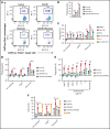
TAK-981 treatment augments rituximab-mediated macrophage phagocytosis and NK cell cytotoxicity in an IFN1-dependent manner. (A) Representative flow cytometry scatter plots. Macrophages are defined as CellTrace Red+ events and Daudi cells as CellTrace Violet+ events. Percent of CellTrace Red+Violet+ in CellTrace Violet+ events corresponds to percent of phagocytosed Daudi cells in total Daudi cells. (B) Phagocytic activity of 1 μM TAK-981–treated hMDM against Daudi cells in the presence of 1 μg/mL human IgG1 (hIgG1) or rituximab at an effector to target (E:T) ratio of 1:1 (mean with SD; n = 3 biological replicates, 2-tailed unpaired Welch's t test). (C) Phagocytic activity of 50 ng/mL IFNγ-, 20 ng/mL IL-4-, and/or 1 μM TAK-981–treated hMDM against Daudi-KILR cells in the presence of hIgG1 or rituximab at an E:T ratio of 4:1 (mean with SD; n = 3 biological replicates, 2-tailed unpaired Welch's t test). (D) Phagocytic activity of 20 μg/mL aIFNAR2- and/or 1 μM TAK-981–treated hMDM against Daudi-KILR cells in the presence of human IgG1 or rituximab at an E:T ratio of 4:1 (mean with SD; n = 3 biological replicates, 2-tailed unpaired Welch's t test). (E) Phagocytic activity of 20 μg/mL aIFNAR2- and/or 1 μM TAK-981–treated hMDM against Daud-KILR cells in the presence of 1 μg/mL human IgG1, daratumumab, or 1 μg/mL rituximab at an E:T ratio of 4:1 (mean with SD; n = 3 biological replicates, 2-tailed unpaired Welch's t test). (F) Cytotoxicity of 20 μg/ml aIFNAR2-, 1 μM TAK-981-, and/or 10 kU/mL IFNα2-treated human NK cells against Daudi-KILR cells in the presence of 1 μg/mL human IgG1 or 1 μg/mL rituximab at an E:T ratio of 4:1 (mean with SD; n = 3 biological replicates, 2-tailed unpaired Welch's t test). *P < .05, **P < .01, ***P < .001. Experiment was repeated using hMDM or human NK cells generated from at least 2 healthy PBMC donors. SD, standard deviation; N.S., not significant (P > .05).
Figure 3
Figure 3
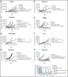
Antitumor activity of TAK-981 combined with monoclonal antibodies in xenograft models. (A) Tumor volumes in Daudi-bearing mice treated with TAK-981 (7.5 mg/kg, biweekly), rituximab (3 mg/kg, weekly), and/or daratumumab (7.5 mg/kg, biweekly) as indicated for 3 weeks (mean with SD; n = 8 mice per group in the representative study, 2-tailed unpaired Welch's t test). The P value of the synergy score was P < .001. (B) Tumor volumes in OCI-Ly10–bearing mice treated with TAK-981 (7.5 mg/kg, biweekly) and/or rituximab (1 mg/kg, weekly) as indicated for 2 weeks (mean with SD; n = 8 mice per group in the representative study, 2-tailed unpaired Welch's t test). The P value of the synergy score was P < .001. (C) Tumor volumes in OCI-Ly10–bearing mice treated with TAK-981 (7.5 mg/kg, biweekly), obinutuzumab (Obi, 1 mg/kg, weekly), and/or rituximab (Rit, 1 mg/kg, weekly) as indicated for 2 weeks (mean with SD; n = 8 mice per group in a single experiment, 2-tailed unpaired Welch's t test). (D) Tumor volumes in TMD8-bearing mice treated with TAK-981 (15 mg/kg, biweekly) and/or rituximab (3 mg/kg, weekly) as indicated for 2 weeks (mean with SD; n = 8 mice per group in the representative study, 2-tailed unpaired Welch's t test). The P value of the synergy score was P = .08 (additive combination effect). (E) Tumor volumes in PHTX-166L–bearing mice treated with TAK-981 (5 mg/kg, weekly) and/or rituximab (5 mg/kg, weekly) as indicated for 2 weeks (mean with SD; n = 6 mice per group in the representative study, 2-tailed unpaired Welch's t test). The P value of the synergy score was P < .05. (F) Tumor volumes in LP-1–bearing mice treated with TAK-981 (7.5 mg/kg, biweekly) and/or daratumumab (2.5 mg/kg, biweekly) as indicated for 5 weeks (mean with SD; n = 8 mice per group in the representative study, 2-tailed unpaired Welch's t test). The P value of the synergy score was P < .05. (G) Tumor volumes in OCI-Ly10–bearing mice treated with TAK-981 (7.5 mg/kg, biweekly), rituximab (3 mg/kg, weekly), and/or rituximab with LALA-PG mutations (3 mg/kg, weekly) as indicated for 2 weeks (mean with SD; n = 8 mice per group in a single experiment, 2-tailed unpaired Welch's t test). (H) Tumor volumes in OCI-Ly10–bearing mice were treated with TAK-981 (7.5 mg/kg, biweekly), rituximab (1 mg/kg, weekly), clodronate liposome (200 µL per mouse at a first dose and 100 µL per mouse at subsequent doses, biweekly), and/or anti-asialo GM1 (aASGM1, 0.25 mg per mouse, weekly) as indicated for 2 weeks (mean with SD; n = 8 mice per group in the representative study). Bar graphs show percent change in tumor volumes from baseline on the indicated day of measurement (individual value; n = 8 mice per group in the representative study, 2-tailed unpaired Welch's t test). *P < .05, **P < .01, ***P < .001. The number of mice achieved CR per the number of total mice in the group is shown. At least 2 similar experiments were performed unless otherwise specified. SD, standard deviation; N.S., not significant (P > .05).
Figure 4
Figure 4
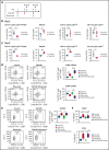
TAK-981 treatment increases M1-like/M2-like ratio in TAM and activates tumor-infiltrated NK cells in OCI-Ly10 xenografts. (A) Schematic diagram of study design. Tumors from 7.5 mg/kg TAK-981 (administered on day 1, 4, and 8)- and/or 1 mg/kg rituximab (administered on day 1 and 8)- treated OCI-Ly10 tumor-bearing mice on day 5 or day 9 were analyzed. (B) Proportion of CD11b+Ly6GLy6ClowF4/80+ TAM, NKp46+ NK cells, CD11b+Ly6GLy6Chigh monocytes, or CD11b+Ly6G+Ly6Cint neutrophils on day 5 (mean with SD; n = 5 mice per group in the representative study, 2-tailed unpaired Welch's t test). (C) Proportion of CD11b+Ly6GLy6ClowF4/80+ TAM, NKp46+ NK cells, CD11b+Ly6GLy6Chigh monocytes, or CD11b+Ly6G+Ly6Cint neutrophils on day 9 (mean with SD; n = 5 mice per group, 2-tailed unpaired Welch's t test). (D) Left, representative flow cytometry scatter plots of CD86/CD206. Right, proportion of M1-like (CD86+CD206) TAM or M2-like (CD86CD206+) TAM in tumors on day 5. (E) Left, representative flow cytometry scatter plots of FCGR4/FSC-H. Right, proportion of FCGR4+ TAM in tumors on day 5. (F) Proportion of CD69+ or IFNγ+ NK cells in tumors on day 5. Box plot center, box, and whiskers correspond to median, IQR (interquartile range), and 1.5 times IQR, respectively (n = 5 mice per group in the representative study, 2-tailed unpaired Welch's t test). Two similar experiments were performed. SD, standard deviation.
Figure 5
Figure 5
Schematic model of TAK-981–mediated antitumor innate immune responses through IFN1 activation. TAK-981 treatment enhances M1 macrophage polarization and NK cell activation through IFN1 signaling, leading to enhanced macrophage phagocytosis and NK cell cytotoxicity against tumor cells, particularly in the presence of therapeutic monoclonal antibodies like rituximab. The induction of activating FCGR levels in macrophages following TAK-981 treatment may also contribute to the enhanced macrophage phagocytosis.

Comment in

  • (SUMO)-wrestling with rituximab.
    Cragg MS. Cragg MS. Blood. 2022 May 5;139(18):2728-2730. doi: 10.1182/blood.2022015912. Blood. 2022. PMID: 35511190 No abstract available.

Similar articles

Cited by

References

    1. Reff ME, Carner K, Chambers KS, et al. Depletion of B cells in vivo by a chimeric mouse human monoclonal antibody to CD20. Blood. 1994;83(2):435–445. - PubMed
    1. Pierpont TM, Limper CB, Richards KL. Past, present, and future of rituximab-the world's first oncology monoclonal antibody therapy. Front Oncol. 2018;8:163. - PMC - PubMed
    1. Cittera E, Leidi M, Buracchi C, et al. The CCL3 family of chemokines and innate immunity cooperate in vivo in the eradication of an established lymphoma xenograft by rituximab. J Immunol. 2007;178(10):6616–6623. - PubMed
    1. Dall'Ozzo S, Tartas S, Paintaud G, et al. Rituximab-dependent cytotoxicity by natural killer cells: influence of FCGR3A polymorphism on the concentration-effect relationship. Cancer Res. 2004;64(13):4664–4669. - PubMed
    1. Uchida J, Hamaguchi Y, Oliver JA, et al. The innate mononuclear phagocyte network depletes B lymphocytes through Fc receptor-dependent mechanisms during anti-CD20 antibody immunotherapy. J Exp Med. 2004;199(12):1659–1669. - PMC - PubMed

Associated data