Review
. 2022 Jul;28(5):1306-1326.
doi: 10.1111/odi.14174. Epub 2022 Mar 5.

Revisiting the embryogenesis of lip and palate development

Affiliations
Review

Revisiting the embryogenesis of lip and palate development

Nigel L Hammond et al. Oral Dis. 2022 Jul.

Abstract

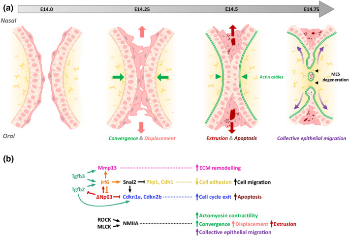
Clefts of the lip and palate (CLP), the major causes of congenital facial malformation globally, result from failure of fusion of the facial processes during embryogenesis. With a prevalence of 1 in 500-2500 live births, CLP causes major morbidity throughout life as a result of problems with facial appearance, feeding, speaking, obstructive apnoea, hearing and social adjustment and requires complex, multi-disciplinary care at considerable cost to healthcare systems worldwide. Long-term outcomes for affected individuals include increased mortality compared with their unaffected siblings. The frequent occurrence and major healthcare burden imposed by CLP highlight the importance of dissecting the molecular mechanisms driving facial development. Identification of the genetic mutations underlying syndromic forms of CLP, where CLP occurs in association with non-cleft clinical features, allied to developmental studies using appropriate animal models is central to our understanding of the molecular events underlying development of the lip and palate and, ultimately, how these are disturbed in CLP.

Keywords: cleft lip; cleft palate; facial development.


Conflict of interest statement

The authors have declared that no conflict of interest exists.

Figures

FIGURE 1
FIGURE 1
Human facial development. (a) Migratory neural crest cells populate the facial processes. (b) The medial and lateral nasal processes (mnp & lnp, respectively) fuse with the maxillary processes (mxp) to form the upper lip. (c) Bilateral cleft lip. (d) The secondary palate develops from the maxillary processes. (e) The paired palatal shelves (ps) grow vertically before elevating to a horizontal position above the tongue and fusing via the midline epithelial seam. Subsequently, the palatal mesenchyme differentiates into bone and muscle forming the hard and soft palate, respectively. (f) Failure of these processes results in cleft palate with the nasal septum (ns) visible
FIGURE 2
FIGURE 2
Development of the lip and palate in mice. (a‐c: scanning electron microscopy ‐ frontal views) The upper lip and primary palate form from a series of facial processes which merge by E12.5. (d) Timeline of secondary palate development. (e–g: scanning electron microscopy ‐ ventral views; h–j: histological analysis) The palatal shelves develop from the maxillary processes and grow vertically lateral to the tongue during E12 and E13 (e, h). (f, i) During E14, the palatal shelves elevate above the tongue and fuse in the midline via the midline epithelial seam (g, j). mnp: medial nasal processes; lnp, lateral nasal processes; mx, maxillary processes; pp, primary palate; p, palatal shelves; t, tongue
FIGURE 3
FIGURE 3
Molecular mechanisms of lip fusion and epithelial seam dissolution. (a) Schematic diagram of the lambdoidal (λ) epithelial seam at E11.5, formed through fusion of the medial nasal and lateral nasal processes. SHH and TGFβ‐mediated Pbx signalling converge on WNT to regulate pathways involved in epithelial seam dissolution. (b) Pbx plays a dual‐role in lip fusion, (1) by regulating a WNT‐p63‐Irf6 cascade to promote epithelial apoptosis; (2) by promoting epithelial‐mesenchymal transformation, cell plasticity/migration through regulation of Snail1. Cross‐talk between both pathways is achieved by post‐translational modification of Gsk3β on Snail1. SHH ensures appropriate p63‐Irf6 signalling by up‐regulating WNT antagonists and restricting Tfap2a signalling. (Adapted from Kurosaka et al., ; Losa et al., 2018). mnp, medial nasal process; lnp, lateral nasal process; EMT, epithelial‐mesenchymal transformation
FIGURE 4
FIGURE 4
Sequential rugae interposition during secondary palate growth and patterning. (a–e) Whole‐mount in situ hybridisation of E12.5‐E15.5 palatal shelves. (a–d) During palatogenesis, Shh expression defines epithelial rugae (r1‐r8) and their sequential interposition on the oral surface. In the posterior palate, Shh is expressed in sensory papillae. (e) Shox2 (anterior) and Meox2 (posterior) show differential gene expression along the anterior‐posterior axis, the boundary of which is defined by R1. r, ruga; sp, sensory papillae
FIGURE 5
FIGURE 5
Molecular regulation of secondary palate growth and patterning. (a, b) Schematic diagram showing molecular regulation and cross‐talk of key molecules involved in secondary palate growth and patterning. (a, b) Shh is a key gene in oral‐nasal patterning, signalling from the oral epithelium to the underlying mesenchyme to positively regulate Osr2, Fgf10 and Foxf1/Foxf2/Foxl1 molecular cascades. Fgf10 and Bmp4 maintain expression of Shh, whereas Dlx5Fgf7 signalling in the nasal mesenchyme restricts Shh. (b) Shox2 and Msx1 expression is restricted to the anterior palate. Outgrowth of the anterior palate is controlled by a network involving Shh, Bmp, Msx1, Wnt5a and Pax9. (c) Meox2, Barx1 and Tbx22 expression is restricted to the posterior palate. Pax9 has a central role in regulating posterior signalling, promoting Osr2, Bmp4 and WNT signalling. Adapted from Li et al., . MEE, medial edge epithelia
FIGURE 6
FIGURE 6
Molecular mechanisms of secondary palate fusion. (a) Schematic diagram illustrating the morphological events in secondary palatal fusion. (b) TGFβ signalling promotes MES degeneration and extracellular matrix remodelling by regulating cascades involving Irf6 and Mmp13, respectively. The transcription factors Irf6 and Tp63 function in a regulatory feedback loop to control medial edge epithelial cell fate. Rho‐kinase (ROCK) and myosin light‐chain kinase (MLCK) converge to activate non‐muscle myosin IIA (NMIIA), driving actomyosin contractility. Together this pathway regulates epithelial convergence, displacement, extrusion and migration to ensure midline epithelial seam dissolution and mesenchymal continuity. Adapted from Kim et al., . MEE, medial edge epithelia; MES, midline epithelial seam

Similar articles

Cited by

References

    1. 1000 Genomes Project Consortium , Durbin, R. M. , Abecasis, G. R. , Altshuler, D. L. , Auton, A. , Brooks, L. D. , Durbin, R. M. , Gibbs, R. A. , Hurles, M. E. , & McVean, G. A. (2010). A map of human genome variation from population‐scale sequencing. Nature, 467, 1061–1073. - PMC - PubMed
    1. Alappat, S. R. , Zhang, Z. , Suzuki, K. , Zhang, X. , Liu, H. , Jiang, R. , Yamada, G. , & Chen, Y. (2005). The cellular and molecular etiology of the cleft secondary palate in Fgf10 mutant mice. Developmental Biology, 277, 102–113. 10.1016/j.ydbio.2004.09.010 - DOI - PubMed
    1. Almaidhan, A. , Cesario, J. , Landin Malt, A. , Zhao, Y. , Sharma, N. , Choi, V. , & Jeong, J. (2014). Neural crest‐specific deletion of Ldb1 leads to cleft secondary palate with impaired palatal shelf elevation. BMC Developmental Biology, 14, 3. 10.1186/1471-213X-14-3 - DOI - PMC - PubMed
    1. Ashique, A. M. , Fu, K. , & Richman, J. M. (2002). Endogenous bone morphogenetic proteins regulate outgrowth and epithelial survival during avian lip fusion. Development, 129, 4647–4660. 10.1242/dev.129.19.4647 - DOI - PubMed
    1. Baek, J. A. , Lan, Y. , Liu, H. , Maltby, K. M. , Mishina, Y. , & Jiang, R. (2011). Bmpr1a signaling plays critical roles in palatal shelf growth and palatal bone formation. Developmental Biology, 350, 520–531. 10.1016/j.ydbio.2010.12.028 - DOI - PMC - PubMed