Fungal Endophthalmitis on Ocular Ultrasound: A Case Report
- PMID: 35226845
- PMCID: PMC8885216
- DOI: 10.5811/cpcem.2021.10.53797
Fungal Endophthalmitis on Ocular Ultrasound: A Case Report
Abstract
Introduction: Endophthalmitis is a rare intraocular infection caused by numerous organisms from several possible sources. Fungal endophthalmitis is a rare subset of this pathology with limited diagnostics available. One of the few options to make this diagnosis is vitreous sampling, which is invasive, and results are not immediately available.
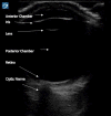
Case report: This case report describes the successful use of point-of-care ultrasound to visualize an intraocular fungal mass in a 60-year-old male who presented to the emergency department (ED) with two weeks of left eye pain and erythema approximately two months postoperative from a cataract extraction surgery.
Conclusion: Fungal endophthalmitis is a rare and challenging diagnosis. Methods of diagnosing this pathology are not readily available in the ED. Point-of-care ultrasound may be a useful adjunct for the prompt diagnosis of fungal endophthalmitis.
Conflict of interest statement
Figures
References
-
- Chang CC, Chen SC. Fungal eye infections: new hosts, novel emerging pathogens but no new treatments? Curr Fungal Infect Rep. 2018;2:66–70.
-
- Gursimrat KH, Dawkins RC, Paul RA, et al. Fundal endophthalmitis: a 20-year experience at a tertiary referral centre. Clin Exp Ophthalmol. 2020;48(7):964–72. - PubMed


