. 2022 Apr 26;13(2):e0316321.
doi: 10.1128/mbio.03163-21. Epub 2022 Mar 1.

Entamoeba histolytica Develops Resistance to Complement Deposition and Lysis after Acquisition of Human Complement-Regulatory Proteins through Trogocytosis

Affiliations

Entamoeba histolytica Develops Resistance to Complement Deposition and Lysis after Acquisition of Human Complement-Regulatory Proteins through Trogocytosis

Hannah W Miller et al. mBio. .

Abstract

Entamoeba histolytica is the cause of amoebiasis. The trophozoite (amoeba) form of this parasite is capable of invading the intestine and can disseminate through the bloodstream to other organs. The mechanisms that allow amoebae to evade complement deposition during dissemination have not been well characterized. We previously discovered a novel complement-evasion mechanism employed by E. histolytica. E. histolytica ingests small bites of living human cells in a process termed trogocytosis. We demonstrated that amoebae were protected from lysis by human serum following trogocytosis of human cells and that amoebae acquired and displayed human membrane proteins from the cells they ingested. Here, we aimed to define how amoebae are protected from complement lysis after performing trogocytosis. We found that amoebae were protected from complement lysis after ingestion of both human Jurkat T cells and red blood cells and that the level of protection correlated with the amount of material ingested. Trogocytosis of human cells led to a reduction in deposition of C3b on the surface of amoebae. We asked whether display of human complement regulators is involved in amoebic protection, and found that CD59 was displayed by amoebae after trogocytosis. Deletion of a single complement-regulatory protein, CD59 or CD46, from Jurkat cells was not sufficient to alter amoebic protection from lysis, suggesting that multiple, redundant complement regulators mediate amoebic protection. However, exogeneous expression of CD46 or CD55 in amoebae was sufficient to confer protection from lysis. These studies shed light on a novel strategy for immune evasion by a pathogen. IMPORTANCE Entamoeba histolytica is the cause of amoebiasis, a diarrheal disease of global importance. While infection is often asymptomatic, the trophozoite (amoeba) form of this parasite is capable of invading and ulcerating the intestine and can disseminate through the bloodstream to other organs. Understanding how E. histolytica evades the complement system during dissemination is of great interest. Here, we demonstrate for the first time that amoebae that have performed trogocytosis (nibbling of human cells) resist deposition of the complement protein C3b. Amoebae that have performed trogocytosis display the complement-regulatory protein CD59. Overall, our studies suggest that acquisition and display of multiple, redundant complement regulators is involved in amoebic protection from complement lysis. These findings shed light on a novel strategy for immune evasion by a pathogen. Since other parasites use trogocytosis for cell killing, our findings may apply to the pathogenesis of other infections.

Keywords: Entamoeba histolytica; complement; immune evasion; trogocytosis.


Conflict of interest statement

The authors declare no conflict of interest.

Figures

FIG 1
FIG 1
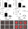
Ingestion of latex beads does not protect amoebae from complement lysis. Amoebae were incubated with 3 × 106 or 1.5 × 107 fluorescent latex beads for 1 h or were incubated without latex beads, and then exposed to human serum. Amoebic viability was determined with Zombie Violet viability dye. Imaging flow cytometry was used to quantify bead ingestion and amoebic viability. (A) The percentage of amoebae that had ingested any number of latex beads. (B) Mean number of ingested beads among amoebae that had ingested beads. (C) Normalized amoeba death following exposure to human serum. Values are normalized to the amoebae that were incubated without latex beads. (D) Quantification of the number of ingested beads per amoeba. Shown are the data plots from one replicate per sample type. Amoebae that did not perform bead ingestion fall into the “Bead −” gate, while amoebae that ingested beads fall into the “Bead +” gate. (G) Representative images of amoebae that were incubated without beads or in the presence of 3 × 106 or 1.5 × 107 beads. Ingested beads are shown in yellow. Data are from four replicates across two independent experiments. ns, no significant difference (P > 0.05); **, P ≤ 0.01; ***, P ≤ 0.001; ****, P ≤ 0.0001.
FIG 2
FIG 2
Amoebic protection from complement following trogocytosis is dose dependent. Amoebae were labeled with CMFDA cytoplasm dye and incubated in the absence of human cells or with increasing concentrations of human Jurkat cells or primary human red blood cells. Human cells were labeled with DiD membrane dye. Following exposure to human serum, amoeba death was assessed with Zombie Violet viability dye, and ingested human cell material was determined by quantifying mean fluorescence intensity (MFI) of DiD present on amoebae. (A) Normalized MFI of DiD on amoebae incubated in the absence of Jurkat cells or with increasing concentrations of Jurkat cells. (B) Normalized death of amoebae from conditions in panel A. (C) Death of amoebae from conditions in panel A, expressed as percent protection. Percent protection was calculated by subtracting the total death of amoebae incubated with human cells from the total death of amoebae incubated in the absence of Jurkat cells. (D) Normalized MFI of DiD on amoebae incubated in the absence of red blood cells or with increasing concentrations of red blood cells. (E) Normalized death of amoebae from conditions in panel D. (F) Death of amoebae from conditions in panel D, expressed as percent protection. (G) Representative images of amoebae incubated with increasing concentrations of Jurkat cells. Amoebae are shown in green, and ingested human cell material is shown in red. Data were analyzed by imaging flow cytometry and are from 6 replicates across 3 independent experiments. (H) Representative images of amoebae incubated with red blood cells. Data were analyzed by imaging flow cytometry and are from 6 replicates across 3 independent experiments. ns, no significant difference (P > 0.05); *, P ≤ 0.05; **, P ≤ 0.01; ***, P ≤ 0.001; ****, P ≤ 0.0001.
FIG 3
FIG 3
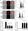
Amoebic trogocytosis of human cells inhibits deposition of complement C3b. Amoebae were incubated in the absence of Jurkat T cells or in the presence of Jurkat cells, and subsequently exposed to human serum. Viability was assessed with Zombie Violet dye. The presence of C3b was detected using a mouse monoclonal antibody to C3b and iC3b. (A) Death of amoebae that were incubated in the absence of Jurkat cells or in the presence of Jurkat cells. (B) Mean fluorescence intensity of deposited C3b on amoebae. (C) Deposited C3b on dead or live amoebae. (D) Deposited C3b on dead or live amoebae that had been incubated in the absence of Jurkat cells (open circles) or in the presence of Jurkat cells (filled circles). (E) Representative images of C3b deposition (red) on live or dead amoebae. Data were analyzed by imaging flow cytometry and are from 6 replicates across 3 independent experiments. *, P ≤ 0.05; ***, P ≤ 0.001; ****, P ≤ 0.0001.
FIG 4
FIG 4
Amoebae acquire and display the complement-regulatory protein CD59 from human cells. Amoebae were allowed to perform trogocytosis on human Jurkat T cells for 5 min or 1 h or were incubated in the absence of Jurkat cells. Human CD59 (red) was detected on the amoebae surface by monoclonal antibody staining. Amoebae were labeled with CMFDA (green), and human cell nuclei were labeled with Hoechst (blue). (A) Representative images from amoebae incubated in the absence of Jurkat cells or amoebae that performed trogocytosis on human Jurkat T cells for 5 min or 1 h. Arrows indicate patches of displayed CD59 on the amoeba surface. (B) 3D rendering of Z-stack images taken from amoebae that were incubated with human Jurkat T cells for 1 h. (C) Zoomed-in image of amoebae that were incubated with human Jurkat T cells for 1 h. Data were analyzed by confocal microscopy. One hundred thirty-six images were collected from 1 independent experiment.
FIG 5
FIG 5
The amount of displayed CD59 increases with increased trogocytosis of human cells. Acquired CD59 molecules were quantified on amoebae that were allowed to perform trogocytosis on human Jurkat T cells for 5 min or 1 h or were incubated in the absence of Jurkat cells. (A) Masking strategy for analysis of displayed CD59 on the amoeba surface. A mask was created in order to allow the detection of CD59 that overlapped with amoebae while excluding CD59 on intact human cells attached to amoebae. The mask is displayed in turquoise, as an overlay on the individual images. Amoebae were labeled with CMFDA (green), human cell nuclei were labeled with Hoechst (blue), and CD59 was detected with a monoclonal antibody (red). Extracellular human cell nucleus fluorescence was removed from the masked analysis area. The excluded area around human cell nuclei was then dilated by 4 pixels to include the entire diameter of the intact extracellular human cells and associated CD59. CD59 was analyzed in the remaining masked analysis area of each image to allow analysis of displayed patches of acquired CD59 on amoebae. (B) Normalized mean fluorescence intensity of CD59 on amoebae after 5 min or 1 h of trogocytosis or amoebae that were incubated in the absence of Jurkat cells. To normalize the data, samples were normalized to the condition using 1 h of trogocytosis. (C) Representative images of amoebae that had performed trogocytosis on human Jurkat T cells for 5 min or 1 h. Arrows indicate displayed CD59. Data were analyzed by imaging flow cytometry and are from 5 replicates across 3 independent experiments. The no-primary-control condition was used in 2 of 3 independent experiments, and data are from 3 replicates. **, P ≤ 0.01; ***, P ≤ 0.001; ****, P ≤ 0.0001.
FIG 6
FIG 6
Removal of CD59 and CD46 is not sufficient to sensitize amoebae to complement lysis. (A to D) Human Jurkat T cells deficient in CD59 or CD46 were constructed using CRISPR/Cas9. Immunofluorescence and imaging flow cytometry were used to quantify CD59 or CD46. (A) Representative images of CD59 antibody staining (red) in vector control human cells or CD59 mutants. (B) Intensity of CD59 antibody staining in vector control human cells (gray) or CD59 mutants (black). Of CD59 mutants, 99.5% were in the CD59-negative gate (−CD59), while 0.75% of vector control cells were in this gate. (C) Representative images of CD46 antibody staining (red) in vector control human cells or CD46 mutants. (D) Intensity of CD46 antibody staining in vector control human cells (gray) or CD46 mutants (black). Of CD46 mutants, 99.5% were in the CD46-negative gate (−CD46), while 0.27% of vector control cells were in this gate. (E and F) Amoebae were labeled with CMFDA cytoplasm dye and incubated in the absence of Jurkat cells or with Jurkat cells. Human cell lines were either vector control cells, CD59 knockout (KO) mutants, or CD46 KO mutants. Human cells were labeled with DiD membrane dye. Following exposure to human serum, amoeba death was assessed with Zombie Violet viability dye and ingested human cell material was determined by quantifying mean fluorescence intensity of DiD present on amoebae. (E) Normalized death of amoebae. (F) Normalized mean fluorescence intensity of DiD on amoebae. Data were analyzed by imaging flow cytometry and are from 6 replicates across 3 independent experiments. ns, no significant difference (P > 0.05); ****, P ≤ 0.0001.
FIG 7
FIG 7
Amoebae transfected with expression plasmids display human CD46 or CD55. Amoebae were transfected with expression constructs for human CD46 or human CD55 or the expression plasmid backbone as a negative control. (A) RT-PCR analysis with primers specific for human CD46, human CD55, or amoebic GAPDH. Samples were incubated with or without reverse transcriptase (RT) to control for DNA contamination. (B to J) Amoebae were labeled with a cytoplasmic dye (CMFDA), and immunofluorescence was performed without permeabilization. (B) Mean fluorescence intensity of human CD46 antibody staining. Amoebae were stably transfected with a plasmid for expression of human CD46 (red) or vector backbone (beige) and were stained using a CD46 primary antibody and a far-red secondary antibody. Amoebae expressing human CD46 were also incubated without primary antibody (gray), or without any antibodies (black). (C) Mean fluorescence intensity of human CD55 antibody staining. Amoebae were stably transfected with a plasmid for expression of human CD55 (blue) or vector backbone (beige) and were stained using a CD46 primary antibody and a far-red secondary antibody. Amoebae expressing human CD55 were also incubated without primary antibody (gray) or without any antibodies (black). (D) Mean fluorescence intensity of amoebic Gal/GalNAc lectin antibody staining. Amoebae were stably transfected with a plasmid for expression of human CD46 (red), human CD55 (blue), or vector backbone (beige) and were stained using a Gal/GalNAc lectin primary antibody and a far-red secondary antibody. Amoebae were also incubated without primary antibody, or without any antibodies. (E to G) Histograms corresponding to the mean fluorescence intensity data shown in panels B to D. (G) Antibody staining of vector control amoebae, stained with both antibodies (beige), without primary antibody (gray), or without any antibodies (black). (H to J) Representative images corresponding to the analysis shown in panels B to G. Data shown in panel A are representative of two replicates. Data shown in panels B to J were analyzed by imaging flow cytometry are from 3 or 4 replicates across 2 independent experiments; the data from one independent experiment are shown.
FIG 8
FIG 8
Display of human CD46 or CD55 is sufficient to protect amoebae from complement lysis. Amoebae were stably transfected with CD46 or CD55 expression constructs or vector backbone. Following exposure to either media or human serum, amoeba viability was assessed using Zombie Violet viability dye and imaging flow cytometry. Amoebic death was normalized to vector control amoebae that were incubated with human serum. These data are from 8 replicates across 4 independent experiments. ns, no significant difference (P > 0.05); *, P ≤ 0.05; ***, P ≤ 0.001; ****, P ≤ 0.0001.

Similar articles

Cited by

References

    1. Wang H, Naghavi M, Allen C, Barber RM, Bhutta ZA, Carter A, Casey DC, Charlson FJ, Chen AZ, Coates MM, Coggeshall M, Dandona L, Dicker DJ, Erskine HE, Ferrari AJ, Fitzmaurice C, Foreman K, Forouzanfar MH, Fraser MS, Fullman N, Gething PW, Goldberg EM, Graetz N, Haagsma JA, Hay SI, Huynh C, Johnson CO, Kassebaum NJ, Kinfu Y, Kulikoff XR, Kutz M, Kyu HH, Larson HJ, Leung J, Liang X, Lim SS, Lind M, Lozano R, Marquez N, Mensah GA, Mikesell J, Mokdad AH, Mooney MD, Nguyen G, Nsoesie E, Pigott DM, Pinho C, Roth GA, Salomon JA, Sandar L, Silpakit N, et al.. 2016. Global, regional, and national life expectancy, all-cause mortality, and cause-specific mortality for 249 causes of death, 1980–2015: a systematic analysis for the Global Burden of Disease Study 2015. Lancet 388:1459–1544. doi:10.1016/S0140-6736(16)31012-1. - DOI - PMC - PubMed
    1. Marie C, Petri WA. 2013. Amoebic dysentery. BMJ Clin Evid 2013:e0918. - PMC - PubMed
    1. Alvarado-Esquivel C, Hernandez-Tinoco J, Sanchez-Anguiano LF. 2015. Seroepidemiology of Entamoeba histolytica infection in general population in rural Durango, Mexico. J Clin Med Res 7:435–439. doi:10.14740/jocmr2131w. - DOI - PMC - PubMed
    1. Gilchrist CA, Petri SE, Schneider BN, Reichman DJ, Jiang N, Begum S, Watanabe K, Jansen CS, Elliott KP, Burgess SL, Ma JZ, Alam M, Kabir M, Haque R, Petri WA. 2016. Role of the gut microbiota of children in diarrhea due to the protozoan parasite Entamoeba histolytica. J Infect Dis 213:1579–1585. doi:10.1093/infdis/jiv772. - DOI - PMC - PubMed
    1. Haque R, Huston CD, Hughes M, Houpt E, Petri WA. 2003. Amebiasis. N Engl J Med 348:1565–1573. doi:10.1056/NEJMra022710. - DOI - PubMed

Publication types

Substances