. 2022 May 1;205(9):1016-1035.
doi: 10.1164/rccm.202112-2742SO.

Autoimmune Pulmonary Alveolar Proteinosis

Affiliations

Autoimmune Pulmonary Alveolar Proteinosis

Cormac McCarthy et al. Am J Respir Crit Care Med. .

Abstract

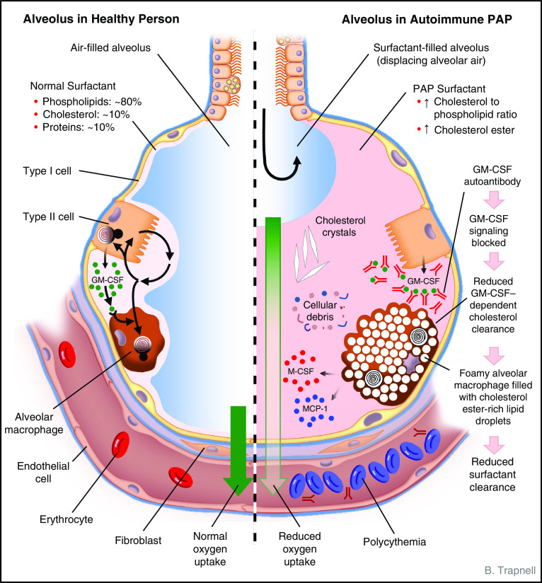
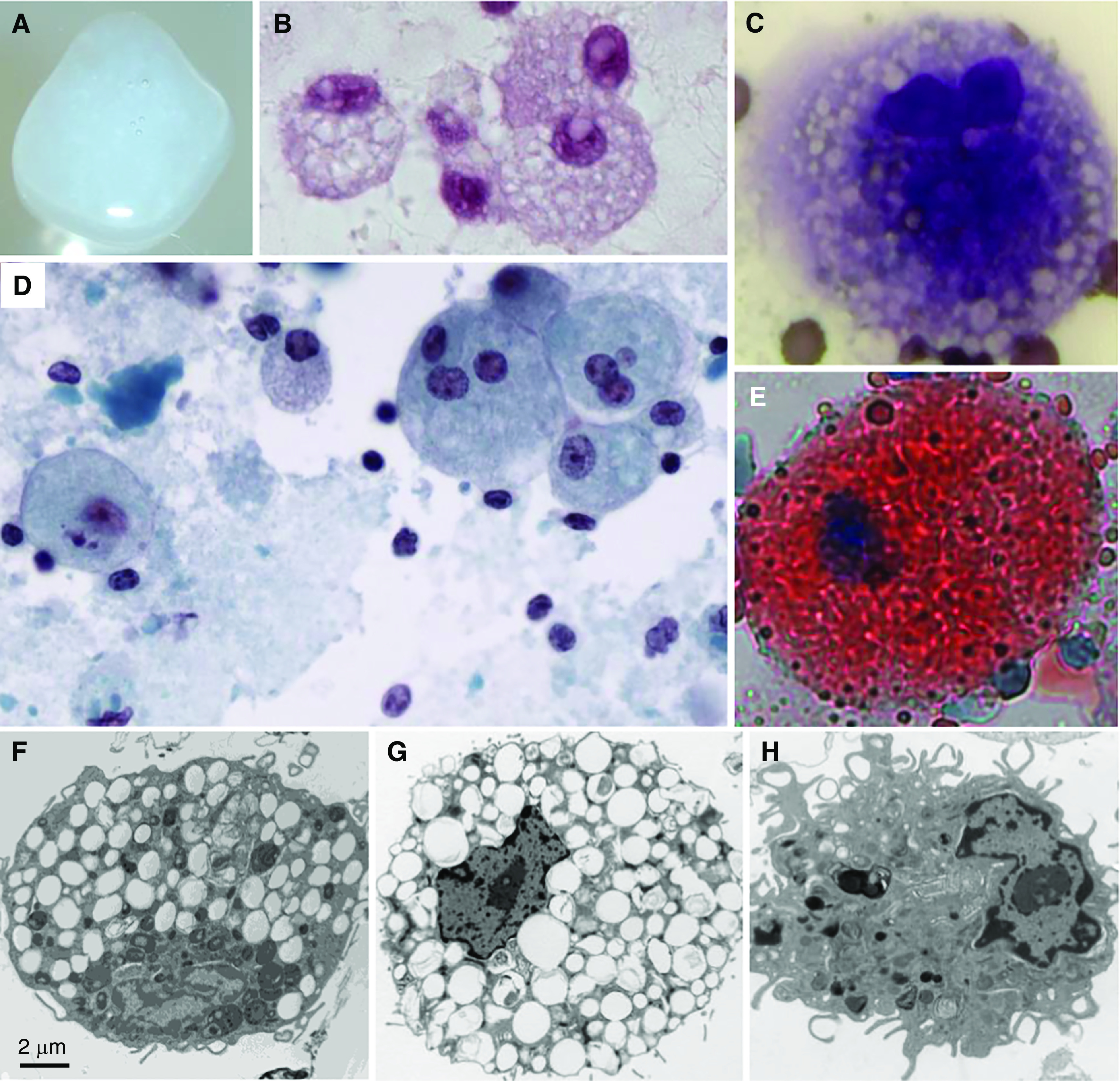
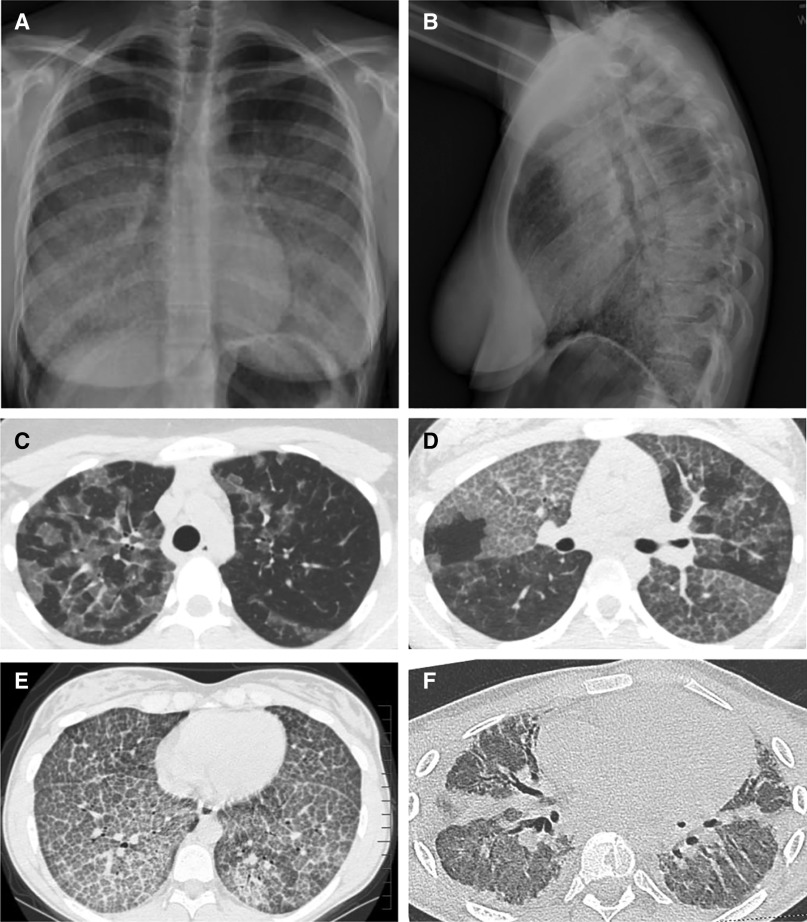
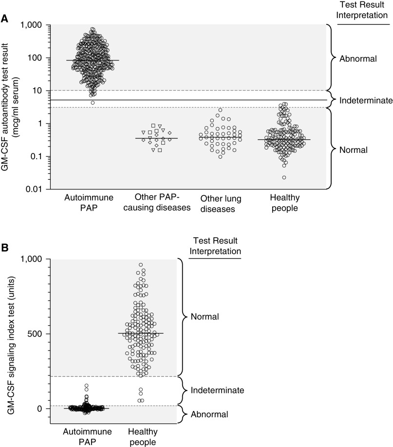
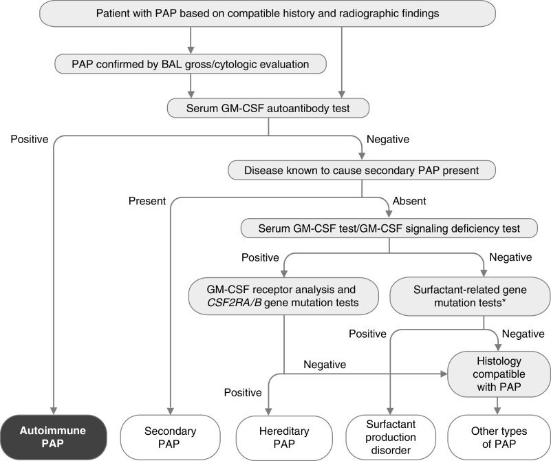
Autoimmune pulmonary alveolar proteinosis (PAP) is a rare disease characterized by myeloid cell dysfunction, abnormal pulmonary surfactant accumulation, and innate immune deficiency. It has a prevalence of 7-10 per million; occurs in individuals of all races, geographic regions, sex, and socioeconomic status; and accounts for 90% of all patients with PAP syndrome. The most common presentation is dyspnea of insidious onset with or without cough, production of scant white and frothy sputum, and diffuse radiographic infiltrates in a previously healthy adult, but it can also occur in children as young as 3 years. Digital clubbing, fever, and hemoptysis are not typical, and the latter two indicate that intercurrent infection may be present. Low prevalence and nonspecific clinical, radiological, and laboratory findings commonly lead to misdiagnosis as pneumonia and substantially delay an accurate diagnosis. The clinical course, although variable, usually includes progressive hypoxemic respiratory insufficiency and, in some patients, secondary infections, pulmonary fibrosis, respiratory failure, and death. Two decades of research have raised autoimmune PAP from obscurity to a paradigm of molecular pathogenesis-based diagnostic and therapeutic development. Pathogenesis is driven by GM-CSF (granulocyte/macrophage colony-stimulating factor) autoantibodies, which are present at high concentrations in blood and tissues and form the basis of an accurate, commercially available diagnostic blood test with sensitivity and specificity of 100%. Although whole-lung lavage remains the first-line therapy, inhaled GM-CSF is a promising pharmacotherapeutic approach demonstrated in well-controlled trials to be safe, well tolerated, and efficacious. Research has established GM-CSF as a pulmonary regulatory molecule critical to surfactant homeostasis, alveolar stability, lung function, and host defense.

Keywords: BAL; alveolar macrophages; autoantibodies; granulocyte/macrophage colony–stimulating factor; surfactant.


Figures

Figure 1.
Figure 1.
Schematic depicting the anatomy and biology of the alveolus in health (left) and autoimmune pulmonary alveolar proteinosis (PAP) (right). Normally, alveolar structure and function are maintained by a surfactant layer sufficiently thick (about one to three molecules) to reduce surface tension and thin enough to permit adequate diffusion of oxygen across the alveolar wall. Homeostasis is maintained by balanced secretion of surfactant by alveolar type II cells and clearance by type II cells and by alveolar macrophages, which require GM-CSF (granulocyte/macrophage colony–stimulating factor) to export cholesterol normally. In autoimmune PAP, autoantibodies bind and block GM-CSF from activating receptors on alveolar macrophages (and other cells, e.g., neutrophils). Because surfactant uptake by alveolar macrophages is GM-CSF independent and export of surfactant-derived cholesterol is GM-CSF dependent, cholesterol accumulates within the cytoplasm. Alveolar macrophages esterify and sequester cholesterol within intracytoplasmic vesicles (as a cell-protective mechanism), resulting in foamy-appearing cells with secondary impairment of multiple macrophage functions, including phagocytosis and surfactant clearance. Over time, alveoli fill with insoluble surfactant sediment, cellular debris, cholesterol crystals, and several cytokines (indicated; see text). A thickened surfactant layer and surfactant-filled alveoli contribute to reduced oxygen delivery, resulting in hypoxemia, dyspnea, and in severe cases polycythemia, respiratory failure, and death. Pulmonary fibrosis (not shown) also occurs in some patients. See text for further details. MCP-1 = monocyte chemoattractant protein-1; M-CSF = macrophage colony–stimulating factor.
Figure 2.
Figure 2.
Sputum, sputum cytology, and alveolar macrophage ultrastructure in autoimmune pulmonary alveolar proteinosis (PAP). (A) Gross appearance of freshly expectorated sputum from a patient with autoimmune PAP. (B–E) Microscopic appearance of sputum cytology after staining with periodic acid–Schiff reagent (B); Diff-Quick, showing a foamy alveolar macrophage (C); Papanicolaou reagent (D); or oil red O, showing an oil red O–positive alveolar macrophage (E). (F) Alveolar macrophage obtained by BAL from an 18-year-old man with autoimmune PAP several months after lung transplantation performed as therapy for pulmonary fibrosis. Note the numerous, single membrane–delimited, intracytoplasmic lipid droplets. Scale bar, 2 μm. (G and H) Uranyl acetate staining. Alveolar macrophages obtained from a nonhuman primate passively immunized with highly purified, human GM-CSF (granulocyte/macrophage colony–stimulating factor) autoantibodies derived from patients with autoimmune PAP, showing intracytoplasmic lipid droplets (G), or a saline-injected primate, showing a normal macrophage appearance (H).
Figure 3.
Figure 3.
Appearance of the chest radiograph and chest computed tomography scan in autoimmune pulmonary alveolar proteinosis (PAP). (A and B) Posterior–anterior (A) and lateral (B) chest radiographs of a 19-year-old woman with autoimmune PAP showing diffuse ground-glass opacification of the lung parenchyma. (C–F) Representative images from computed tomography of the chest showing the diversity of radiographic findings in autoimmune PAP in a 45-year-old man (C), a 58-year-old woman (D), a 15-year-old girl (E), and an 18-year-old man (F). (C) Image showing ground-glass opacification involving some but not all secondary lobules resulting in a distinctive “geographic” pattern. Also, note the disproportionate involvement of the left and right lung parenchyma. (D) Image showing a distinctive pattern of interlobular septal thickening superimposed on ground-glass opacification, often referred to as “crazy paving.” Also, note the sharply demarcated differences in the degree of involvement between adjacent lung lobes. (E) Image revealing a homogeneous pattern of crazy paving throughout all regions of the lung parenchyma. (F) Image revealing extensive pulmonary fibrosis with parenchymal distortion from traction bronchiectasis. This patient underwent bilateral lung transplantation for pulmonary fibrosis and respiratory failure shortly after this image was obtained.
Figure 4.
Figure 4.
Blood-based tests used to diagnose autoimmune pulmonary alveolar proteinosis (PAP). (A) Measurement of serum GM-CSF (granulocyte/macrophage colony–stimulating factor) autoantibody concentration by the serum GM-CSF autoantibody test. Shown are results for individuals with the indicated diseases and interpretive ranges for the test results. Each symbol represents the test result for one individual determined by ELISA using a patient-derived, affinity-purified, polyclonal GM-CSF autoantibody reference standard as previously reported (155). The reference ranges used for test result interpretation (normal, ⩽3.1 μg/ml; indeterminate, >3.1 to <10.2 μg/ml; and abnormal, ⩾10.2 μg/ml) were determined according to guidelines established by the Clinical and Laboratory Standards Institute (234), and updated on April 5, 2019, on the basis of results for 153 healthy individuals (median, 0.33 μg/ml; 90% confidence interval [CI], 0.3–0.4 μg/ml) and 339 patients with autoimmune PAP (median, 84 μg/ml; 90% CI, 10.2–499 μg/ml). Test results within the indeterminate range are typically confirmed by evaluation of GM-CSF signaling. (B) Measurement of GM-CSF signaling by the GM-CSF signaling index (GM-CSF-SI) test. Shown are results for individuals with autoimmune PAP and healthy people and the interpretive ranges for the test results. Each symbol represents the test result for one individual determined by incubating heparinized whole blood with recombinant human GM-CSF (10 ng/ml, 30 min) followed by flow cytometry to quantify phosphorylated STAT5. Results are expressed as GM-CSF-SI, calculated as the mean fluorescence intensity (MFI) in GM-CSF–stimulated cells minus the MFI of unstimulated cells divided by the MFI of unstimulated cells and multiplied by 100, as previously reported (75). The reference ranges used for test result interpretation (normal, ⩾216 units; indeterminate, >20 to <216 units; abnormal, ⩽20 units) were determined according to guidelines established by the Clinical and Laboratory Standards Institute (234) and updated on April 8, 2019, on the basis of results for 77 healthy individuals (median, 506 units; 90% CI, 483–564 units) and 80 patients with autoimmune PAP (median, 2.2 units; 90% CI, 0–4.2 units). STAT5 = signal transducer and activator of transcription 5.
Figure 5.
Figure 5.
Algorithm for diagnosis of pulmonary alveolar proteinosis (PAP). PAP is suspected on the basis of history and radiographic findings and can be confirmed by the gross and microscopic appearance of BAL fluid. A serum GM-CSF (granulocyte/macrophage colony–stimulating factor) autoantibody test is performed and is highly sensitive and specific for diagnosis of autoimmune PAP. Secondary PAP is diagnosed in individuals with a negative test result who have a condition known to cause PAP. Those with negative test results and no known PAP-causing disease should undergo blood GM-CSF signaling testing and serum GM-CSF testing to identify hereditary PAP caused by CSF2RA or CSF2RB gene mutations, followed by appropriate confirmatory genetic testing. Patients with normal concentrations of serum GM-CSF (e.g., low/undetectable) and normal GM-CSF signaling test results (e.g., detectable) should undergo genetic testing used to identify surfactant production disorders (also known as congenital PAP), including mutational analysis of SFTPC, SFTPB, ABCA3, and NKX2.1 (*). A transbronchial or surgical lung biopsy may be needed if all test results are negative, and further genetic testing may be needed to identify other ultrarare PAP-causing diseases (i.e., methionine-transfer RNA mutations [235], lysinuric protein intolerance [236], or lymphoid cell deficiency–related PAP [237]) Adapted from Reference .

References

    1. Rosen SH, Castleman B, Liebow AA. Pulmonary alveolar proteinosis. N Engl J Med . 1958;258:1123–1142. - PubMed
    1. Trapnell BC, Whitsett JA, Nakata K. Pulmonary alveolar proteinosis. N Engl J Med . 2003;349:2527–2539. - PubMed
    1. Seymour JF, Presneill JJ. Pulmonary alveolar proteinosis: progress in the first 44 years. Am J Respir Crit Care Med . 2002;166:215–235. - PubMed
    1. Trapnell BC, Nakata K, Bonella F, Campo I, Griese M, Hamilton J, et al. Pulmonary alveolar proteinosis. Nat Rev Dis Primers . 2019;5:16. - PubMed
    1. Borie R, Danel C, Debray MP, Taille C, Dombret MC, Aubier M, et al. Pulmonary alveolar proteinosis. Eur Respir Rev . 2011;20:98–107. - PMC - PubMed

Publication types

MeSH terms

Substances

Supplementary concepts