. 2022 Feb 28;23(1):64.
doi: 10.1186/s13059-022-02638-6.

LAP2α preserves genome integrity through assisting RPA deposition on damaged chromatin

Affiliations

LAP2α preserves genome integrity through assisting RPA deposition on damaged chromatin

Kaiwen Bao et al. Genome Biol. .

Abstract

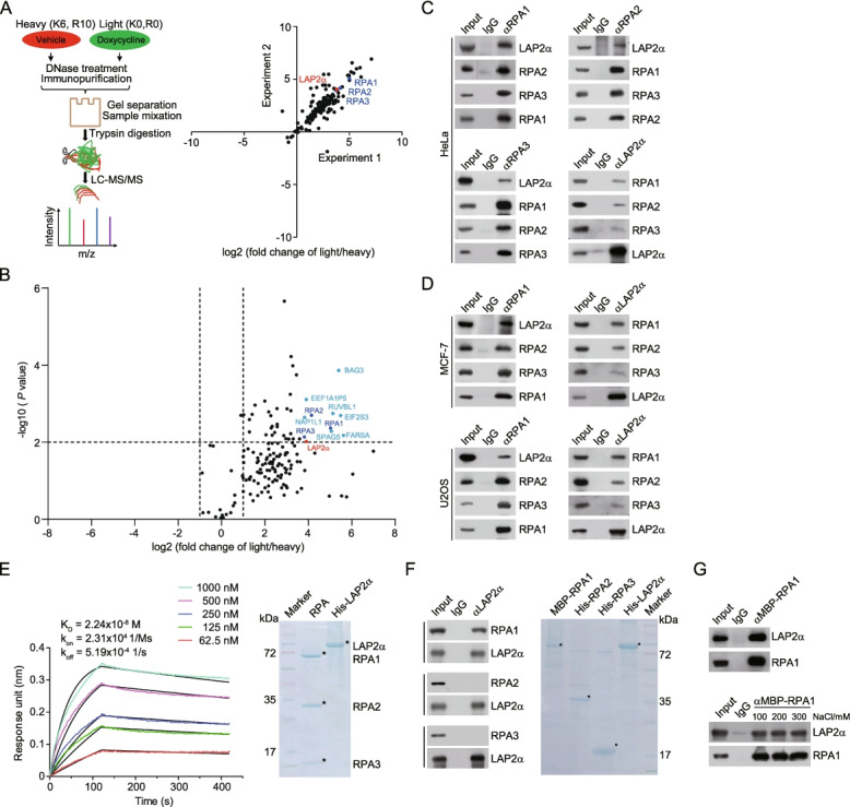
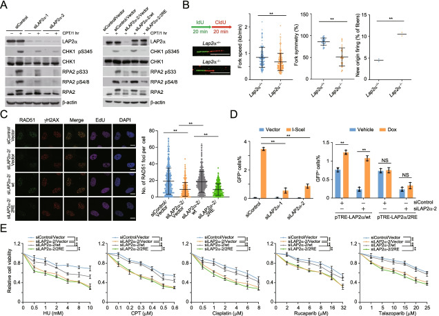
Background: Single-stranded DNA (ssDNA) coated with replication protein A (RPA) acts as a key platform for the recruitment and exchange of genome maintenance factors in DNA damage response. Yet, how the formation of the ssDNA-RPA intermediate is regulated remains elusive.

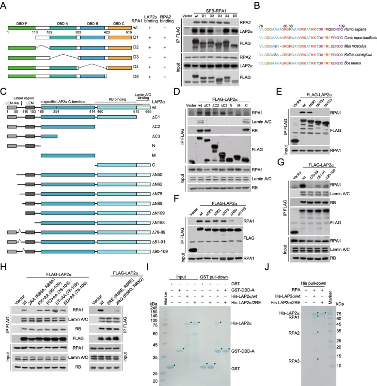
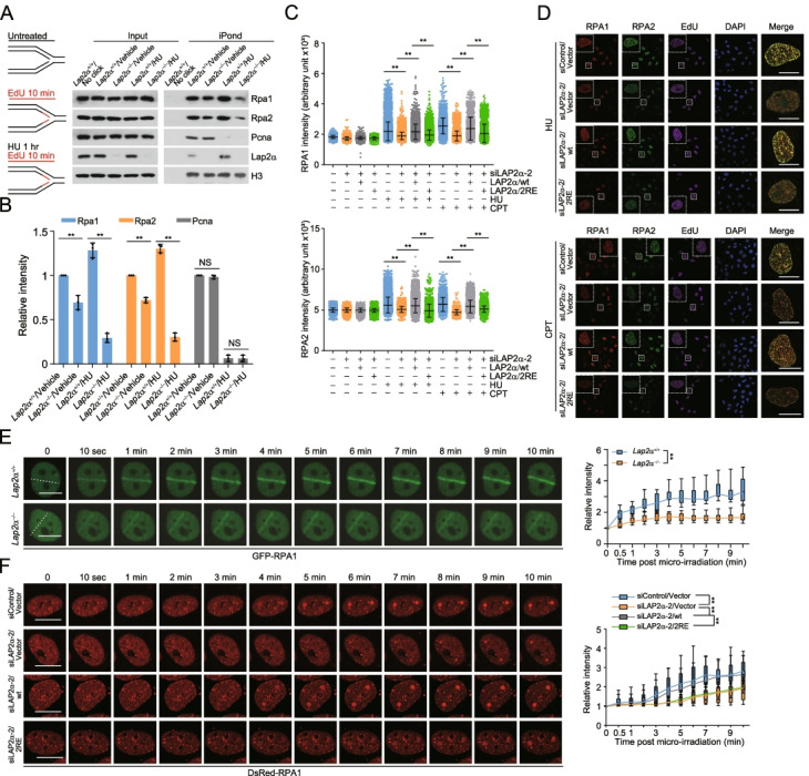
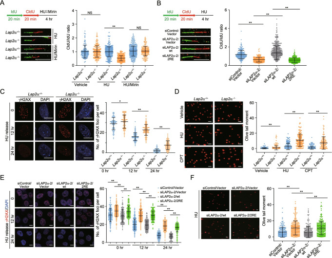
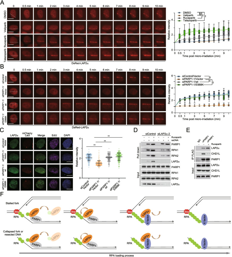
Results: Here, we report that the lamin-associated protein LAP2α is physically associated with RPA, and LAP2α preferentially facilitates RPA deposition on damaged chromatin via physical contacts between LAP2α and RPA1. Importantly, LAP2α-promoted RPA binding to ssDNA plays a critical role in protection of replication forks, activation of ATR, and repair of damaged DNA. We further demonstrate that the preference of LAP2α-promoted RPA loading on damaged chromatin depends on poly ADP-ribose polymerase PARP1, but not poly(ADP-ribosyl)ation.

Conclusions: Our study provides mechanistic insight into RPA deposition in response to DNA damage and reveals a genome protection role of LAP2α.

Keywords: ATR activation; Genome stability; Homologous recombination; LAP2α; PARP1; RPA loading.


Conflict of interest statement

The authors declare that they have no competing interests.

Figures

Fig. 1
Fig. 1
RPA is physically associated with LAP2α. A SILAC-based quantitative mass spectrometry analysis of RPA1-containing protein complexes with HeLa cells that allow doxycycline (Dox)-inducible expression of stably integrated FLAG-RPA1. Control cells were labelled with “heavy isotopic lysine and arginine” (K6R10) and the cells under Dox (1 ng/μl) treatment were labelled with “light isotopic lysine and arginine” (K0R0). DNase (300 U/ml)-treated cellular extracts were immunopurified with anti-FLAG affinity beads and eluted with FLAG peptide. The eluates were desalted by gel separation and mixed for digestion followed by mass spectrometry analysis. Biological duplicate experiments were performed. B Volcano plot showing the relative enrichment of RPA1-containing protein complex from SILAC-based quantitative mass spectrometry. Each point represents the potential interactor, and the 11 top candidates (fold change > 15 and P value < 0.01) are indicated. C Co-immunoprecipitation analysis of the interaction between LAP2α and RPA. Whole-cell lysates from HeLa cells were pre-treated with DNase followed by immunoprecipitation and immunoblotting with antibodies against the indicated proteins. α, anti-. D Co-immunoprecipitation analysis of the interaction between LAP2α and RPA. Whole-cell lysates from MCF-7 and U2OS cells were pre-treated with DNase followed by immunoprecipitation and immunoblotting with antibodies against the indicated proteins. E Analysis of the binding affinity of recombinant RPA purified from bacteria cells and His-tagged LAP2α purified from insect cells by biolayer interferometry (BLI) assay. The black line indicates fitted curves and the color traces represent raw data. Data are representative of two independent experiments. RPA1, RPA2, and RPA3 of the RPA complex were co-purified and examined by Coomassie brilliant blue staining. F Co-immunoprecipitation analysis of the association of individual recombinant RPA subunit with LAP2α. All proteins were individually purified from insect cells. G Co-immunoprecipitation analysis of the association of recombinant RPA1 with LAP2α in buffer with an increasing amount of ionic strength
Fig. 2
Fig. 2
Key determinants for LAP2α-RPA binding. A Whole-cell lysates from HeLa cells expressing S protein-FLAG-Streptavidin binding peptide (SFB)-tagged RPA1 deletions were immunoprecipitated and immunoblotted with antibodies against the indicated proteins. The RPA1 domain and deletion mutants annotated with residue numbers are shown. DBD represents DNA-binding domain. B Schematic representation of the evolutionarily conserved residues in the linker region of LAP2α. C Schematic representation of the domain structure of LAP2α and its variants. The domains and truncated or deleted variants are marked with the indicated residue numbers. The LEM (LAP2, emerin, MAN1) domain, LEM-like domain, LAP2α-specific C-terminus, and RB or Lamin A/C binding regions are shown. N, N-terminus from amino acid 1 to 185; M, middle region from 186 to 414; C, C-terminus from 415-695. ΔN50, residues from 1-50 are deleted; Δ76-89, residues from 76 to 89 are deleted; and so forth. D–F Whole-cell lysates from HeLa cells expressing FLAG-GFP-tagged LAP2α truncation mutants were immunoprecipitated and immunoblotted with antibodies against the indicated proteins. G Whole-cell lysates from HeLa cells expressing FLAG-GFP-tagged LAP2α deletion mutants were immunoprecipitated and immunoblotted with antibodies against the indicated proteins. H FLAG-tagged LAP2α variants were transfected into HeLa cells followed by co-immunoprecipitation and immunoblotting analysis. RK>AA (90-109) represents that all arginine and lysine residues from amino acid 90 to 109 of LAP2α are simultaneously replaced by alanine, and so forth. I GST pull-down assays with recombinant GST-DBD-A and His-tagged LAP2α variants. GST-DBD-A and His-tagged LAP2α were purified from bacteria cells and insect cells, respectively. J His pull-down assays with recombinant His-tagged LAP2α variants and RPA. RPA (including RPA1, RPA2, and RPA3) and His-tagged LAP2α variants were purified from bacteria cells and insect cells, respectively. The asterisks indicate the recombinant proteins stained by Coomassie brilliant blue
Fig. 3
Fig. 3
LAP2α promotes the loading of RPA onto damaged chromatin. A Proteins associated with replication forks were isolated by iPOND as described in “Methods” and detected by immunoblotting. Lap2α+/+ or Lap2α/ MEFs were EdU-labelled for 10 min and harvested immediately or after a 1-h chase with 2 mM HU. B Quantitative analysis of Rpa1, Rpa2, and Pcna binding to EdU-labelled nascent replication forks with iPOND assays. The relative intensity of chromatin bound factors to histone H3 was normalized against that in vehicle-treated control cells. C High content imaging analysis of foci intensity of RPA1 and RPA2 in U2OS cells. pLenti-vector, LAP2α/wt or LAP2α/2RE stably integrated U2OS cells were transfected with control or LAP2α 3′UTR siRNA (siLAP2α-2) and treated with 2 mM HU for 4 h or 1 μM CPT for 2 h followed by pre-extraction, fixation, immunostaining, and high content microscopy analysis. Before collection, cells were labelled with EdU for 1 h. D Representative images from C collected by confocal microscopy. E Laser micro-irradiation (IR) (50% laser energy) followed by live cell imaging analysis of GFP-RPA1 recruitment kinetics in GFP-RPA1 expressing Lap2α+/+ or Lap2α/ MEFs. Fluorescence intensities in micro-irradiated areas relative to the nuclear background were quantified (n > 20). F pLenti-vector, LAP2α/wt or LAP2α/2RE stably integrated U2OS cells were co-transfected with DsRed-RPA1 and control siRNA or LAP2α 3′UTR siRNA followed by Hoechst sensitization, micro-IR (405 nm) and live cell imaging analysis. Fluorescence intensities in micro-irradiated areas relative to the nuclear background were quantified (n > 20). Data are mean ± SDs for B, E, and F from biological triplicate experiments, and C from biological duplicate experiments. **P < 0.01; NS, not significant; one-way ANOVA for B; Mann-Whitney test for C; two-way ANOVA for E and F. Scale bar, 10 μm
Fig. 4
Fig. 4
LAP2α maintains DNA replication integrity through loading RPA onto ssDNA. A DNA fiber assay with Lap2α+/+ or Lap2α/ MEFs. Cells were sequentially labelled with DNA analog IdU and CldU for the indicated time followed by HU (4 mM, 4 h) treatment in the absence or presence of mirin (100 μM, 4 h). The ratios of CldU and IdU length were calculated in each treatment (n > 150). Scale bar, 10 μm. B pLenti-vector, LAP2α/wt or LAP2α/2RE stably integrated U2OS cells were transfected with control siRNA or LAP2α 3′UTR siRNA and experiments analogous to A were performed (n > 150). Scale bar, 10 μm. C Lap2α+/+ or Lap2α/ MEFs were treated with 2 mM HU for 16 h and released followed by γH2AX staining and confocal microscopy inspection. The foci number of γH2AX per cell in each treatment was quantified (n > 100). Scale bar, 10 μm. D Accumulation of damaged DNA was examined and quantified with alkaline comet assay in HU (2 mM, 4 h) or CPT (1 μM, 2 h) treated Lap2α+/+ or Lap2α/ MEFs (n > 150). Scale bar, 100 μm. E pLenti-vector, LAP2α/wt or LAP2α/2RE stably integrated U2OS cells were transfected with control siRNA or LAP2α 3′UTR siRNA and experiments analogous to C were performed (n > 150). Scale bar, 10 μm. F pLenti-vector, LAP2α/wt or LAP2α/2RE stably integrated U2OS cells were transfected with control siRNA or LAP2α 3′UTR siRNA followed by HU treatment and experiments analogous to D were performed (n > 300). Scale bar, 100 μm. Data are mean ± SDs for A and B from biological duplicate experiments, and C–F from biological triplicate experiments. *P < 0.05; **P < 0.01; NS, not significant; Mann-Whitney test
Fig. 5
Fig. 5
LAP2α-promoted RPA loading is required for ATR activation and homologous recombination. A Immunoblotting analysis of ATR kinase activity. Control U2OS cells and LAP2α/wt or LAP2α/2RE stably integrated U2OS cells were transfected with indicated siRNAs and in the absence or presence of CPT (1 μM, 1 h). The cellular extracts were collected to examine CPT-induced phosphorylation events. B DNA fiber assay with Lap2α+/+ or Lap2α/ MEFs. Cells were sequentially labelled with DNA analog IdU and CldU for the indicated time. Fork speed (n > 120) and fork symmetry (n > 25) were determined by measuring the length of CldU track, and the percentage of new origins was quantified with only CldU staining fibers. C Immunostaining and confocal microscopy analysis of RAD51 foci formation. LAP2α/wt or LAP2α/2RE stably integrated U2OS cells were transfected with LAP2α 3′UTR siRNA, exposed to IR (4 Gy) and cultured for 3 h followed by 1 h EdU labelling before collection. The foci number of RAD51 per EdU-positive cell in each group was quantified (n > 150). D Homologous recombination efficiencies monitored by DR-GFP reporter assays. Control DR-GFP U2OS cells and DR-GFP U2OS cells that allow for Dox-inducible expression of stably integrated pTRE-LAP2α/wt or pTRE-LAP2α/2RE were co-transfected with HA-I-SceI and the indicated siRNAs. Cells were treated with vehicle or Dox (1 ng/ μl) for 48 h to induce the expression of LAP2α/wt or LAP2α/2RE. The proportions of GFP-positive cells were determined by flow cytometry. E Survival analysis of U2OS cells expressing LAP2α 3′UTR siRNA and LAP2α/wt or LAP2α/2RE under different drug treatment. Data are mean ± SDs for B from biological duplicate experiments, and C–E from biological triplicate experiments. **P < 0.01; NS, not significant; Mann-Whitney test for B and C; one-way ANOVA for D; two-way ANOVA for E. Scale bar, 10 μm
Fig. 6
Fig. 6
LAP2α is engaged into damaged chromatin in a PARP1-dependent manner. A Laser micro-IR (35% laser energy) followed by live cell imaging analysis of DsRed-LAP2α recruitment kinetics in PARP1 inhibitor (PARPi)-treated U2OS cells. DsRed-LAP2α expressing cells were pre-treated with different PARPis (10 μM) for 4 h before laser micro-IR. Fluorescence intensities in micro-irradiated areas relative to the nuclear background were quantified (n > 20). B Laser micro-IR (50% laser energy) followed by live cell imaging analysis of DsRed-LAP2α recruitment kinetics in PARP1-knockdwon or -overexpression U2OS cells. pLenti-vector, PARP1/wt, or PARP1/E988K stably integrated U2OS cells were co-transfected with control siRNA or PARP1 3′UTR siRNA (siPARP1-1) and DsRed-LAP2α. Fluorescence intensities in micro-irradiated areas relative to the nuclear background were quantified (n > 20). C U2OS-LacO cells expressing mCherry-LacI, PARP1 variants, and PARP1 3′UTR siRNA were labelled with EdU for 1 h before immunostaining and confocal microscopy analysis. The intensity of LAP2α foci in mCherry-LacI and EdU-positive cells was quantified and normalized to the nuclear background (n > 100). D ssDNA-pull-down analysis of LAP2α accumulation, PARP1 binding, and RPA loading with nuclear extracts from LAP2α-knockdown U2OS cells under rucaparib (10 μM, 4 h) treatment. 5’ biotin-labelled 70-nt ssDNA was used in pull-down assays. E Co-immunoprecipitation analysis of the interaction between LAP2α and PARP1 with cellular extracts from U2OS cells pre-treated with vehicle or rucaparib (10 μM, 4 h). F A proposed regulatory model for PARP1 in directing LAP2α-promoted RPA loading at damaged chromatin. X-factor represents the molecular machinery that may function to retain the longer occupancy of LAP2α on damaged chromatin. Data are mean ± SDs for A–C from biological triplicate experiments. **P < 0.01; NS, not significant; two-way ANOVA for A and B; Mann-Whitney test for C. Scale bar, 10 μm

References

    1. Buisson R, Boisvert JL, Benes CH, Zou L. Distinct but concerted roles of ATR, DNA-PK, and Chk1 in countering replication stress during S phase. Mol Cell. 2015;59:1011–1024. - PMC - PubMed
    1. Cimprich KA, Cortez D. ATR: an essential regulator of genome integrity. Nat Rev Mol Cell Biol. 2008;9:616–627. - PMC - PubMed
    1. Blackford AN, Jackson SP. ATM, ATR, and DNA-PK: the trinity at the heart of the DNA damage response. Mol Cell. 2017;66:801–817. - PubMed
    1. Halazonetis TD, Gorgoulis VG, Bartek J. An oncogene-induced DNA damage model for cancer development. Science. 2008;319:1352–1355. - PubMed
    1. Bartkova J, Horejsi Z, Koed K, Kramer A, Tort F, Zieger K, et al. DNA damage response as a candidate anti-cancer barrier in early human tumorigenesis. Nature. 2005;434:864–870. - PubMed

Publication types

LinkOut - more resources