

Optical coherence tomography in diagnosing polypoidal choroidal vasculopathy. Looking into the future: a systematic review and meta-analysis
- PMID: 35227320
- PMCID: PMC8883730
- DOI: 10.1186/s40942-022-00365-5
Optical coherence tomography in diagnosing polypoidal choroidal vasculopathy. Looking into the future: a systematic review and meta-analysis
Abstract
Background: Polypoidal choroidal vasculopathy (PCV) is an exudative maculopathy with features similar to wet age macular degeneration. The incidence of PCV is known to be higher in the Asian population compared to Caucasians. Imaging modality is needed to make the diagnosis of PCV. Although Indocyanine green angiography (ICGA) is still the gold standard, it is not routinely performed in vitreoretinal practice. Thus another imaging modality is currently a popular research area. Spectral domain optical coherence tomography (SD-OCT) has emerged as a new imaging modality mostly available in clinics. Some studies have reported the sensitivity and specificity of SD-OCT in diagnosing PCV with different results and thresholds.
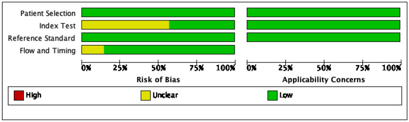
Methods: Relevant studies from PubMed, Science Direct and Google Scholar databases were systematically searched. In random effect models using STATA 14 software, a meta-analysis was performed to determine the pooled diagnostic accuracy. QUADAS 2 was used to evaluate the risk of bias of each study by Revman 5.4 software.
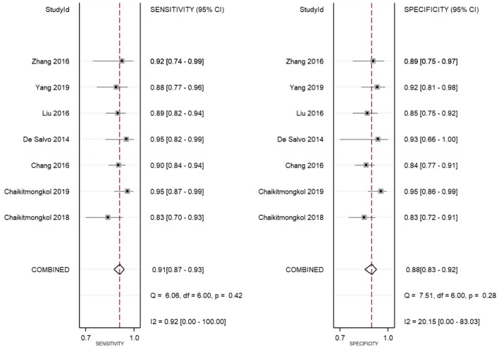
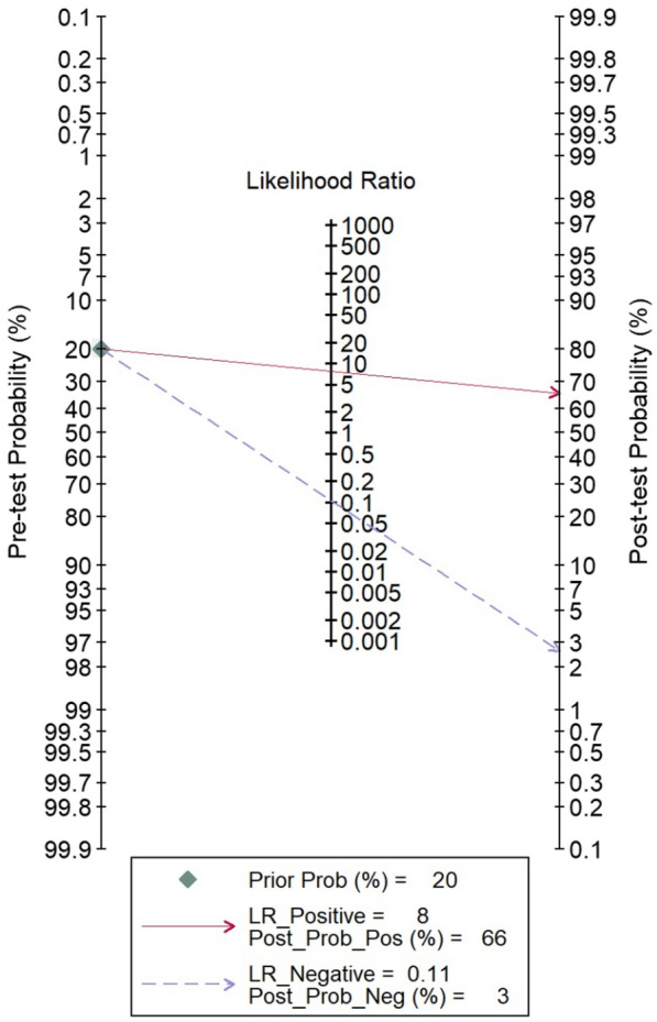
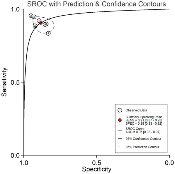
Results: Seven eligible studies which met the inclusion and exclusion criteria were enrolled in this study. A total of 911 eyes were included to investigate the diagnostic accuracy of SD-OCT. As a result, the pooled sensitivity was 0.91 (95% CI 0.87-0.93), specificity 0.88 (95% 0.83-0.92), positive likelihood ratio 8, negative likelihood ratio 11, the area under the summary receiver operating characteristic curve 0.95 (95% CI 0.93-0.97), and diagnostic odds ratio 71.81 (95% CI 38.89-132.74).
Conclusion: SD-OCT provided a high diagnostic value for detecting PCV. Sharply peaked pigment epithelial detachment (PED), notched PED, bubble sign, multiple PED, and double-layer sign were the most common features found in PCV.
Keywords: Diagnosis; Indocyanine green angiography (ICGA); Meta-analysis; Optical coherence tomography (OCT); Polypoidal choroidal vasculopathy.
© 2022. The Author(s).
Conflict of interest statement
We would like to give the following facts which may be considered as potential conflict to this work. We wish to confirm that there are no known conflict of interest in our publication, therefore we ensures that the publisher has the author’s license of copyright and permission to publish the studies.
Figures


References
-
- Cheung CMG, Lai TYY, Teo K, Ophth M, Ruamviboonsuk P, Chen S, et al. Polypoidal choroidal vasculopathy consensus nomenclature and none indocyanine green angiograph diagnostic criteria from the Asia-Pacific Ocular Imaging Society PCV Workgroup. Ophthalmology. 2020;128:443–452. doi: 10.1016/j.ophtha.2020.08.006. - DOI - PubMed
Publication types
LinkOut - more resources
Full Text Sources
Research Materials
Miscellaneous
