Observational Study
. 2022 Apr:173:91-100.
doi: 10.1016/j.resuscitation.2022.02.017. Epub 2022 Feb 25.

Magnetic resonance imaging adds prognostic value to EEG after pediatric cardiac arrest

Affiliations
Observational Study

Magnetic resonance imaging adds prognostic value to EEG after pediatric cardiac arrest

Alyssa E Smith et al. Resuscitation. 2022 Apr.

Abstract

Aim: To investigate how combined electrographic and radiologic data inform outcomes in children after cardiac arrest.

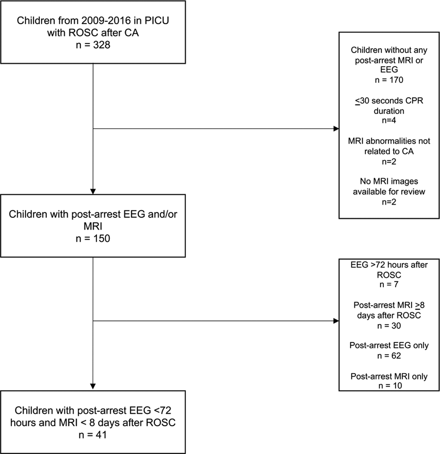
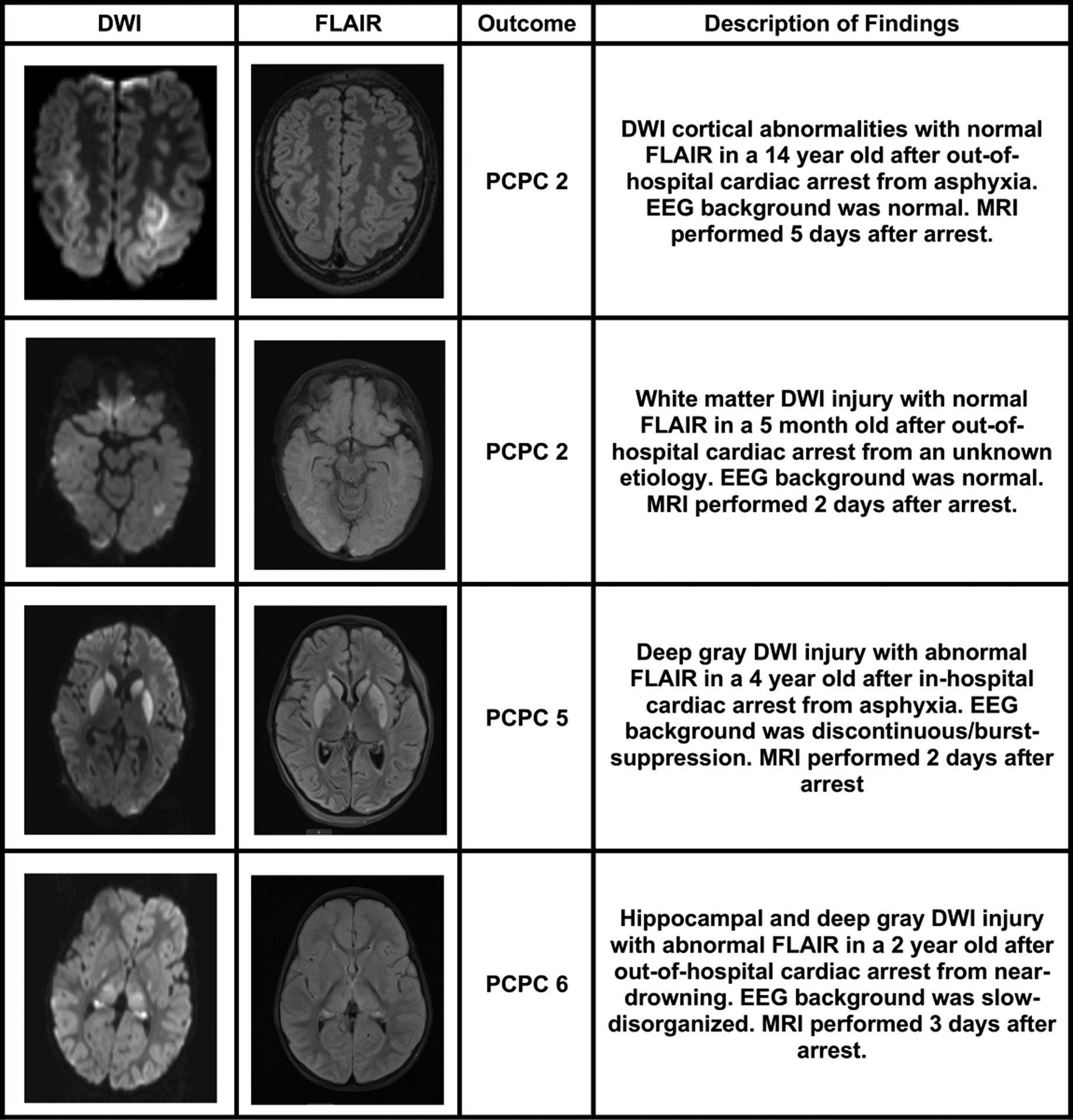
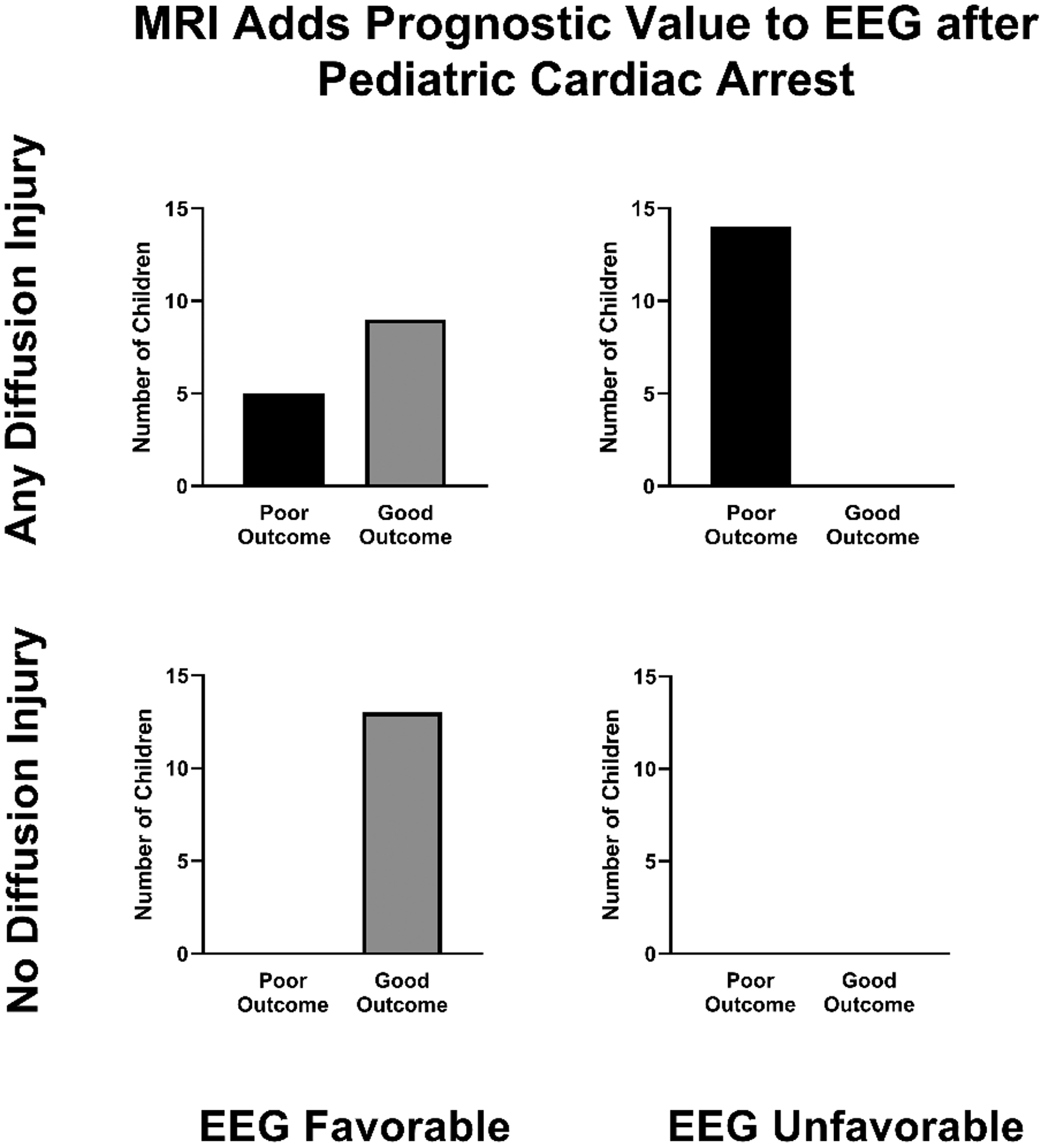
Methods: Retrospective observational study of children admitted to the pediatric intensive care unit (PICU) of a tertiary children's hospital with diagnosis of cardiac arrest from 2009 to 2016. The first 20 min of electroencephalogram (EEG) background was blindly scored. Presence and location of magnetic resonance imaging (MRI) diffusion-weighted image (DWI) abnormalities were correlated with T2-weighted signal. Outcomes were categorized using Pediatric Cerebral Performance Category (PCPC) scores at hospital discharge, with "poor outcome" reflecting a PCPC score of 4-6. Logistic regression models examined the association of EEG and MRI variables with outcome.

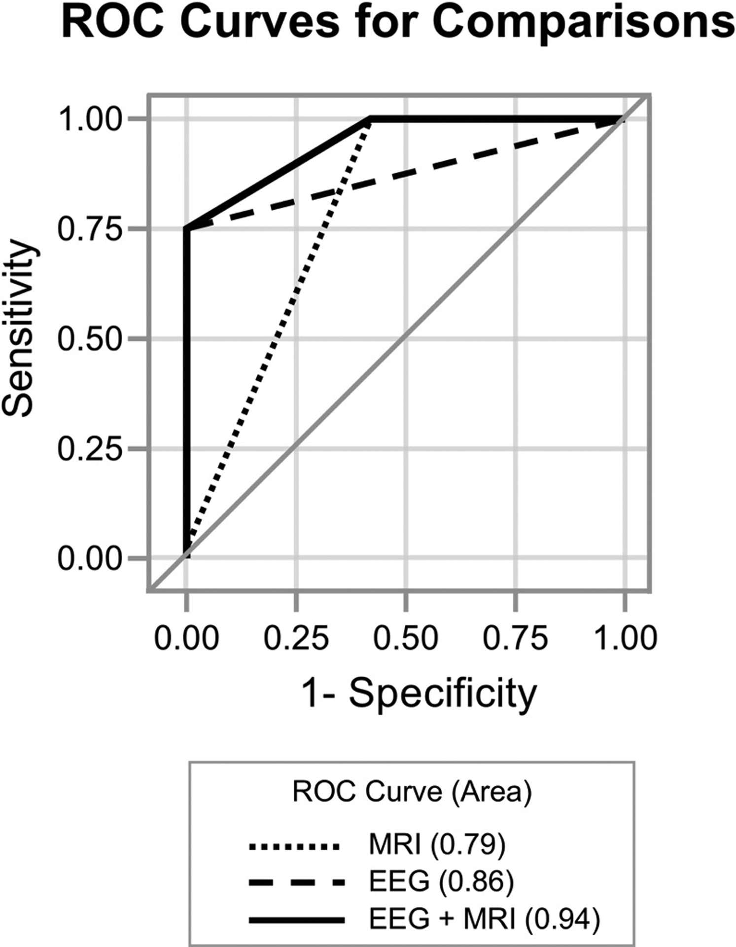
Results: 41 children met inclusion criteria and had both post-arrest EEG monitoring within 72 hours after ROSC and brain MRI performed within 8 days. Among the 19 children with poor outcome, 10 children did not survive to discharge. Severely abnormal EEG background (p < 0.0001) and any diffusion restriction (p < 0.0001) were associated with poor outcome. The area under the ROC curve (AUC) for identifying outcome based on EEG background alone was 0.86, which improved to 0.94 with combined EEG and MRI data (p = 0.02).

Conclusion: Diffusion abnormalities on MRI within 8 days after ROSC add to the prognostic value of EEG background in children surviving cardiac arrest.

Keywords: EEG; MRI; Outcome; Pediatric cardiac arrest.


Conflict of interest statement

Declaration of Competing Interest The authors declare that they have no known competing financial interests or personal relationships that could have appeared to influence the work reported in this paper.

Figures

Fig. 1:
Fig. 1:. Inclusion and Exclusion.
PICU=pediatric intensive care unit; ROSC=return of spontaneous circulation; CA=cardiac arrest; EEG=electroencephalogram; MRI= magnetic resonance imaging.
Figure 2:
Figure 2:. Examples of post-arrest neuroimaging abnormalities
Four children with MRI performed after cardiac arrest are highlighted. MRI= magnetic resonance imaging; DWI=diffusion-weighted imaging; FLAIR=fluid-attenuated inversion recovery; PCPC=Pediatric Cerebral Performance Category.
Fig. 3:
Fig. 3:
MRI diffusion abnormalities with EEG background in relation to outcome at hospital discharge. All children with poor outcome had MRI diffusion abnormalities, of which more had an unfavorable EEG background than favorable. No child with unfavorable EEG background had normal diffusion imaging. Favorable EEG background includes normal or slow-disorganized and unfavorable EEG background includes discontinuous/burst-suppression or attenuated-featureless. EEG=electroencephalogram; MRI= magnetic resonance imaging.
Fig. 4
Fig. 4. Multimodal test results improve ability to discriminate poor outcome after pediatric cardiac arrest.
ROC curves depicting EEG suppressed background (Category 3 or 4, AUC=0.86), MRI diffusion abnormalities (AUC=0.79) and combined (EEG+MRI, AUC=0.94) were compared. A model combining EEG suppressed background with MRI diffusion abnormalities is better able to distinguish poor outcome than either EEG (p=0.02) or MRI alone (p=0.0008). EEG=electroencephalogram performed less than 72 hours after cardiac arrest; MRI=magnetic resonance imaging, post-arrest; AUC=area under the ROC curve.

References

    1. Suominen P, Olkkola KT, Voipio V, Korpela R, Palo R, Räsänen J. Utstein style reporting of in-hospital paediatric cardiopulmonary resuscitation. Resuscitation. 2000;45(1):17–25. doi:10.1016/S0300-9572(00)00167-2 - DOI - PubMed
    1. Reis AG, Nadkarni V, Perondi MB, Grisi S, Berg RA. A Prospective Investigation Into the Epidemiology of In-Hospital Pediatric Cardiopulmonary Resuscitation Using the International Utstein Reporting Style. PEDIATRICS. 2002;109(2):200–209. doi:10.1542/peds.109.2.200 - DOI - PubMed
    1. Berg RA, Nadkarni VM, Clark AE, et al. Incidence and Outcomes of Cardiopulmonary Resuscitation in PICUs: Crit Care Med. Published online December 2015:1. doi:10.1097/CCM.0000000000001484 - DOI - PMC - PubMed
    1. Atkins DL, Everson-Stewart S, Sears GK, et al. Epidemiology and Outcomes From Out-of-Hospital Cardiac Arrest in Children: The Resuscitation Outcomes Consortium Epistry–Cardiac Arrest. Circulation. 2009;119(11):1484–1491. doi:10.1161/CIRCULATIONAHA.108.802678 - DOI - PMC - PubMed
    1. Ichord R, Silverstein FS, Slomine BS, et al. Neurologic outcomes in pediatric cardiac arrest survivors enrolled in the THAPCA trials. Neurology. 2018;91(2):e123–e131. doi:10.1212/WNL.0000000000005773 - DOI - PMC - PubMed

Publication types