Review
. 2022 May;17(5):637-650.
doi: 10.1016/j.jtho.2022.01.021. Epub 2022 Feb 25.

Thymic Carcinomas-A Concise Multidisciplinary Update on Recent Developments From the Thymic Carcinoma Working Group of the International Thymic Malignancy Interest Group

Affiliations
Review

Thymic Carcinomas-A Concise Multidisciplinary Update on Recent Developments From the Thymic Carcinoma Working Group of the International Thymic Malignancy Interest Group

Anja C Roden et al. J Thorac Oncol. 2022 May.

Abstract

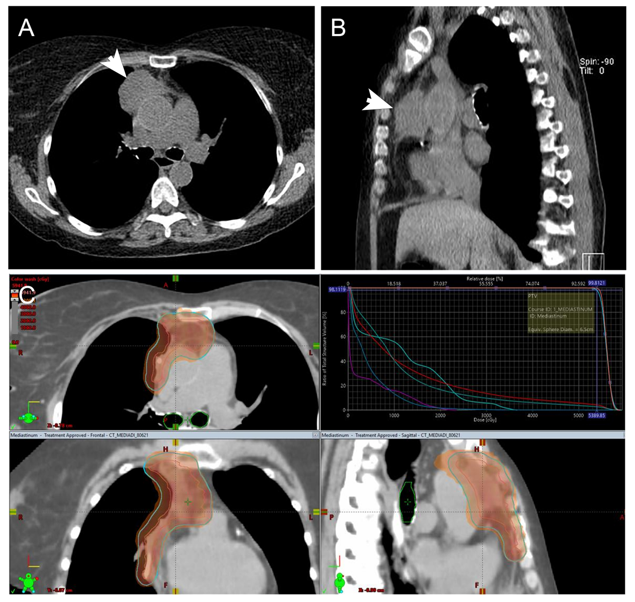
Thymic carcinomas are rare malignancies that in general arise in the prevascular (anterior) mediastinum. These tumors are usually invasive, often present at advanced stages, and typically behave aggressively. Studies are hampered by the paucity of these tumors, the large variety of carcinoma subtypes, and the lack of unique morphologic and immunophenotypic features. Despite these challenges, advances in diagnostic imaging, surgical approaches, systemic therapies, and radiation therapy techniques have been made. The WHO classification of thymic epithelial tumors has been updated in 2021, and the eighth tumor nodal metastasis staging by the American Joint Committee on Cancer/Union for International Cancer Control included thymic carcinomas in 2017. Molecular alterations that provide more insight into the pathogenesis of these tumors and that potentially permit use of novel targeted therapies are increasingly being identified. New approaches to radiation therapy, chemotherapy, and immunotherapy are under evaluation. International societies, including the International Thymic Malignancy Interest Group, European Society of Thoracic Surgeons, and Japanese, Chinese, and Korean thymic associations, have been critical in organizing and conducting multi-institutional clinical studies. Herein, we review contemporary multidisciplinary perspectives in diagnosis and management of thymic carcinoma.

Keywords: ITMIG; International Thymic Malignancy Interest Group; TNM staging; Thymic carcinoma; WHO.


Figures

Figure 1:
Figure 1:
Relationship between Masaoka Koga staging and TNM staging.
Figure 2.
Figure 2.
Thymic squamous cell carcinoma in a 28-year-old woman. A. CT imaging from an FDG PET-CT study shows an 11 cm prevascular necrotic appearing mass (m) with a small right pleural effusion (e), right hilar (curved arrow) and a subcarinal smaller than 1 cm lymph node (straight arrow). B. Fused image shows the mass is markedly FDG avid with an SUVmax of 12.2. The lymph nodes have an SUVmax of up to 4. Thymic carcinoma is typically markedly FDG avid. This FDG uptake is useful in identifying metastatic disease which may be overlooked with morphological imaging (CT or MRI), as seen with the subcarinal lymph node which was smaller than 1cm yet showed FDG uptake and was proven to be due to metastatic disease, N2, upstaging the patient to stage IVb.
Figure 3:
Figure 3:
Micronodular thymic carcinoma with lymphoid hyperplasia. A. A circumscribed lesion is comprised of scattered nodular areas (*) in a background of lymphoid follicles with germinal centers (arrow). B. Lymphoid follicles with germinal centers (arrow) are in between paler cellular nodules (*). C. The nodules are comprised of neoplastic cells that are characterized by a polygonal shape, prominent nucleoli and occasional mitotic figures (arrow). The neoplastic cells are positive for keratin (pankeratin) which also highlights the nodular arrangement of the neoplastic cells (D), CD5 (E), and CD117 (F). Magnification, H&E x 1.25 (A), x 10 (B), x 40 (C), pankeratin scanning power (D), CD5 x 400 (E), CD117 x 400 (F).
Figure 4:
Figure 4:
Thymic carcinoma entities described during the last 5 years: A.-C. Hyalinizing clear cell carcinoma A. Nests of tumor cells with clear cytoplasm in hyalinized stroma. B. P40 staining labels all tumor cell nuclei. C. CK5/6 stain reveals heterogeneous positivity of tumor cells (“mosaic pattern”). D.-F. Sebaceous carcinoma D. Epidermoid tumor cells and clear sebocytes. E. P40 staining of epidermoid cell nuclei but not sebocyte nuclei. F. Adipophilin staining restricted to sebocytes. Androgen receptor was also positive in the sebaceous carcinoma (not shown). Magnification, H&E x 50 (A), x 200 (D), p40 x 100 (B, E), CK5/6 x 100 (C), adipophilin x 100 (F)
Figure 5:
Figure 5:
A 62-year-old woman with TNM stage II thymic carcinoma (arrows on non-contrast enhanced CT chest; A, axial slices, B, sagittal slices) underwent wedge resection of the right upper lobe lung and pericardectomy (R1 resection with positive anterior and posterior margins and invasion of the pericardium) (pT2N0M0). No adjuvant chemotherapy was given due to stage IV chronic kidney disease. She was going to be treated with post-operative radiotherapy for R1 resection. C. Volumetric modulated arc therapy radiation treatment plan to 5400 cGy in 30 fractions. The red line indicates gross tumor volumes; the blue line is the planning target and the color wash overlay is the 100% isodose level.

Similar articles

Cited by

References

    1. Koizumi T, Otsuki K, Tanaka Y, et al.: National incidence and initial therapy for thymic carcinoma in Japan: based on analysis of hospital-based cancer registry data, 2009-2015. Jpn J Clin Oncol 2020, 50:434–9. - PubMed
    1. Roden AC, Fang W, Shen Y, et al.: Distribution of Mediastinal Lesions Across Multi-Institutional, International, Radiology Databases. J Thorac Oncol 2020, 15:568–79. - PubMed
    1. Shepherd A, Riely G, Detterbeck F, et al.: Thymic Carcinoma Management Patterns among International Thymic Malignancy Interest Group (ITMIG) Physicians with Consensus from the Thymic Carcinoma Working Group. J Thorac Oncol 2017, 12:745–51. - PMC - PubMed
    1. Radovich M, Pickering CR, Felau I, et al.: The Integrated Genomic Landscape of Thymic Epithelial Tumors. Cancer Cell 2018, 33:244–58 e10. - PMC - PubMed
    1. Padda SK, Yao X, Antonicelli A, et al.: Paraneoplastic Syndromes and Thymic Malignancies: An Examination of the International Thymic Malignancy Interest Group Retrospective Database. J Thorac Oncol 2018, 13:436–46. - PMC - PubMed

Publication types