. 2022 Jun 1;68(3):198-208.
doi: 10.1262/jrd.2021-128. Epub 2022 Feb 26.

Polarized epithelium-sperm co-culture system reveals stimulatory factors for the secretion of mouse epididymal quiescin sulfhydryl oxidase 1

Affiliations

Polarized epithelium-sperm co-culture system reveals stimulatory factors for the secretion of mouse epididymal quiescin sulfhydryl oxidase 1

Yu-Syuan Wei et al. J Reprod Dev. .

Abstract

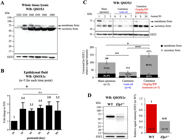
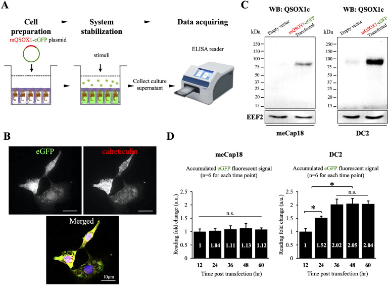
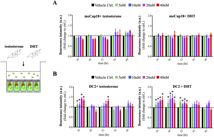
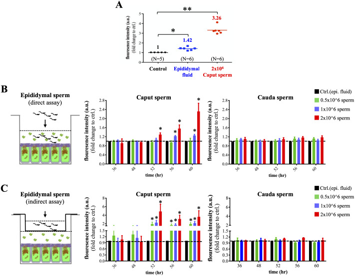
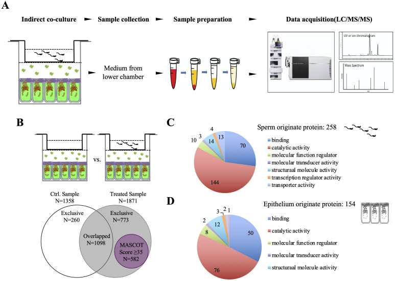
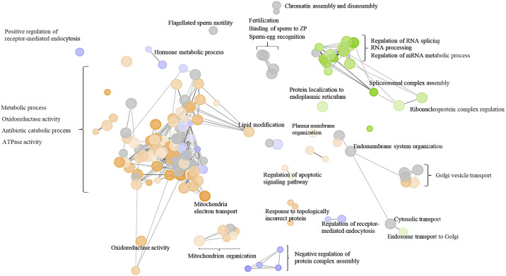
Spermatozoa acquire fertilization ability through post-translational modifications. These membrane surface alterations occur in various segments of the epididymis. Quiescin sulfhydryl oxidases, which catalyze thiol-oxidation reactions, are involved in disulfide bond formation, which is essential for sperm maturation, upon transition and migration in the epididymis. Using castration and azoospermia transgenic mouse models, in the present study, we showed that quiescin sulfhydryl oxidase 1 (QSOX1) protein expression and secretion are positively correlated with the presence of testosterone and sperm cells. A two-dimensional in vitro epithelium-sperm co-culture system provided further evidence in support of the notion that both testosterone and its dominant metabolite, 5α-dihydrotestosterone, promote epididymal QSOX1 secretion. We also demonstrated that immature caput spermatozoa, but not mature cauda sperm cells, exhibited great potential to stimulate QSOX1 secretion in vitro, suggesting that sperm maturation is a key regulatory factor for mouse epididymal QSOX1 secretion. Proteomic analysis identified 582 secretory proteins from the co-culture supernatant, of which 258 were sperm-specific and 154 were of epididymal epithelium-origin. Gene Ontology analysis indicated that these secreted proteins exhibit functions known to facilitate sperm membrane organization, cellular activity, and sperm-egg recognition. Taken together, our data demonstrated that testosterone and sperm maturation status are key regulators of mouse epididymal QSOX1 protein expression and secretion.

Keywords: Epididymis; Epithelium; Fertility; Quiescin sulfhydryl oxidase; Spermatozoa.


Conflict of interest statement

The authors declare no conflicts of interest that could prejudice the impartiality of the research reported.

Figures

Fig. 1.
Fig. 1.
Effects of testosterone and spermatozoa on QSOX1 protein expression and secretion in the mouse epididymis. (A) Epididymal tissue from 20- to 80-postnatal day mice were analyzed using western blot, and a representative blot has been presented. Postnatal epididymal QSOX1 expression coincided with the presence of spermatozoa and 1st testosterone surge at day 30. (B) Epididymal fluid collected from 20 to 80-postnatal day mice was subjected to western blot analysis for the detection of secretory QSOX1 (QSOX1c). A significant 2.4-fold increase in QSOX1c was detected at postnatal day 30, following which the signal became steady. For each postnatal time-point, epididymal fluid from 3 individual animals was collected, for quantitative analysis. (C) Epididymal tissues from loss- and gain-of-function mouse castration models were homogenized to examine the effect of testosterone on QSOX1 protein expression. Ten ICR mice were randomly allocated into sham operation (n = 3), castration+corn oil (n = 4, IP 100 μl corn oil), and castration+testosterone (n = 3, IP 5 mg/kg body weight in 100 μl dissolved in corn oil) groups. Significant deceases in both membrane and secretory forms of epididymal QSOX1 were detected after castration; addition of exogenous testosterone rescued epididymal QSOX1 protein expression. (D) Azoospermia mouse model supported a positive association between the presence of sperm and epididymal QSOX1 protein expression, as a pronounced decline (–59%) in QSOX1c was detected in the absence of sperm cells. Four animals for each phenotype (wild-type or Vasa-Cre:Elp–/–) were used for western blot quantification analysis, and a representative blot has been presented. Statistical analysis was performed as described in the Materials and Methods section. Bars represent mean +/– standard deviation. Statistical significance was considered at P < 0.05 (*). N.S indicates a non-significant difference.
Fig. 2.
Fig. 2.
Validation of mouse QSOX1-eGFP transfection and secretion in epididymal epithelial cell lines. (A) Mouse caput epididymal epithelium were transiently transfected with mouse QSOX1-eGFP plasmid. After the designed experimental procedures, the culture supernatant was collected for ELISA detection of the eGFP signal. (B) Indirect immunofluorescent staining against eGFP was used to validate the presence of transfected mQSOX1-eGFP fusion protein. The perinuclear eGFP staining was similar to the known cellular localization of endogenous QSOX1. The eGFP signal largely overlapped with calreticulin, an ER marker, indicating that eGFP-QSOX1 is involved in the ER-mediated secretory pathway. Representative images from DC2 cells have been presented. (C) Western blot analysis for QSOX1c confirmed the presence and overexpression of transfected mQSOX1-eGFP, at the size of ~97 kDa, both in meCap18 and DC2 cells. Representative western blot images have been presented. (D) Culture supernatants from transfected meCap18 and DC2 cells were collected at intervals of 12 h, following which the accumulated eGFP signals were measured. No apparent changes were detected from the meCap18 culture supernatants. An elevated signal was detected in the DC2 culture supernatants at 24 h and 36 h post-transfection; the fact that the signal became steady after 36 h indicated a minimal spontaneous secretion activity after this time-point. Statistical analysis was performed, as described in the Materials and Methods section. Bars represent mean +/– standard deviation. Statistical significance was considered at P < 0.05 (*). N.S. indicates a non-significant difference.
Fig. 3.
Fig. 3.
Testosterone and 5α-dihydrotestosterone (DHT) stimulate QSOX1 secretion in vitro. (A) Upon transfection of meCap18 with mQSOX1-eGFP, neither testosterone nor DHT stimulated QSOX1 secretion, as no significant changes were detected in the eGFP signal, at all time-points measured. (B) When DC2 was used, testosterone exerted a dose-dependent stimulatory effect at 36 h. In comparison to testosterone, DHT showed persistent stimulatory effects on QSOX1 secretion, until 52 h. Statistical analysis was performed as described in the Materials and Methods section. Six experimental repeats were performed for each concentration tested, at each time-point. Bars represent mean +/– standard deviation. Statistical significance was considered at P < 0.05 (*).
Fig. 4.
Fig. 4.
In vitro evaluation of sperm as a factor for QSOX1 secretion. (A) Transfected DC2 was used to examine the effects of exogenous stimuli on the secretion of QSOX1. Epididymal fluid exerted significant stimulatory effect (1.42-fold) on QSOX1 secretion. A 3.26-fold increase in eGFP signal was detected when 2 × 10⁶ caput sperm cells were co-cultured with DC2, which indicated that the caput sperm was more effective in stimulating epididymal QSOX1 secretion than luminal factors. (B) In the direct stimulation assay, we observed dose-dependent stimulatory effects on QSOX1 secretion from the caput sperm, but not the cauda sperm, at 52 h. (C) In the indirect stimulation assay, we observed dose-dependent effects from the caput sperm, but not the cauda sperm, on promoting QSOX1 secretion, at 52 h. Moreover, the level of stimulation was more apparent (2- to 6-fold), as compared to that upon direct stimulation (1.2- to 2.6-fold). For each concentration tested at each time-point, 6 experimental repeats were performed. Bars represent mean +/– standard deviation. Statistical significance was considered at P < 0.05. Asterisks indicate levels of significance, * P < 0.05; ** P < 0.01.
Fig. 5.
Fig. 5.
Proteomic identification of sperm-epithelium interactomes responsible for QSOX1 secretion. (A) To identify sperm-releasing factors that might be responsible for stimulating QSOX1 secretion, supernatant from the lower chamber of the indirect co-culture (sperm-DC2) system was collected at 52 h, for proteomics analysis. (B) After subtracting protein IDs from the control group (without the presence of sperm cells), 773 proteins were found to be exclusively present in the stimulated group (in grey). Based on MASCOT proteomic criteria described in the Materials and Methods section, 582 proteins were considered reliable IDs (in purple circle). (C) Further analyses showed that among the 582 identified IDs, 258 were sperm-origin proteins. The majority of sperm-origin proteins (55.8%) had known function for catalytic activity, while around 27% were responsible for the cellular binding process. (D) Analysis of 154 epididymal epithelium proteins indicated that two major functions for epithelium secretomes were related to catalytic activity (49.4%) and cellular binding process (32.5%).
Fig. 6.
Fig. 6.
Panther Gene Ontology (GO) analyses of proteomics data based on biological processes and molecular functions. Reliable protein IDs (n = 582) were processed for Panther GO analysis. Subcategories were created and are shown using different colors. Associated molecular functions have also been indicated accordingly. The majority of the identified proteins exhibited a known function related to metabolic process, oxidoreductase activity, and antibiotic catabolic process. Other proteins identified were also highly related to sperm maturation processes, such as sperm oocyte recognition, sperm-oocyte binding, chromatin assembly, plasma membrane organization, and lipid modification. Another group of proteins were found to have function in regulating Golgi vesicle transport, which might be responsible for the ER-mediated cellular secretory pathway of epididymal QSOX1.

Similar articles

Cited by

References

    1. Kuo YW, Li SH, Maeda K, Gadella BM, Tsai PS. Roles of the reproductive tract in modifications of the sperm membrane surface. J Reprod Dev 2016; 62: 337–343. - PMC - PubMed
    1. Nixon B, De Iuliis GN, Hart HM, Zhou W, Mathe A, Bernstein IR, Anderson AL, Stanger SJ, Skerrett-Byrne DA, Jamaluddin MFB, Almazi JG, Bromfield EG, Larsen MR, Dun MD. Proteomic Profiling of Mouse Epididymosomes Reveals their Contributions to Post-testicular Sperm Maturation. Mol Cell Proteomics 2019; 18(Suppl 1): S91–S108. - PMC - PubMed
    1. Browne JA, Leir SH, Yin S, Harris A. Transcriptional networks in the human epididymis. Andrology 2019; 7: 741–747. - PMC - PubMed
    1. Sipilä P, Björkgren I. Segment-specific regulation of epididymal gene expression. Reproduction 2016; 152: R91–R99. - PubMed
    1. Sullivan R, Légaré C, Lamontagne-Proulx J, Breton S, Soulet D. Revisiting structure/functions of the human epididymis. Andrology 2019; 7: 748–757. - PubMed

Substances