

miR-32 promotes MYC-driven prostate cancer
- PMID: 35228520
- PMCID: PMC8885642
- DOI: 10.1038/s41389-022-00385-8
miR-32 promotes MYC-driven prostate cancer
Abstract
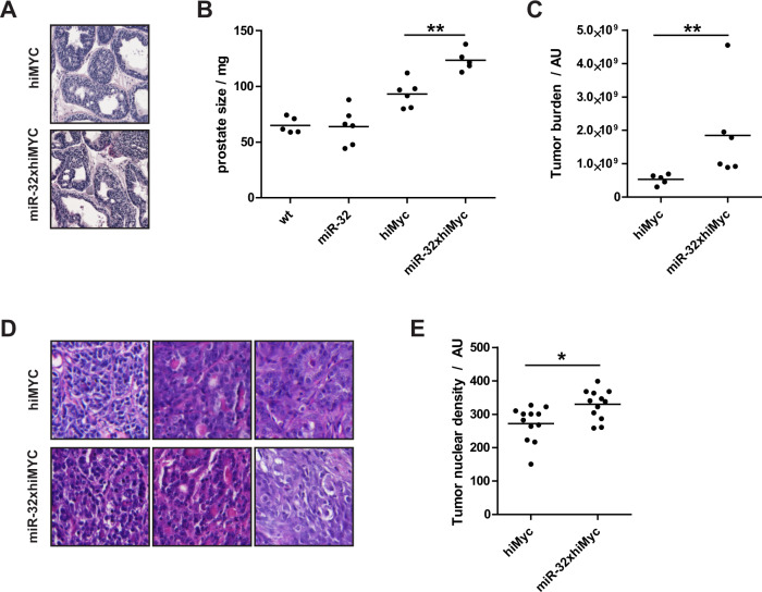
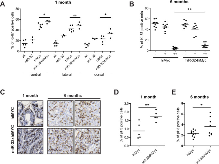
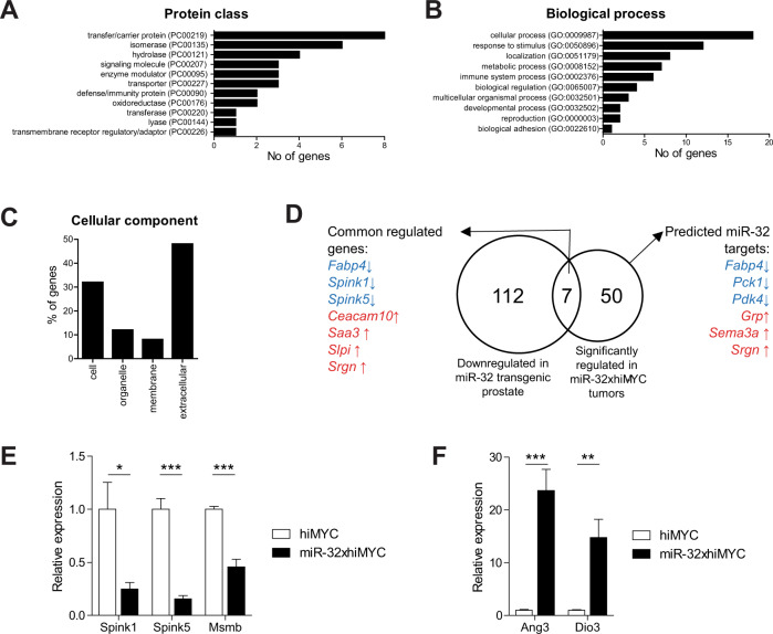
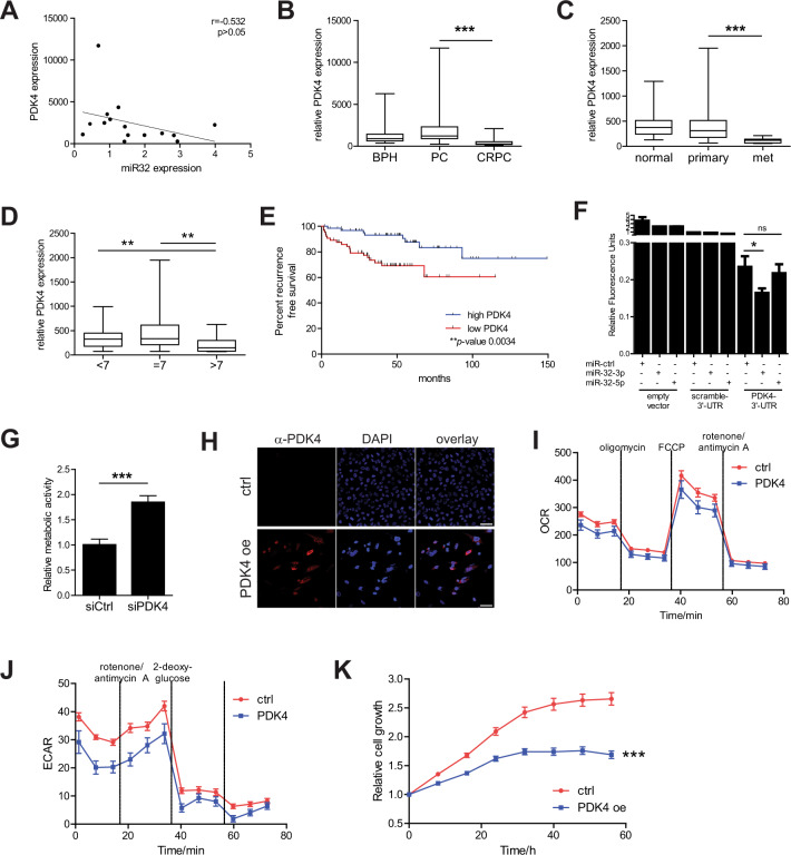
miR-32 is an androgen receptor (AR)-regulated microRNA, expression of which is increased in castration-resistant prostate cancer (PC). We have previously shown that overexpression of miR-32 in the prostate of transgenic mice potentiates proliferation in prostate epithelium. Here, we set out to determine whether increased expression of miR-32 influences growth or phenotype in prostate adenocarcinoma in vivo. We studied transgenic mice expressing MYC oncogene (hiMYC mice) to induce tumorigenesis in the mouse prostate and discovered that transgenic overexpression of miR-32 resulted in increased tumor burden as well as a more aggressive tumor phenotype in this model. Elevated expression of miR-32 increased proliferation as assessed by Ki-67 immunohistochemistry, increased nuclear density, and higher mitotic index in the tumors. By gene expression analysis of the tumorous prostate tissue, we confirmed earlier findings that miR-32 expression regulates prostate secretome by modulating expression levels of several PC-related target genes such as Spink1, Spink5, and Msmb. Further, we identified Pdk4 as a tumor-associated miR-32 target in the mouse prostate. Expression analysis of PDK4 in human PC reveals an inverse correlation with miR-32 expression and Gleason score, a decrease in castration-resistant and metastatic tumors compared to untreated primary PC, and an association of low PDK4 expression with a shorter recurrence-free survival of patients. Although decreased PDK4 expression induces the higher metabolic activity of PC cells, induced expression of PDK4 reduces both mitotic respiration and glycolysis rates as well as inhibits cell growth. In conclusion, we show that miR-32 promotes MYC-induced prostate adenocarcinoma and identifies PDK4 as a PC-relevant metabolic target of miR-32-3p.
© 2022. The Author(s).
Conflict of interest statement
The authors declare no competing interests.
Figures
Similar articles
-
NDRG2 acts as a negative regulator downstream of androgen receptor and inhibits the growth of androgen-dependent and castration-resistant prostate cancer.Cancer Biol Ther. 2015;16(2):287-96. doi: 10.1080/15384047.2014.1002348. Cancer Biol Ther. 2015. PMID: 25756511 Free PMC article.
-
lncRNA PCAT19 promotes the proliferation of laryngocarcinoma cells via modulation of the miR-182/PDK4 axis.J Cell Biochem. 2019 Aug;120(8):12810-12821. doi: 10.1002/jcb.28552. Epub 2019 Mar 13. J Cell Biochem. 2019. PMID: 30868666
-
In Vivo Expression of miR-32 Induces Proliferation in Prostate Epithelium.Am J Pathol. 2017 Nov;187(11):2546-2557. doi: 10.1016/j.ajpath.2017.07.012. Epub 2017 Aug 19. Am J Pathol. 2017. PMID: 28827140
-
Comprehensive proteomic profiling identifies the androgen receptor axis and other signaling pathways as targets of microRNAs suppressed in metastatic prostate cancer.Oncogene. 2016 May 5;35(18):2345-56. doi: 10.1038/onc.2015.295. Epub 2015 Sep 14. Oncogene. 2016. PMID: 26364608 Free PMC article.
-
Resolving the Coffey Paradox: what does the androgen receptor do in normal vs. malignant prostate epithelial cells?Am J Clin Exp Urol. 2018 Apr 1;6(2):55-61. eCollection 2018. Am J Clin Exp Urol. 2018. PMID: 29666833 Free PMC article. Review.
Cited by
-
Role of mir-32-3p in the diagnosis and risk assessment of osteoporotic fractures.J Orthop Surg Res. 2024 Nov 1;19(1):709. doi: 10.1186/s13018-024-05206-9. J Orthop Surg Res. 2024. PMID: 39487541 Free PMC article.
-
Recent progress in microRNA research for prostate cancer.Discov Oncol. 2024 Sep 27;15(1):480. doi: 10.1007/s12672-024-01376-4. Discov Oncol. 2024. PMID: 39331237 Free PMC article. Review.
-
Modulation of Small RNA Signatures by Astrocytes on Early Neurodegeneration Stages; Implications for Biomarker Discovery.Life (Basel). 2022 Oct 27;12(11):1720. doi: 10.3390/life12111720. Life (Basel). 2022. PMID: 36362875 Free PMC article. Review.
-
The effect of neural network architecture on virtual H&E staining: Systematic assessment of histological feasibility.Patterns (N Y). 2023 Apr 7;4(5):100725. doi: 10.1016/j.patter.2023.100725. eCollection 2023 May 12. Patterns (N Y). 2023. PMID: 37223268 Free PMC article.
-
Molecular landscape for risk prediction and personalized therapeutics of castration-resistant prostate cancer: at a glance.Front Endocrinol (Lausanne). 2024 Jun 3;15:1360430. doi: 10.3389/fendo.2024.1360430. eCollection 2024. Front Endocrinol (Lausanne). 2024. PMID: 38887275 Free PMC article. Review.
References
-
- Torre LA, Bray F, Siegel RL, Ferlay J, Lortet-Tieulent J, Jemal A. Global cancer statistics, 2012. CA Cancer J Clin. 2015;65:87–108. - PubMed
-
- Schröder FH, Hugosson J, Roobol MJ, Tammela TL, Ciatto S, Nelen V, et al. Screening and prostate-cancer mortality in a randomized European study. N. Engl J Med. 2009;360:1320–8. - PubMed
-
- Ha M, Kim VN. Regulation of microRNA biogenesis. Nat Rev Mol Cell Biol. 2014;15:509–24. - PubMed
-
- Hausser J, Zavolan M. Identification and consequences of miRNA-target interactions-beyond repression of gene expression. Nat Rev Genet. 2014;15:599–612. - PubMed
Grants and funding
LinkOut - more resources
Full Text Sources
Research Materials
