. 2022 Feb 28;13(1):1076.
doi: 10.1038/s41467-022-28724-8.

HPV infection alters vaginal microbiome through down-regulating host mucosal innate peptides used by Lactobacilli as amino acid sources

Affiliations

HPV infection alters vaginal microbiome through down-regulating host mucosal innate peptides used by Lactobacilli as amino acid sources

Alizee Lebeau et al. Nat Commun. .

Abstract

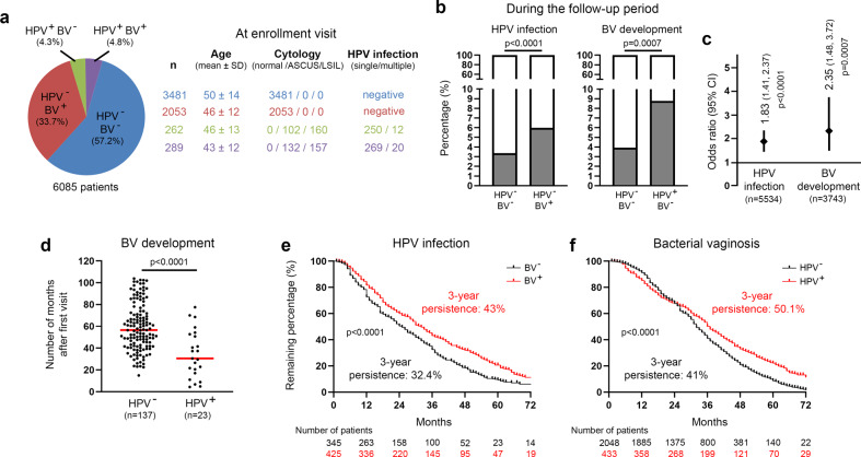
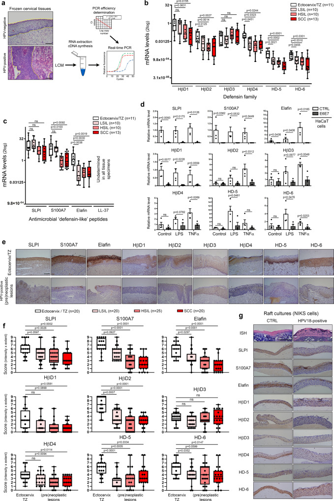
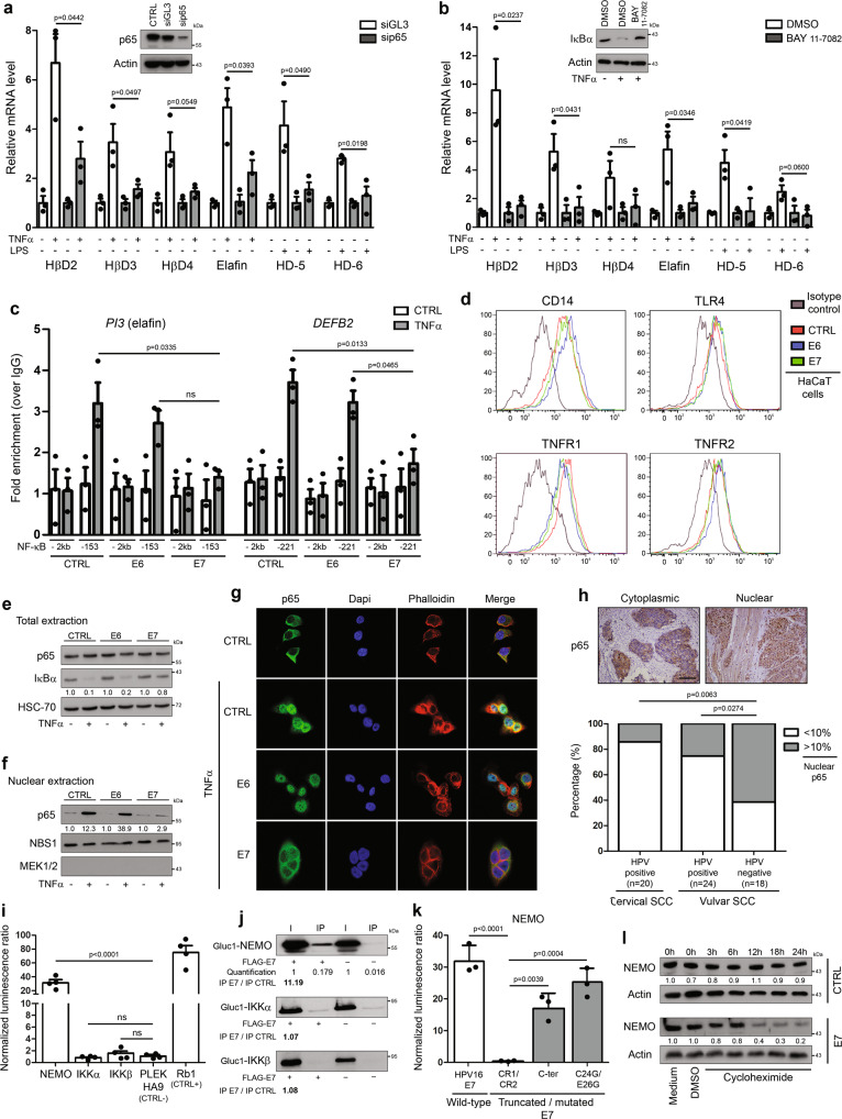
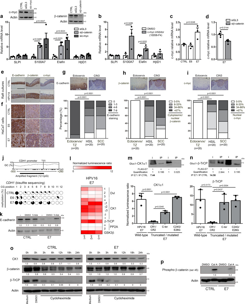
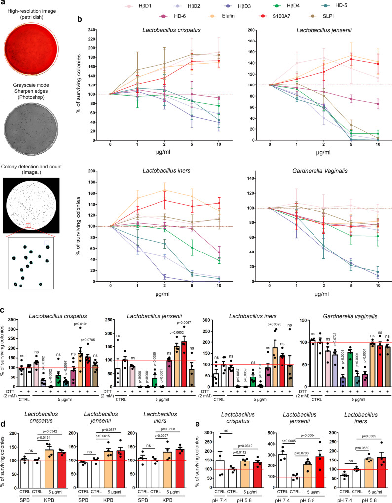
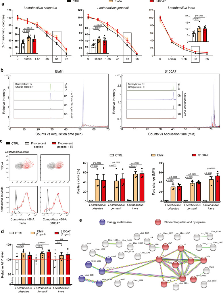
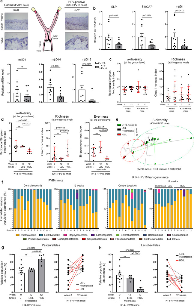
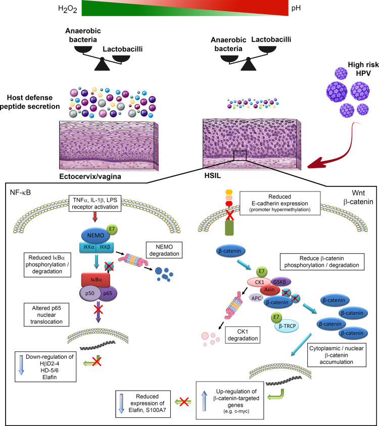
Despite the high prevalence of both cervico-vaginal human papillomavirus (HPV) infection and bacterial vaginosis (BV) worldwide, their causal relationship remains unclear. While BV has been presumed to be a risk factor for HPV acquisition and related carcinogenesis for a long time, here, supported by both a large retrospective follow-up study (n = 6,085) and extensive in vivo data using the K14-HPV16 transgenic mouse model, we report a novel blueprint in which the opposite association also exists. Mechanistically, by interacting with several core members (NEMO, CK1 and β-TrCP) of both NF-κB and Wnt/β-catenin signaling pathways, we show that HPV E7 oncoprotein greatly inhibits host defense peptide expression. Physiologically secreted by the squamous mucosa lining the lower female genital tract, we demonstrate that some of these latter are fundamental factors governing host-microbial interactions. More specifically, several innate molecules down-regulated in case of HPV infection are hydrolyzed, internalized and used by the predominant Lactobacillus species as amino acid source sustaining their growth/survival. Collectively, this study reveals a new viral immune evasion strategy which, by its persistent/negative impact on lactic acid bacteria, ultimately causes the dysbiosis of vaginal microbiota.


Conflict of interest statement

The authors declare no competing interests.

Figures

Fig. 1
Fig. 1. Retrospective clinical follow-up analysis evaluating the relationship between genital HPV infection and BV occurrence/persistence.
a General characteristics of the study population (n = 6,085). b Probability of high-risk HPV infection or BV development during the follow-up period according to the status for the other gynecological disorder at first (enrollment) visit. c Forest plot showing the odds ratio (OR) and 95% confidence intervals (CI) for developing one pathological condition when the other one was preceding in time [HPV infection (OR: 1.83, 95% CI: 1.41–2.37); BV development (OR: 2.35, 95% CI: 1.48–3.72)]. d Number of months for BV occurrence in HPV-positive and -negative patients. e Kaplan-Meier estimates for the persistence of HPV infection according to the BV status (negative versus positive). The clearance of HPV infections was ascertained by both cytology (Bethesda system) and PCR-based HPV test (Abbott High-risk HPV assay). f Kaplan–Meier curve for the persistence of BV according to the HPV status. Two consecutive negative results (using Hay/Ison grading system) at least 12 months apart were required to consider a patient really/durably cured of BV. P values were determined using two-sided Fisher’s exact test b, c, two-sided unpaired t-tests (d) and log-rank (Mantel-Cox) test e, f. Source data are provided as a Source Data file.
Fig. 2
Fig. 2. HPV oncoproteins impair both the constitutive and inducible innate (antimicrobial) peptide expression.
a Schematic illustration of the steps involved in gene expression analysis using microdissected frozen specimens. mRNA expression of epithelial-specific members of the defensin b and “defensin-like” c families was measured by RT-qPCR in HPV-positive (pre)neoplastic lesions (LSIL, n = 10; HSIL, n = 10; SCC, n = 13). Uninfected squamous samples [ectocervix, vagina, transformation zone (TZ)] (n = 11) were used as control. Gene expression was normalized using four calibrator genes (HPRT, GAPDH, 18S and TBP). Box limits: 25th to 75th percentiles; line: median; whiskers: minimum to maximum. d RT-qPCR analysis of SLPI, S100A7, elafin, HβD1-4 and HD-5/6 expression in immortalized keratinocytes stably transduced or not with HPV16 E6/E7 oncoproteins and stimulated with TNFα or LPS. Each experiment was normalized to the amount of HPRT mRNA from the same sample. Results represent the means ± SEM of four independent experiments. e HPV-negative and positive tissue specimens stained for antimicrobial peptides expressed by the squamous epithelium lining the lower part of the female reproductive tract. A reduced immunoreactivity for most of these innate factors was clearly observed in HPV-infected (pre)neoplastic lesions. f Semi-quantitative evaluation of innate peptide expression (intensity and extent of the immunostainings) in both normal epithelium from HPV-negative samples (n = 20) and (pre)neoplastic lesions (LSIL, n = 20; HSIL, n = 25; SCC, n = 20). Box limits: 25th to 75th percentiles; line: median; whiskers: minimum to maximum. g Representative control and HPV18-positive organotypic raft culture sections stained for all analyzed host defense (antimicrobial) peptides. As a control, HPV DNA was detected by in situ hybridization and, consistent with an episomal infection, a diffuse punctate pattern was observed. Images are representative of three independent experiments. The scale bar represents 100 μm. P values were determined using one-way ANOVA followed by Dunnett’s multiple comparison post-hoc test b, c, two-sided unpaired t-tests d and ANOVA Kruskal–Wallis test followed by Dunn post-hoc test f. ns: not significant (p > 0.1). Source data are provided as a Source Data file.
Fig. 3
Fig. 3. E7 viral oncoprotein reduces drastically TNFα/LPS-induced innate peptide expression through inducing NEMO degradation and impairing subsequent p65 nuclear translocation.
TNFα/LPS-induced defensin/elafin expression was analyzed by RT-qPCR in non-infected cells transfected with siRNA targeting p65 a or treated with 5 µM BAY 11-7082 b. Cells transfected with control siRNA (siGL3) or treated with DMSO were used as negative controls. Each experiment was normalized to the amount of HPRT mRNA from the same sample. Results represent the means ± SEM of three independent experiments. c Occupancy of NF-κB binding sites on PI3 (-153 bp) and DEFB2 (-221 bp) promoters was evaluated by ChIP in control, HPV16 E6-positive and E7-positive cells. Primers targeting a region with no putative NF-κB sites (-2 kb) in both promoters served as negative controls. Results represent the means ± SEM of three independent experiments. d Analysis of cell surface expression of TNFα (TNFR1-2) and LPS (CD14 and TLR4) receptors by control, HPV16 E6 and E7-transduced cells using flow cytometry. Keratinocytes stably transduced or not with HPV16 E6 or E7 were first treated with TNFα and both the degradation of IκBα e and the nuclear translocation of p65 was then analyzed by Western blot f or immunofluorescence g. The absence of colocalization between p65 and DAPI in E7-transduced cells (cytoplasmic sequestration of p65) should be noticed. h Anti-p65 immunostaining in both HPV-positive (n = 44) and negative SCC (n = 18). Low densities of epithelial cells displaying a nuclear p65 immunoreactivity were observed in virus-related tumors. Of note, tissue specimens of vulvar cancer were included due to the impossibility of analyzing HPV-negative SCC arising from the uterine cervix. i Binding of protein members of the IKK kinase complex with HPV16 E7 oncoprotein assessed by Gaussia princeps luciferase complementation assay (GPCA). Retinoblastoma-associated protein (Rb1) and PLEKHA9 were used as positive and negative control, respectively. Results represent the means ± SEM of four independent experiments. j Co-IP of NEMO, IKKα and IKKβ with HPV16 E7 oncoprotein. k GPCA analyzing the binding of truncated/mutated forms of HPV16 E7 with NEMO. Results represent the means ± SEM of three independent experiments. l NEMO stability in both control and HPV16 E7-transduced cells following treatment with 100 µM cycloheximide. The scale bar represents 100 μm. P values were determined using two-sided unpaired t-tests a, b, one-way ANOVA followed by Dunnett’s multiple comparison post-hoc test c, i, Fisher’s exact test h and one-way ANOVA followed by Bonferroni post-hoc test k. ns: not significant (p > 0.1). The Western blot and co-IP analyses were independently performed three times and twice, respectively. One representative experiment is shown. Source data are provided as a Source Data file.
Fig. 4
Fig. 4. E7 viral oncoprotein inhibits constitutive innate peptide expression (elafin and S100A7) through promoting β-catenin stabilization/signaling and subsequent c-myc expression.
The reactivation of so called “constitutive” peptide (SLPI, S100A7, elafin and HβD1) expression was analyzed by RT-qPCR in HPV16 E6E7-positive cells transfected with siRNA targeting c-myc or β-catenin a or treated with 40 µM 10058-F4 b. Cells transfected with control siRNA (siGL3) or treated with DMSO were used as negative controls. Each experiment was normalized to the amount of HPRT mRNA from the same sample. Results represent the means ± SEM of three b or four a independent experiments. The level of c-myc mRNA was determined by RT-qPCR in control, HPV16 E6 and E7-transduced cells c. Results represent the means ± SEM of three independent experiments. The effect of knockdown of β-catenin on c-myc expression in HPV16 E7-positive cells was assessed by RT-qPCR d. Results represent the means ± SEM of four independent experiments. e Representative pictures of control and HPV18-positive organotypic raft culture sections stained for E-cadherin, β-catenin and c-myc. f Representative examples of anti-E-cadherin, β-catenin and c-myc immunoreactivities displayed by control, E6-positive and E7-positive keratinocytes. Anti-E-cadherin g, anti-β-catenin h and anti-c-myc immunostainings i in both HPV-negative specimens [ectocervix/transformation zone (TZ), n = 20] and HPV-positive (pre)neoplastic lesions (HSIL, n = 20; SCC, n = 20). Note the reduced expression of E-cadherin associated with cytoplasmic/nuclear β-catenin immunoreactivity observed in HPV-positive tissues. Regarding c-myc, its expression was almost exclusively detected in HPV-positive (pre)neoplastic lesions. j The methylation status of E-cadherin gene (CDH1) promoter in control, HPV16 E6 and E7-transduced cells was assessed by bisulfite genomic sequencing. The variation in GC content within CDH1 promoter as well as the percentage of methylation of each individual CpG island contained in the PCR fragment analyzed by bisulfite sequencing are shown. k The reactivation of hypermethylated CDH1 gene was determined by Western blot following 5-aza-deoxycytidine (5-Aza, 5 µM) treatment. l Binding of protein members of the β-catenin degradation complex with HPV16 E7 oncoprotein was assessed by Gaussia princeps luciferase complementation assay (GPCA). Heatmap representing the score (normalized luminescence ratio) of each analyzed protein for potential interaction with HPV E7 is shown. Three independent experiments were performed. The interactions between HPV16 E7 oncoprotein and CK1α1 m or β-TrCP n were validated by both Co-IP and GPCA using the truncated/mutated forms of E7. Results represent the means ± SEM of three independent experiments. o CK1, β-catenin and β-TrCP stability in both control and HPV16 E7-transduced cells following treatment with cycloheximide (100 µM). p CK1-dependent phosphorylation of β-catenin (ser45) in both control and E7-positive cells following treatment with calyculin A (50 nM). The scale bar represents 100 μm. P values were determined using one-way ANOVA followed by Dunnett’s multiple comparison post-hoc test a, c, two-sided unpaired t-tests b, d, χ2 test g, h, i and one-way ANOVA followed by Bonferroni post-hoc test m, n. The Western blot and co-IP analyses were independently performed three times and twice, respectively. One representative experiment is shown. Source data are provided as a Source Data file.
Fig. 5
Fig. 5. Innate peptides abundantly and constitutively secreted by the squamous mucosa lining the lower gynecologic tract exhibit a positive effect on Lactobacillus survival.
a Schematic illustration of the different steps involved in bacterial colony detection and count. b Systematic analysis of both defensin and “defensin-like” peptide activity on predominant bacterial species detected in normal (L. crispatus, L. jensenii and L. iners) and pathological (BV) (G. vaginalis) conditions. Results represent the means ± SEM of three (L. crispatus, L. jensenii and L. iners) or five (G. vaginalis) independent experiments. c Effect of reducing environment on antimicrobial/protective activity of tested innate peptides. Results represent the means ± SEM of four (L. jensenii and G. vaginalis), five (L. iners) or six (L. crispatus) independent experiments. The positive effect on Lactobacillus survival displayed by the most abundant innate peptides secreted by cervical/vaginal mucosa (elafin and S100A7) was also assessed in both potassium phosphate buffer (KPB) (d) and acidic (pH 5.8) conditions (e). For each experiment, peptides were incubated with bacteria for 6 h. Results represent the means ± SEM of four independent experiments. P values were determined using one-way ANOVA followed by Dunnett’s multiple comparison post-hoc test (c, e). ns: not significant (p > 0.1). Source data are provided as a Source Data file.
Fig. 6
Fig. 6. Host defense peptides are metabolized and used as amino acid source by Lactobacillus species.
a Elafin and S100A7 were incubated with Lactobacillus species for 45 min to 9 h in the absence of nutrients (starvation assay) and the percentage of surviving colonies was determined. Results (each time point) represent the means ± SEM of six (L. iners: CTRL, elafin and S100A7), eight (L. crispatus/L. jensenii: elafin and S100A7) or eleven (L. crispatus/L. jensenii: CTRL) independent experiments. b The hydrolysis of host defense peptides (elafin and S100A7) by the dominant Lactobacillus species constituting the vaginal microbiome was evaluated at different time points (0 h, 3 h and 6 h) by mass spectrometry. Note the disappearance of native proteins after 3 to 6 h. c The internalization of fluorescent-labeled peptides within the cytoplasm of bacteria was then assessed by flow cytometry. Both the percentage of positive cells and mean fluorescence intensity (MFI) are shown. Results represent the means ± SEM of three independent experiments. d ATP production after 6 h of incubation between peptides and bacteria. Results represent the means ± SEM of five independent experiments. e STRING analysis of the bacterial proteins that have incorporated 13C615N2-labeled lysines (from exogenous elafin). All proteins identified by mass spectrometry in L. crispatus, L. jensenii or L. iners were pooled. P values were determined using two-way ANOVA followed by Bonferroni post-hoc test a and one-way ANOVA followed by Dunnett’s multiple comparison post-hoc test c, d. ns: not significant (p > 0.1). Source data are provided as a Source Data file.
Fig. 7
Fig. 7. Estrogen-induced cervical/vaginal carcinogenesis in transgenic mice expressing HPV16 induces an imbalance in the vaginal microflora.
a Schematic representation of the mouse reproductive tract. The morphology of the squamous epithelium lining the cervix/vagina and vulva in K14-HPV16 and control (FVB/n) mice is shown. Both the increased percentage of proliferative (Ki-67-positive) cells and the thickening of the epithelium in case of HPV16 oncogene expression should be noticed. b mRNA level of SLPI, S100A7, mouse orthologs of HβD1-3 (mβD1, mβD4, mβD14) and HD-5/6 (mβD12, mβD15) was measured by RT-qPCR. Microdissected frozen squamous epithelia from FVB/n and K14-HPV16 mice were analyzed. Each experiment was normalized to the amount of both HPRT and GAPDH mRNAs from the same sample. Results represent the means ± SEM of ten independent experiments. c Bacterial intrinsic diversity (reciprocal Simpson biodiversity index) and richness (deduced from Chao1 index). The reported values (at the genus level) for each individual mouse in the four defined groups [FVB/n (week 0 versus week 12) and K14-HPV16 (week 0 versus week 12)] are shown (n = 12 per group). The means ± SD are represented. d Bacterial α-diversity, genus richness and evenness (Simpson index) for K14-HPV16 mice. Data were separated depending on 17β-estradiol treatment duration [week 0 (n = 12) versus week 12 (n = 12)] and preneoplastic lesion grade [hyperplasia/LSIL (n = 6) versus HSIL (n = 6)]. The means ± SD are represented. e β-diversity of the vaginal microbial profile in K14-HPV16 mice was visualized using a Bray-Curtis dissimilarity matrix-based non-parametric dimensional scaling (NMDS) model (three dimensions). f Stacked bar charts depicting the relative abundance of the twelve main bacterial orders detected in control (FVB/n) and K14-HPV16 mice by 16 S V5-V6 amplicon sequencing. Relative abundance of bacteria belonging to the order of Pasteurellales g and Lactobacillales h in K14-HPV16 mice depending on 17β-estradiol treatment duration [week 0 (n = 12) versus week 12 (n = 12)] and HPV-related lesion grade [hyperplasia/LSIL (n = 6) versus HSIL (n = 6)]. The means ± SEM are represented. The comparison of relative abundance of bacteria in paired (week 0 versus week 12) lavage samples is also shown. The scale bar represents 100 μm. P values were determined using two-sided unpaired t-tests b and non-parametric Kruskal-Wallis test corrected with a two-stage linear step-up procedure of Benjamini, Krieger and Yekutieli c, d, g, h. ns: not significant (p > 0.1). Source data are provided as a Source Data file.
Fig. 8
Fig. 8. Schematic representation of the proposed model.
Left In HPV-positive cells, E7 oncoprotein impairs pro-inflammatory-induced innate peptide expression through inducing NEMO degradation and subsequent p65 cytoplasmic sequestration. Right In parallel, E7 reduces E-cadherin expression and alters β-catenin degradation complex by interacting with both CK1 and β-TrCP. As a result, defense peptides (e.g., elafin and S100A7) which are used by Lactobacillus species as amino acid source are greatly down-regulated by the β-catenin target gene c-myc. An imbalance in the vaginal flora is, therefore, achieved as a consequence of HPV persistence and subsequent immune evasion.

References

    1. de Sanjose S, et al. Worldwide prevalence and genotype distribution of cervical human papillomavirus DNA in women with normal cytology: a meta-analysis. Lancet Infect. Dis. 2007;7:453–459. - PubMed
    1. de Martel C, Plummer M, Vignat J, Franceschi S. Worldwide burden of cancer attributable to HPV by site, country and HPV type. International journal of cancer. J. Int. du cancer. 2017;141:664–670. - PMC - PubMed
    1. Schiffman M, et al. Carcinogenic human papillomavirus infection. Nat. Rev. Dis. Prim. 2016;2:16086. - PubMed
    1. Luo X, et al. HPV16 drives cancer immune escape via NLRX1-mediated degradation of STING. J. Clin. Investig. 2020;130:1635–1652. - PMC - PubMed
    1. Chang YE, Laimins LA. Microarray analysis identifies interferon-inducible genes and Stat-1 as major transcriptional targets of human papillomavirus type 31. J. Virol. 2000;74:4174–4182. - PMC - PubMed

Publication types