. 2022 Feb 28;12(1):3343.
doi: 10.1038/s41598-022-07287-0.

Surgical treatment of diffuse and multi-lobes involved glioma with the assistance of a multimodal technique

Affiliations

Surgical treatment of diffuse and multi-lobes involved glioma with the assistance of a multimodal technique

Jiayu Liu et al. Sci Rep. .

Abstract

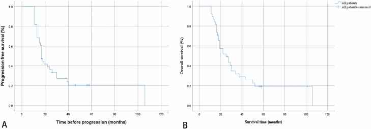
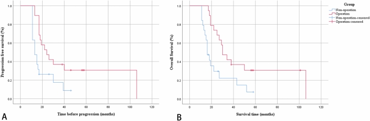
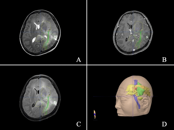
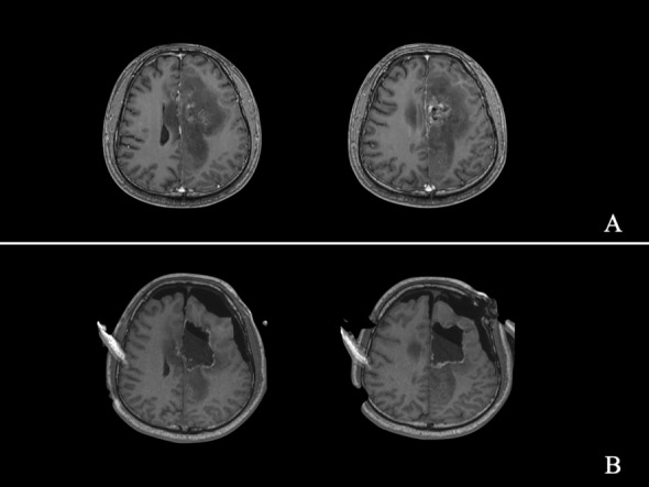
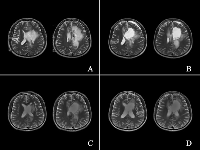
Diffuse and multi-lobes involved glioma (DMG) is a rare disease, and the aim of this study was to assess the role of multimodal-assisted surgical resection of tumours combined with chemoradiotherapy and identify prognosis. Clinical data were collected from 38 patients with a diagnosis of DMG. Nineteen patients received multimodal-assisted surgical resection of tumours combined with chemoradiotherapy, and another 19 patients underwent chemoradiotherapy alone after stereotactic puncture biopsy. The clinical characteristics, magnetic resonance imaging (MRI) findings, histopathological diagnosis, progression-free survival, and overall survival of DMG patients were retrospectively analysed. Twenty-six males and 12 females were included, and the age of the participants ranged from 10 to 80 years (46.34 ± 15.61). The median overall survival in our study was 25 months, and the progression-free survival was 17 months. The extent of resection was 50.10-73.60% (62.54% ± 7.92%). The preoperative and the postoperative KPS score of the patients in the operation group showed no statistically significant difference. The results of logistic regression demonstrated that overall survival was positively associated with operative treatment + chemoradiotherapy (p = 0.003) but negatively associated with age and corpus callosal involvement (p = 0.028 and 0.022, respectively). Kaplan-Meier analyses showed that those who underwent surgical treatment had a significant progression-free and overall survival benefit compared to those who did not undergo surgical treatment (log-rank test; p = 0.011 and 0.008, respectively). Older age and involvement of the corpus callosum represent a poor prognosis in DMG patients. Multimodal-assisted surgical resection of tumours combined with chemoradiotherapy might be a treatment option for DMG. Further research is needed to obtain the clear evidence of the effect of surgical treatment.


Conflict of interest statement

The authors declare no competing interests.

Figures

Figure 1
Figure 1
Survival Analysis in Total. Data were available in all 38 patients. Kaplan–Meier analyses: (A) Median progression-free survival was 17 months. (B) Median overall survival was 25 months.
Figure 2
Figure 2
Survival analysis between groups. Kaplan–Meier analyses: (A) the progression-free survival was 24 months in the comprehensive treatment group and 13 months in the nonoperation group. Patients who received comprehensive treatment had a significant progression-free survival benefit compared to those who did not receive comprehensive treatment (log-rank test, p = 0.011). (B) The overall survival was 30 months in the comprehensive treatment group and 16 months in the nonoperation group. Patients who received comprehensive treatment had a significant overall survival benefit compared to those who did not receive comprehensive treatment (log-rank test, p = 0.008).
Figure 3
Figure 3
Illustration of the multimodal technique in Case 1. Preoperative MRI and multimodal reconstruction of a 41-year-old man with diffuse and multi-lobes involved glioma. A preoperative axial (AC) magnetic resonance imaging (MRI) scan showed a giant glioma. (D) Three-dimensional profile of the tumour. The arcuate tract is shown in brown. The optical radiation is shown in yellow. The pyramidal tract is shown in purple. The Broca area is shown in red. A tumour is shown in green.
Figure 4
Figure 4
Illustrative Case 2-a. A 33-year-old man was diagnosed with diffuse and multi-lobes involved glioma, which was successfully partially removed. Preoperative axial (A) MRI scan showed a giant tumour. An intraoperative axial (B) MRI scan showed partial tumour removal.
Figure 5
Figure 5
Illustrative Case 2-b. Preoperative axial (A) T2-weighted MRI scan; axial (B) MRI scan before discharge after surgery; axial (C) MRI scan at 12 months after surgery; axial (D) MRI scan at 41 months after surgery.
Figure 6
Figure 6
Illustrative Case 2-c. This patient received 2400 cGy CyberKnife therapy at 6 months after surgery.

Similar articles

Cited by

References

    1. Nevin S. Gliomatosis cerebri. Brain. 1938;61:170–191. doi: 10.1093/brain/61.2.170. - DOI
    1. Louis DN, et al. The 2007 WHO classification of tumours of the central nervous system. Acta Neuropathol. 2007;114:97–109. doi: 10.1007/s00401-007-0243-4. - DOI - PMC - PubMed
    1. Louis DN, et al. The 2016 World Health Organization classification of tumors of the central nervous system: A summary. Acta Neuropathol. 2016;131:803–820. doi: 10.1007/s00401-016-1545-1. - DOI - PubMed
    1. Bonomi R, et al. Multimodal neuroimaging of gliomatosis cerebri: A case series of four patients. Acta Radiol. Open. 2020;9:2058460120942789. doi: 10.1177/2058460120942789. - DOI - PMC - PubMed
    1. Ranjan S, Warren KE. Gliomatosis cerebri: Current understanding and controversies. Front. Oncol. 2017;7:165. doi: 10.3389/fonc.2017.00165. - DOI - PMC - PubMed

MeSH terms