Review
. 2022 Jun;23(6):407-427.
doi: 10.1038/s41580-022-00452-3. Epub 2022 Feb 28.

Human topoisomerases and their roles in genome stability and organization

Affiliations
Review

Human topoisomerases and their roles in genome stability and organization

Yves Pommier et al. Nat Rev Mol Cell Biol. 2022 Jun.

Abstract

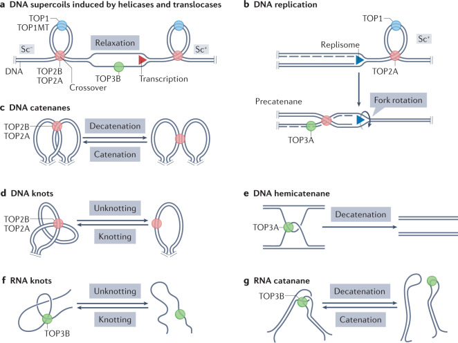
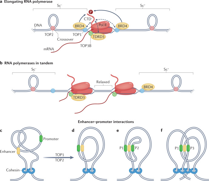
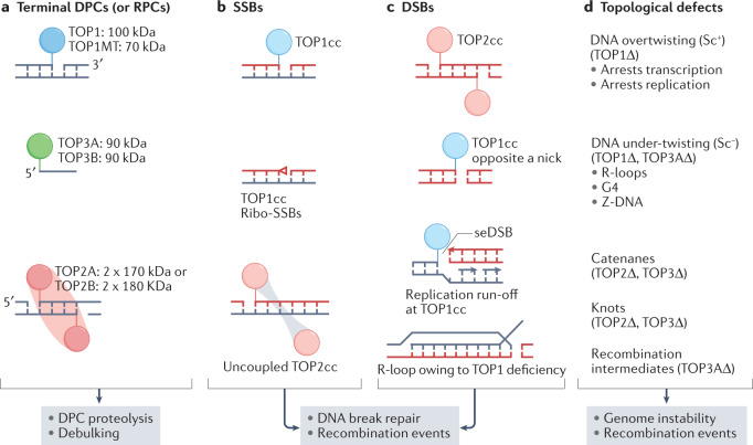
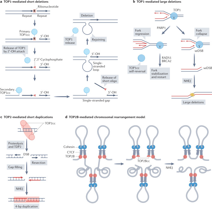
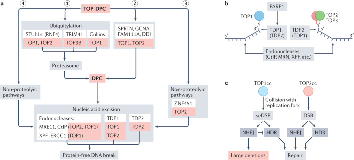
Human topoisomerases comprise a family of six enzymes: two type IB (TOP1 and mitochondrial TOP1 (TOP1MT), two type IIA (TOP2A and TOP2B) and two type IA (TOP3A and TOP3B) topoisomerases. In this Review, we discuss their biochemistry and their roles in transcription, DNA replication and chromatin remodelling, and highlight the recent progress made in understanding TOP3A and TOP3B. Because of recent advances in elucidating the high-order organization of the genome through chromatin loops and topologically associating domains (TADs), we integrate the functions of topoisomerases with genome organization. We also discuss the physiological and pathological formation of irreversible topoisomerase cleavage complexes (TOPccs) as they generate topoisomerase DNA-protein crosslinks (TOP-DPCs) coupled with DNA breaks. We discuss the expanding number of redundant pathways that repair TOP-DPCs, and the defects in those pathways, which are increasingly recognized as source of genomic damage leading to neurological diseases and cancer.


Conflict of interest statement

The authors declare no competing interests.

Figures

Fig. 1
Fig. 1. Topological problems solved by human topoisomerases.
Sites of action of topoisomerases (red, blue and green circles), duplex nucleic acids (parallel lines) without their double-helix structure and DNA segments whose ends are not free to rotate on their helical axis due to steric and physical constraints (parallel pegs) representing topological domains. a | Twin supercoiled domain model. Opening the DNA duplex by helicases and ATPase translocases within a topological domain during transcription and chromatin remodelling generates positive DNA supercoiling (Sc+) ahead of the moving helicase (or translocase) and negative supercoiling (Sc) behind it. Excessive supercoiling generates writhe that brings together distant regions of DNA that form crossovers. Topoisomerase 1 (TOP1), mitochondrial TOP1 (TOP1MT), TOP2A and TOP2B remove both Sc+ and Sc by incising double-stranded DNA; TOP3B (and TOP3A) relax hyper-negative supercoiling by nicking and closing single-stranded DNA segments. b | Replication forks generate Sc+ in front of the translocating replisome, which is removed by TOP1 and TOP2A. If the replisome swivels due to the twisting force, Sc+ diffuses behind the replisome and generates precatenanes, which are removed by TOP2A. TOP3A may also remove precatenanes if they include single-stranded DNA segments. c | TOP2A and TOP2B decatenate topological domains by passing one DNA molecule through the double-stranded DNA break made in the other DNA molecule (double-strand passage). d | TOP2A and TOP2B resolve DNA knots by double-strand passage. e | TOP3A in association with the Bloom syndrome protein (BLM)–TOP3A–RecQ-mediated genome instability proteins (RMI1/2) (BTR) dissolvasome complex (not shown) resolves DNA hemicatenanes arising during replication and recombination by passing a single strand of DNA through a break made in another DNA strand (single-strand passage),. f | TOP3B is the only RNA-only topoisomerase; it resolves intramolecular RNA intertwines (knots) by single-strand passage. g | TOP3B can also resolve RNA catenanes by single-strand passage. See Supplementary Fig. 1 for biochemical, molecular and structural details.
Fig. 2
Fig. 2. Functions of topoisomerases in transcription.
a | Transcription induces positive DNA supercoiling (Sc+) ahead of RNA polymerase II (Pol II) and negative supercoiling (Sc) behind it. Topoisomerase 1 (TOP1) directly binds the carboxyl terminus domain (CTD) of Pol II (dashed arrows) and is activated by bromodomain-containing protein 4 (BRD4), which phosphorylates the Pol II CTD (P). This interaction can efficiently remove Sc+ and allow translocation of Pol II. Behind the transcription complex, excessive Sc must be removed to prevent formation of R-loops and alternative DNA structures (not shown). Recruitment of TOP3B by Tudor domain-containing protein 3 (TDRD3), which interacts with the Pol II CTD, suppresses R-loops. TOP1 deficiency also leads to increased levels of R-loops owing to Sc accumulation behind Pol II. b | In theory, polymerases transcribing in tandem could cancel supercoiling between them, which would facilitate transcript elongation. cf | Removal of topological constraints by TOP2 and TOP1 (part c) facilitates close interactions between enhancers and promoters: enhancer–promoter interaction within a topologically associating domain (TAD) (part d); a single enhancer activating two promoters (P1 and P2) in the same TAD (part e); and activation of two promoters (P1 and P3) in different TADs (part f).
Fig. 3
Fig. 3. Functions of topoisomerases in genome organization.
ad | The loop extrusion model, with proposed roles for topoisomerase 1 (TOP1) and TOP2B.: cohesin holds two ends of a chromatin loop containing an enhancer, TOP1 preferentially removes positive DNA supercoiling induced by enhancer RNA (eRNA) synthesis, the resulting negative DNA supercoiling is proposed to pull the ends of DNA through the cohesin complex, and TOP2B bound to CCCTC-binding factor (CTCF) may allow this translocation by removing topological obstacles such as knots and supercoils (part a); as eRNA transcription continues, the extruded loop increases in size, and extrusion of one end (left) is arrested when cohesin encounters CTCF (part b); following further loop extrusion, the enhancer comes into contact with a promoter, and mRNA synthesis begins (part c); CTCF at the second end of the loop comes into contact with the cohesin complex and the chromatin loop is fully extruded, with TOP1 shown acting next to the promoter and TOP2 at DNA crossovers (part d). eg | Proposed roles of cohesins, TOP1 and TOP2B in assembly of chromatin loops and topologically associating domains (TADs): cohesin and condensin complexes are loaded onto DNA during G1 phase of cell cycle, along with transcription resumption after mitosis (part e); transcription-driven negative DNA supercoiling is proposed to extrude chromatin loops (parts ad) and form TADs, with TOP2B removing associated topological barriers such as DNA crossovers and catenanes (part f), resulting in TAD formation (part g). h | During mitosis, TOP2A is part of the chromosome scaffold comprising condensin complexes, whereas TOP1 is present in loop domains. i | Transversal axial view of chromatin scaffolded around TOP2A and condensins, with TOP1 in loop domains to remove supercoiling tension. Pol II, polymerase II. Sc, supercoiling.
Fig. 4
Fig. 4. Genotoxic and pathogenic topoisomerase lesions.
Catalytic intermediates of topoisomerases are normally transient because topoisomerase cleavage complexes (TOPccs) are self-reversible (Supplementary Fig. 1). Irreversible TOPccs are generated by trapping of TOPccs by anticancer drugs (Supplementary Box 1) and by pre-existing DNA alterations,. TOPccs produce complex nucleic acid alterations, including DNA–protein crosslinks (DPCs) and RNA–protein crosslinks (RPCs), DNA breaks and topological defects. a | DPCs form either at 3′ DNA ends (topoisomerase 1 (TOP1) or mitochondrial TOP1 (TOP1MT)) or 5′ DNA ends (TOP2A, TOP2B, TOP3A or TOP3B). They also form at 5′ RNA ends for TOP3B. TOP1, TOP1MT, TOP2A and TOP2B bind double-stranded DNA; TOP3A and TOP3B bind single-stranded DNA or RNA. DPCs and RPCs generally need to be proteolysed or debulked (denatured) before their excision,. b | DNA single-strand breaks (SSBs) formed by stalled or irreversible TOP1ccs (top). TOP1 can also generate SSBs by converting ribonucleotides incorporated by DNA polymerases into nicks with 2′,3′-cyclophosphate blocking ends (red triangle) (middle),,. Cleavage of DNA by TOP2 can be asymmetrical, with only one component of the TOP2 homodimer forming a TOP2cc (bottom). This situation is commonly observed following treatment with etoposide,. c | DNA double-strand breaks (DSBs) formed by trapping of TOP2A and TOP2B,, following their proteasomal degradation (top),. TOP1 can also generate DSBs, when it nicks the DNA opposite to a nick, or when collision with a replisome produces ‘replication run-off’ with a single-ended DSB (seDSB) (middle). R-loops forming due to insufficient TOP1 activity induce DSBs (bottom). d | Insufficient topoisomerase activity can result in excessive positive DNA supercoiling (Sc+) that arrests transcription and replication, and in negative supercoiling (Sc) that induces formation of R-loops and alternative DNA structures, including G quadruplexes (G4) and Z-DNA,. Catenanes and knots, which also stop DNA (and possibly RNA) transactions, increase in conditions of TOP2 and TOP3 deficiency. Unresolved recombination intermediates owing to TOP3A deficiency lead to sister chromatid exchanges and genomic instability.
Fig. 5
Fig. 5. Topoisomerase-induced mutagenesis and recombination events.
a | Model for formation of topoisomerase 1 (TOP1)-mediated deletions at sites of ribonucleotide incorporation within short tandem repeats. Incorporation of ribonucleotides by DNA polymerases is one of the most common abnormalities in the DNA. A ‘primary’ TOP1 cleavage complex (TOP1cc) forms on a ribonucleotide. The 2′-hydroxyl of the ribose sugar reverses the bond with TOP1 (not shown) and generates a nick with a 2′,3′-cyclophosphate end. A ‘secondary’ TOP1 forms a TOP1cc 5′ of the nick, and the resulting short oligonucleotide bearing the 2′,3′-cyclophosphate is released. The 5′ end of DNA is captured by TOP1, which is followed by rejoining of the two ends and release of TOP1, thus generating a short deletion,,. The ribonuclease activity of TOP1 has been linked with the ‘Indel Signature 4′ (ID4) in the Catalogue of Somatic Mutations in Cancer (COSMIC) database, which consists of 2–5 base pair deletions. The ID4 signature has been proposed to be named the ID-TOP1 mutational signature. b | TOP1-mediated large deletions. A replication fork collides with a TOP1cc on the leading (bottom) strand (Y represents the covalently linked catalytic Tyr at the 3′ end of the break). Replication fork regression induced by poly(ADP-ribose) polymerase 1 (PARP1) promotes TOP1cc self-reversal or fork stabilization and replication restart with RAD51 and breast cancer-associated type 2 (BRCA2). Alternative to fork regression, ‘replication run-off’ generates a single-ended DNA double-strand break (seDSB), and ligation of two distant seDSBs by non-homologous end joining (NHEJ) produces large deletions. c | TOP2-mediated short duplications. Processing of TOP2cc by proteolysis and tyrosyl-DNA phosphodiesterase 2 (TDP2) produce DSBs. The 3′ ends of the break can undergo resection followed by gap filling. Ligation through NHEJ results in 4-bp duplications. An indel signature consisting of 2–4 base pair duplications and due to a TOP2A mutation (K473N) that traps TOP2A has been found in patient tumours and proposed to be named as the ID-TOP2A signature. d | Simplified model of TOP2B-mediated chromosomal rearrangements based on the loop extrusion model. Schematic depicts chromatin loops with cohesin, CCCTC-binding factor (CTCF) and TOP2B at base of loops. Stalled or irreversible TOP2Bccs generate DSBs that disjoin the loops. Rejoining of two adjacent DSBs produces a translocation.
Fig. 6
Fig. 6. Main repair pathways for trapped topoisomerases in humans.
a | Overall scheme for conversion of topoisomerase DNA–protein crosslinks (TOP-DPCs) into protein-free DNA breaks. Association of TOP-DPCs with replication or transcription complexes and phase of the cell cycle (S phase versus G1 phase) are likely determinants of pathway choice. Debulking of TOP-DPCs by the three ubiquitin–proteasome pathways includes the conserved SUMO-targeted ubiquitin ligase (STUbL) pathway, in which RNF4 is the human E3 ubiquitin ligase for TOP1-DPCs, TOP2A-DPCs and TOP2B-DPCs; the TRIM41 E3 ligase pathway for crosslinks between TOP3B and DNA or RNA; and the cullin pathway for TOP1-DPCs, (step 1). Non-proteasomal TOP-DPC proteolytic pathways. The proteases Spartan (SPRTN), GCNA (also known as ACRC), FAM111A and DDI debulk TOP1-DPCs and TOP2-DPCs (step 2). Non-proteolytic pathway for TOP2, driven by SUMO E3 ligase ZNF451 (ref.) (step 3). Nucleic-acid excision pathways for TOP1 and/or TOP2 include excision by the endonucleases MRE11, CtIP and XPF–ERCC1, or excision by tyrosyl-DNA phosphodiesterase 1 (TDP1) and TDP2 (step 4). b | TDP1 is activated by poly(ADP-ribose) polymerase 1 (PARP1),,, and upon cleaving DNA leaves a 3′-phosphate that is further processed by polynucleotide kinase phosphatase (not shown). TDP2 leaves a 5′-phosphate that can be directly ligated or extended by DNA polymerases (not shown). Both TDP1 and TDP2 require debulking of TOP-DPCs to gain access to the tyrosyl–DNA links. Additional excision pathways involve endonucleases. c | Differential roles of non-homologous end joining (NHEJ) and homology-directed repair (HDR) in repair of TOP1-induced single-ended DNA double-strand breaks (seDSBs) and TOP2-DPC-induced DSBs. Whereas seDSB repair by NHEJ is toxic, possibly by inducing large deletions owing to illegitimate end joining of distant seDSBs (Fig. 5b), NHEJ is crucial for repair of TOP2-DPCs. TOP1cc, TOP1 cleavage complex.

References

    1. Pommier Y, Sun Y, Huang SN, Nitiss JL. Roles of eukaryotic topoisomerases in transcription, replication and genomic stability. Nat. Rev. Mol. Cell Biol. 2016;17:703–721. doi: 10.1038/nrm.2016.111. - DOI - PMC - PubMed
    1. Saha S, et al. DNA and RNA cleavage complexes and repair pathway for TOP3B RNA- and DNA–protein crosslinks. Cell Rep. 2020;33:108569. doi: 10.1016/j.celrep.2020.108569. - DOI - PMC - PubMed
    1. Capranico G, Marinello J, Chillemi G. Type I DNA topoisomerases. J. Med. Chem. 2017;60:2169–2192. doi: 10.1021/acs.jmedchem.6b00966. - DOI - PubMed
    1. Lee SK, Wang W. Roles of topoisomerases in heterochromatin, aging, and diseases. Genes. 2019;10:884. doi: 10.3390/genes10110884. - DOI - PMC - PubMed
    1. Bjornsti MA, Kaufmann SH. Topoisomerases and cancer chemotherapy: recent advances and unanswered questions. F1000Res. 2019;8:1704. doi: 10.12688/f1000research.20201.1. - DOI - PMC - PubMed

Publication types