

Synovial fluid proteome profile of surgical versus chemical induced osteoarthritis in rabbits
- PMID: 35228907
- PMCID: PMC8881915
- DOI: 10.7717/peerj.12897
Synovial fluid proteome profile of surgical versus chemical induced osteoarthritis in rabbits
Abstract
Background: Animal models are significant for understanding human osteoarthritis (OA). This study compared the synovial fluid proteomics changes in surgical and chemical induced OA models.
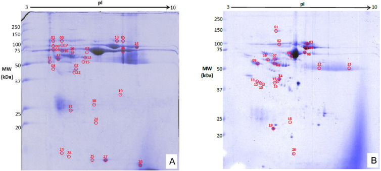
Methods: Thirty rabbits either had anterior cruciate ligament transection (ACLT) procedure or injected intra-articularly with monosodium iodoacetate (MIA, 8 mg) into the right knee. The joints were anatomically assessed, and the synovial fluid proteins analyzed using two-dimensional polyacrylamide gel electrophoresis (2DGE) and MALDI TOF/TOF mass spectrometry analysis at 4, 8 and 12 weeks. The proteins' upregulation and downregulation were compared with control healthy knees.
Results: Seven proteins (histidine-rich glycoprotein, beta-actin-like protein 2 isoform X1, retinol-binding protein-4, alpha-1-antiproteinase, gelsolin isoform, serotransferrin, immunoglobulin kappa-b4 chain-C-region) were significantly expressed by the surgical induction. They characterized cellular process (27%), organization of cellular components or biogenesis (27%), localization (27%) and biological regulation (18%), which related to synovitis, increased cellularity, and subsequently cartilage damage. Three proteins (apolipoprotein I-IV precursor, serpin peptidase inhibitor and haptoglobin precursor) were significantly modified by the chemical induction. They characterized stimulus responses (23%), immune responses (15%), biological regulations (15%), metabolism (15%), organization of cellular components or biogenesis (8%), cellular process (8%), biological adhesions (8%) and localization (8%), which related to chondrocytes glycolysis/death, neovascularization, subchondral bone necrosis/collapse and inflammation.
Conclusions: The surgical induced OA model showed a wider range of protein changes, which were most upregulated at week 12. The biological process proteins expressions showed the chemical induced joints had slower OA progression compared to surgical induced joints. The chemical induced OA joints showed early inflammatory changes, which later decreased.
Keywords: Chemical; MALDI TOF/TOF mass spectrometry; Osteoarthritis; Proteomics; Rabbits; Surgical; Synovial fluid; Two-dimensional polyacrylamide gel electrophoresis.
©2022 Syed Sulaiman et al.
Conflict of interest statement
The authors declare there are no competing interests.
Figures
References
-
- Botter SM, Glasson SS, Hopkins B, Clockaerts S, Weinans H, Van Leeuwen JPTM, Van Osch GJVM. ADAMTS5-/- mice have less subchondral bone changes after induction of osteoarthritis through surgical instability: implications for a link between cartilage and subchondral bone changes. Osteoarthritis and Cartilage. 2009;17(5):636–645. doi: 10.1016/j.joca.2008.09.018. - DOI - PubMed
-
- Burr DB, Gallant MA. Bone remodelling in osteoarthritis. Nature Reviews Rheumatology. 2012;8(11):665–673. - PubMed
-
- Campos WNS, Souza MA, Ruiz T, Peres TP, Néspoli PB, Marques ATC, Colodel EM, De Souza RL. Experimental osteoarthritis in rabbits: lesion progression 1. 2013;32(3):279–285.
Publication types
MeSH terms
Substances
LinkOut - more resources
Full Text Sources
Medical
Research Materials
