. 2022 Mar 28;2(3):100181.
doi: 10.1016/j.crmeth.2022.100181. Epub 2022 Feb 24.

Development of a hybrid alphavirus-SARS-CoV-2 pseudovirion for rapid quantification of neutralization antibodies and antiviral drugs

Affiliations

Development of a hybrid alphavirus-SARS-CoV-2 pseudovirion for rapid quantification of neutralization antibodies and antiviral drugs

Brian Hetrick et al. Cell Rep Methods. .

Abstract

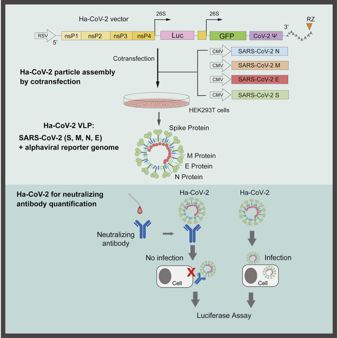
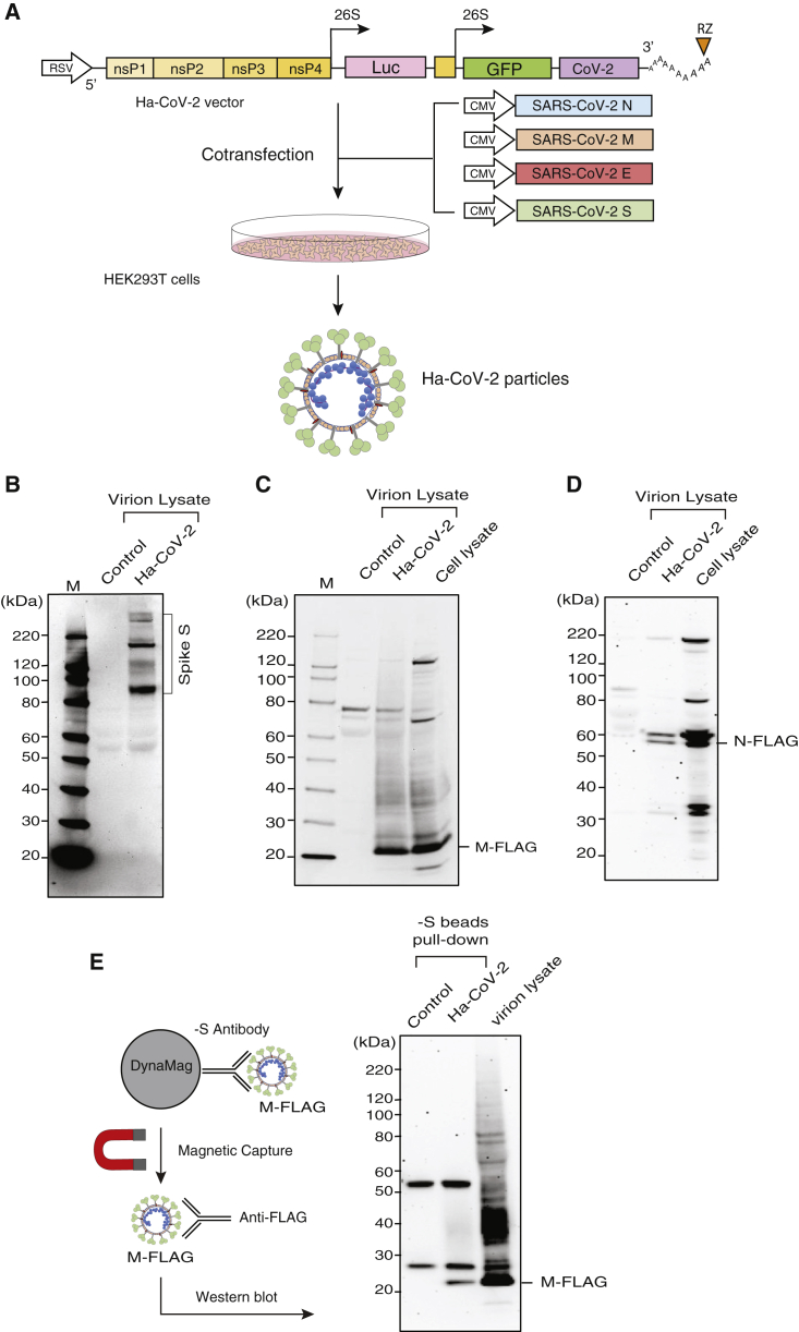
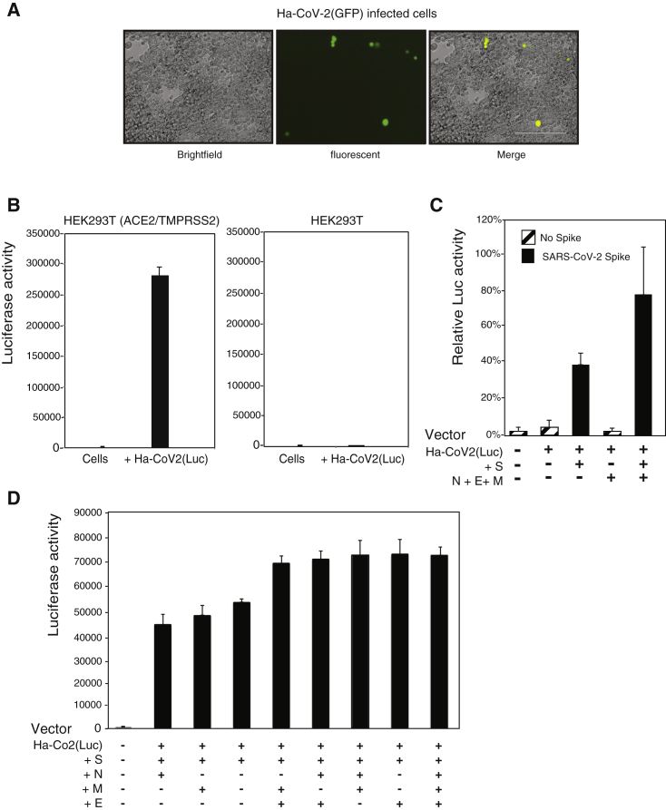
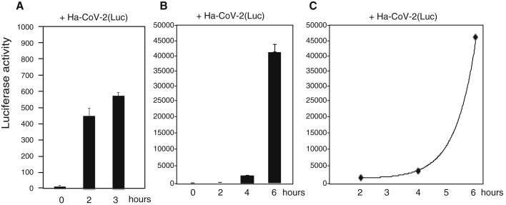
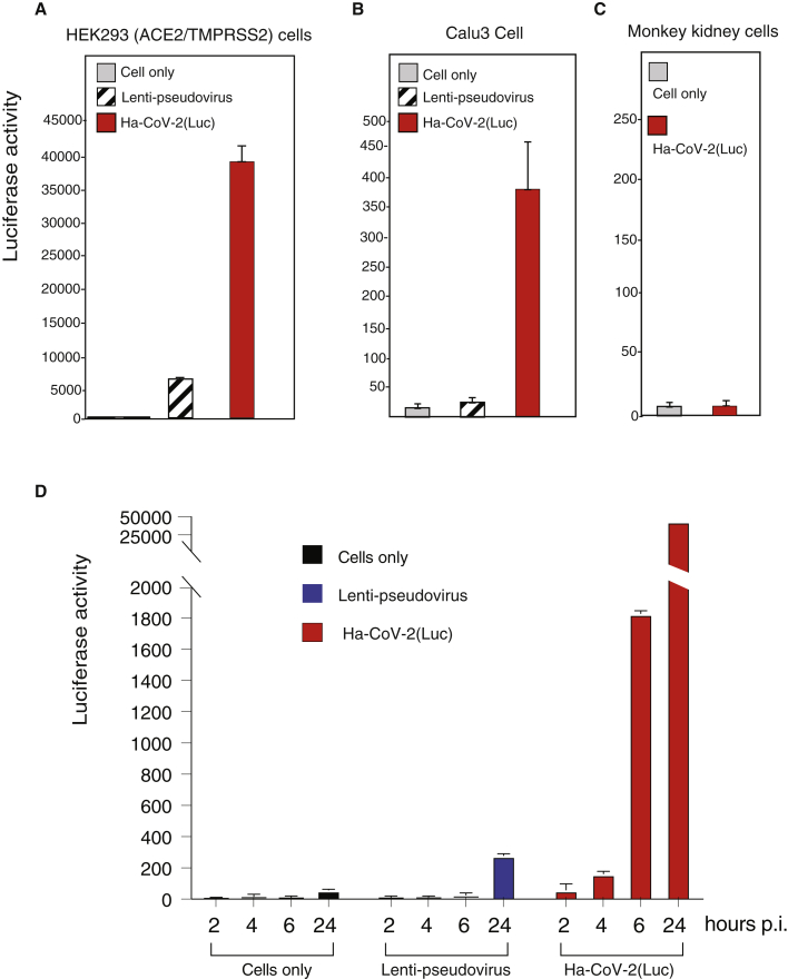
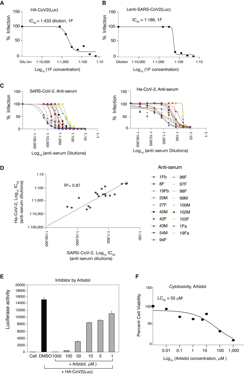
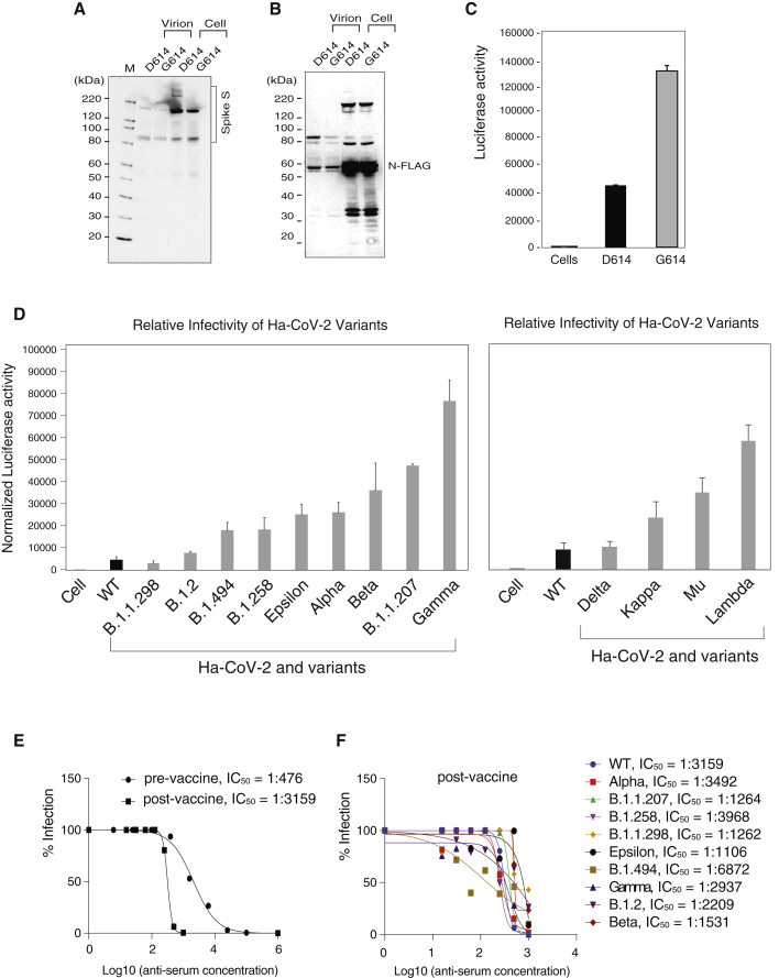
Severe acute respiratory syndrome coronavirus 2 (SARS-CoV-2) spike protein (S)-pseudotyped viruses are commonly used for quantifying antiviral drugs and neutralizing antibodies. Here, we describe the development of a hybrid alphavirus-SARS-CoV-2 (Ha-CoV-2) pseudovirion, which is a non-replicating SARS-CoV-2 virus-like particle composed of viral structural proteins (S, M, N, and E) and an RNA genome derived from a fast-expressing alphaviral vector. We validated Ha-CoV-2 for rapid quantification of neutralization antibodies, antiviral drugs, and viral variants. In addition, as a proof of concept, we used Ha-CoV-2 to quantify the neutralizing antibodies from an infected and vaccinated individual and found that the one-dose vaccination with Moderna mRNA-1273 greatly increased the anti-serum titer by approximately 6-fold. The post-vaccination serum can neutralize all nine variants tested. These results demonstrate that Ha-CoV-2 can be used as a robust platform for the rapid quantification of neutralizing antibodies against SARS-CoV-2 and its emerging variants.

Keywords: COVID-19; Ha-CoV-2; SARS-CoV-2; SARS-CoV-2 variants; alphavirus; antiviral drug; coronavirus; lentivirus; mRNA vaccine; neutralizing antibody; pseudovirus.


Conflict of interest statement

A patent application has been filed by George Mason University and is licensed for product development. Y.W. is a founder of Virongy Biosciences, Inc., and a member of its advisory board. T.C. is the chief scientific officer of Polaris Genomics, Inc.

Figures

None
Graphical abstract
Figure 1
Figure 1
Design and assembly of Ha-CoV-2 particles (A) Illustration of the design of Ha-CoV-2 vector. The vector contains an RSV promoter that transcribes the full-length viral RNA genome to be packaged into Ha-CoV-2 particles. Shown is the 5′ untranslated region followed by open-reading frames coding for nonstructural proteins (nsps) 1–4 from Semliki Forest virus (SFV), viral subgenomic promoters for Luc and GFP reporter expression, the 3′ untranslated region, and a poly(A) tail that is self-cleaved by the hepatitis delta virus ribozyme (RZ). The SARS-CoV-2 packaging signal is inserted in front of the 3′ untranslated region. To assemble viral particles, HEK293T cells were co-transfected with Ha-CoV-2 and the vectors expressing the 4 structural proteins of SARS-CoV-2 (S, M, E, and N). (B) Ha-CoV-2 particles in the supernatant were harvested at 48 h, purified, lysed, and then analyzed by western blot using antibodies for the SARS-CoV-2 S protein. The control is the supernatant from cells transfected with the Ha-CoV-2 vector alone. (C and D) Particles were also assembled using FLAG-tagged M and N. Particles were analyzed with western blot using an antibody against FLAG. (E) Particles in the supernatant were also captured with magnetic beads conjugated with the anti-S antibody and then analyzed with western blot using the antibody again FLAG for FLAG-tagged M protein in the particles.
Figure 2
Figure 2
SARS-CoV-2 S protein and ACE2-dependent infection of target cells by Ha-CoV-2 (A) HEK293T(ACE2/TMPRSS2) cells were infected with Ha-CoV-2(GFP) particles. GFP expression was observed 48 h post infection. (B) ACE2-dependent infection of target cells by Ha-CoV-2(Luc). HEK293T(ACE2/TMPRSS2) and HEK293T cells were infected with Ha-CoV-2(Luc) particles. Luciferase expression was quantified at 24 h post infection. (C) SARS-CoV-2 S-protein-dependent infection of target cells by Ha-CoV-2(Luc). Particles were assembled in the presence or absence of S or M + E + N. Luciferase expression was quantified at 4 h post infection. (D) Requirements of M, E, and N for optimal infectivity of Ha-CoV(Luc). Particles were assembled in the presence of S and in combinations of individual proteins of M, E, and N. Luciferase expression was quantified. Infection and luciferase assays in (B)–(D) were performed 3 times, and the mean and standard deviation (SD) are shown.
Figure 3
Figure 3
Rapid time course of reporter gene expression in Ha-CoV-2(Luc) infection Time courses of 3 and 6 h of luciferase expression following infection of HEK293T(ACE2/TMPRSS2) cells with Ha-CoV-2(Luc) particles. (A) Cells were infected with Ha-CoV-2(Luc) for 2 and 3 h, washed, and then lysed and analyzed for Luc expression. (B and C) Cells were infected with Ha-CoV-2(Luc) for 2 h, washed, cultured in fresh medium, and then lysed and analyzed for Luc expression at different time points. The addition of virus to cells was defined as time “0”. Infection and luciferase assays were performed 3 times, and the mean and SD are shown.
Figure 4
Figure 4
Comparison of the infection time course of Ha-CoV-2 with that of SARS-CoV-2 S-pseudotyped lentivirus (A–C) HEK293T(ACE2/TMPRSS2) and Calu-3 cells were infected with an equal volume of viral particles, Lenti-CoV-2(Luc), or Ha-CoV-2(Luc). Relative infection was quantified by luciferase assay at 72 h post infection. Primary monkey kidney cells were also infected with Ha-CoV-2 for comparison. (D) Comparison of lenti-pseudovirus and Ha-CoV-2 in an infection time course. HEK293T(ACE2/TMPRSS2) cells were infected with an equal volume of viral particles, lenti-CoV-2(Luc), or Ha-CoV-2(Luc). Relative Luc reporter expression was quantified by luciferase assay from 2 to 24 h post infection. Infection and luciferase assays were performed 3 times, and the mean and SD are shown.
Figure 5
Figure 5
Validation of Ha-CoV-2 particles for rapid screening and quantification of neutralizing antibodies (A) Quantification of neutralizing antibodies with Ha-CoV-2 particles. Shown are the concentration-dependent inhibition of Ha-CoV-2(Luc) by the anti-serum 1F and the 1F inhibition curve. 1F was serially diluted and incubated with Ha-CoV-2(Luc) particles for 1 h at 37°C. The Ha-CoV-2(Luc)-antibody complex was used to infect HEK293T(ACE2/TMPRSS2) cells. Neutralization activities were quantified by luciferase assay at 5 h post addition of virus to cells. Control serum was from healthy, uninfected donors. The IC50 was calculated using the relative percentage of infection versus serum concentration. (B) For comparison, the anti-serum 1F was also similarly quantified using a SARS-CoV-2 S-protein-pseudotyped lentivirus, lenti-CoV-2(Luc). Neutralization activities were quantified with luciferase assay at 72 h post infection. (C and D) Correlation of serum neutralization activities quantified with Ha-CoV-2(Luc) and SARS-CoV-2. Convalescent plasma from 19 donors was quantified using infectious SARS-CoV-2 and plaque assays or Ha-CoV-2(Luc). Neutralization activities were plotted, and the IC50 values were calculated. The correlation in IC50 was plotted. (E and F) Rapid quantification of the anti-SARS-CoV-2 activity of Arbidol. HEK293T(ACE2/TMPRSS2) cells were pretreated for 1 h with Arbidol. Cells were infected with Ha-CoV-2(Luc) in the presence of Arbidol. Viral entry inhibition was quantified by luciferase assay at 5 h. An MTT cytotoxicity assay of Abidol was also performed on cells (F).
Figure 6
Figure 6
Quantification of the relative infectivity of Ha-CoV-2 variants and their responses to neutralizing antibodies (A and B) Ha-CoV-2(Luc) particles bearing the G614 mutation S or the parent D614 S were assembled and analyzed for the incorporation of S and N in virions. (C) Ha-CoV-2(Luc)(G614) or Ha-CoV-2(D614) was used to infect target cells, and Luc expression was quantified at 5 h. An equal level of viral particles was used for infection. Infection and luciferase assays were performed 3 times, and the mean and SD are shown. (D) A panel of S protein mutants from SARS-CoV-2 variants were used to assemble Ha-CoV-2(Luc) particles and then to infect target cells. The relative infectivity was quantified and normalized with the genomic RNA copies of individual Ha-CoV-2(Luc) variants. Wild type (WT) refers to Ha-CoV-2 derived from the original SARS-CoV-2 strain. Infection and luciferase assays were performed 3 times, and the mean and SD are shown. (E and F) Quantification of anti-serum against Ha-CoV-2(Luc) and its variants. Convalescent plasma from an infected blood donor, before and after one-dose vaccination, was quantified for inhibition of Ha-CoV-2(Luc) infection. Neutralization activities were quantified by luciferase assay at 12 h post infection. The IC50 was calculated using the relative percentage of infection versus serum concentration (E). The post-vaccination anti-serum was similarly quantified for the inhibition of Ha-CoV-2(Luc) variants (F).

References

    1. Beigel J.H., Tomashek K.M., Dodd L.E., Mehta A.K., Zingman B.S., Kalil A.C., Hohmann E., Chu H.Y., Luetkemeyer A., Kline S., et al. Remdesivir for the treatment of Covid-19 - final report. N. Engl. J. Med. 2020;383:1813–1826. - PMC - PubMed
    1. Boriskin Y.S., Leneva I.A., Pecheur E.I., Polyak S.J. Arbidol: a broad-spectrum antiviral compound that blocks viral fusion. Curr. Med. Chem. 2008;15:997–1005. - PubMed
    1. Cai Y., Zhang J., Xiao T., Peng H., Sterling S.M., Walsh R.M., Rawson S., Rits-Volloch S., Chen B. Distinct conformational states of SARS-CoV-2 spike protein. Science. 2020;369:1586–1592. - PMC - PubMed
    1. Case J.B., Rothlauf P.W., Chen R.E., Liu Z., Zhao H., Kim A.S., Bloyet L.-M., Zeng Q., Tahan S., Droit L., et al. Neutralizing antibody and soluble ACE2 inhibition of a replication-competent VSV-SARS-CoV-2 and a clinical isolate of SARS-CoV-2. Cell Host Microbe. 2020;28:475–485.e5. - PMC - PubMed
    1. Ciccarone V.C., Jessee J.A., Liljeström P. The SFV gene expression system. Methods Mol. Med. 1998;13:237–255. - PubMed

Publication types