. 2022 May;157(5):513-524.
doi: 10.1007/s00418-022-02087-z. Epub 2022 Feb 28.

Angiotensin II type 1 receptor localizes at the blood-bile barrier in humans and pigs

Affiliations

Angiotensin II type 1 receptor localizes at the blood-bile barrier in humans and pigs

Galyna Pryymachuk et al. Histochem Cell Biol. 2022 May.

Abstract

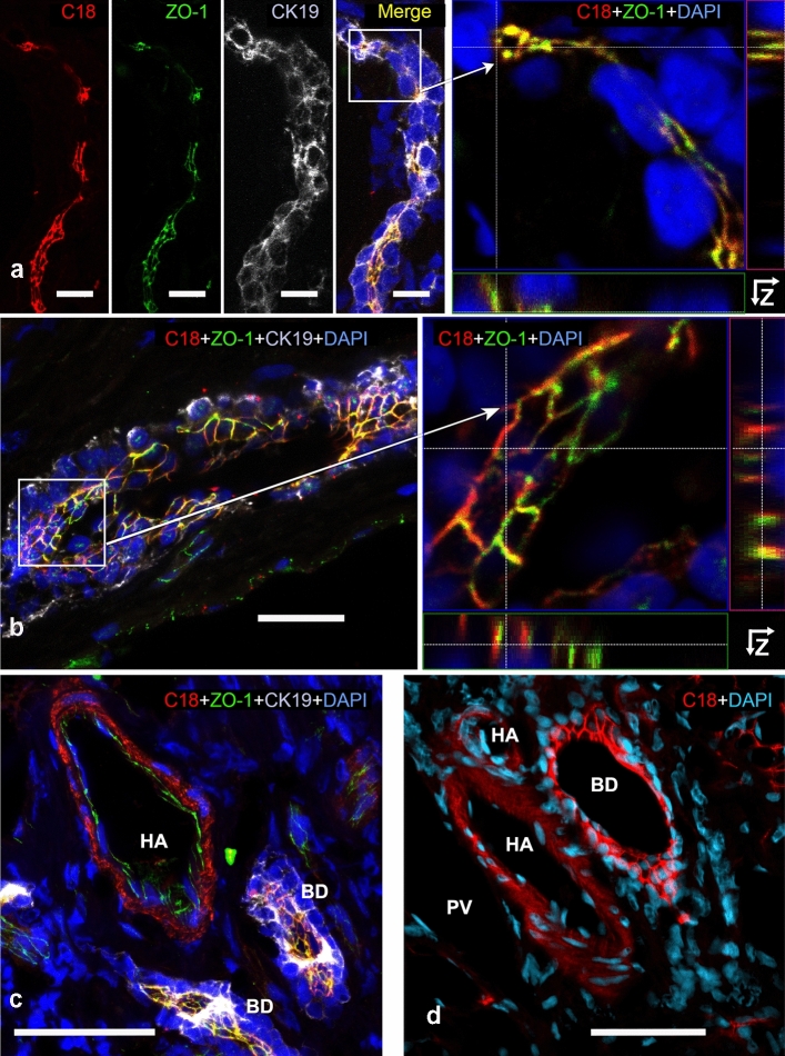
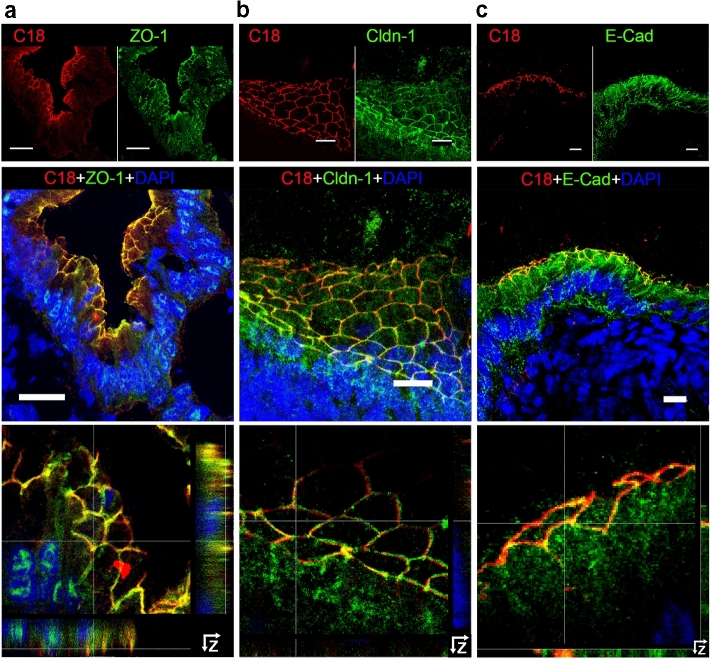
Animal models and clinical studies suggest an influence of angiotensin II (AngII) on the pathogenesis of liver diseases via the renin-angiotensin system. AngII application increases portal blood pressure, reduces bile flow, and increases permeability of liver tight junctions. Establishing the subcellular localization of angiotensin II receptor type 1 (AT1R), the main AngII receptor, helps to understand the effects of AngII on the liver. We localized AT1R in situ in human and porcine liver and porcine gallbladder by immunohistochemistry. In order to do so, we characterized commercial anti-AT1R antibodies regarding their capability to recognize heterologous human AT1R in immunocytochemistry and on western blots, and to detect AT1R using overlap studies and AT1R-specific blocking peptides. In hepatocytes and canals of Hering, AT1R displayed a tram-track-like distribution, while in cholangiocytes AT1R appeared in a honeycomb-like pattern; i.e., in liver epithelia, AT1R showed an equivalent distribution to that in the apical junctional network, which seals bile canaliculi and bile ducts along the blood-bile barrier. In intrahepatic blood vessels, AT1R was most prominent in the tunica media. We confirmed AT1R localization in situ to the plasma membrane domain, particularly between tight and adherens junctions in both human and porcine hepatocytes, cholangiocytes, and gallbladder epithelial cells using different anti-AT1R antibodies. Localization of AT1R at the junctional complex could explain previously reported AngII effects and predestines AT1R as a transmitter of tight junction permeability.

Keywords: AT1R; Canals of Hering; Gallbladder; Human liver; Porcine liver; Tight junctions.


Conflict of interest statement

The authors have no conflicts of interest to declare that are relevant to the content of this article.

Figures

Fig. 1
Fig. 1
Distribution of AT1R in porcine (a) and human (b) liver. Liver cryosections were incubated with anti-AT1R-C18 (red), anti-CK-19 (green (a) or gray (b)), DAPI (blue, nuclei), and in (b), additionally, with anti-ZO-1 (green). CK-19 indicated the presence of cholangiocytes. Where hepatocyte plates formed bile canaliculi, AT1R appeared as tram-track-like pattern which, similar to ZO-1, continued in the canal of Hering. Partial overlap of anti-AT1R and anti-ZO-1 signals are shown in yellow (b). Confocal microscopes a Zeiss LSM880; b Leica DMI 6000B. Scale bars 20 µm (a, b)
Fig. 2
Fig. 2
AT1R localization in the human (ac) and porcine (d) intrahepatic biliary tree. Liver cryosections incubated with anti-AT1R-C18 (red), and DAPI (blue, nuclei) (ad), and additionally with anti-ZO-1 (green, TJ), anti-CK-19 (gray, cholangiocytes) in (ac); partial signal overlap (anti-AT1R and anti-ZO-1) in yellow. AT1R and ZO-1 localize towards the apical membrane of cholangiocytes (ac). Small (a) and larger (bd) bile ducts are shown. HA hepatic artery, BD bile duct. (a, left), (b, left), c and d are shown as MIP; (a, right) and (b, right) are Z-stack analyses. Confocal microscopes ac Leica DMI 6000B; d Zeiss LSM880. Scale bars 20 µm (a, b), 50 µm (c, d)
Fig. 3
Fig. 3
AT1R colocalizes in gallbladder epithelial cells with TJ proteins ZO-1 and claudin-1 and closely associates with AJ protein E-cadherin. Porcine gallbladder cryosections were incubated with anti-AT1R-C18 (red) (ac), anti-ZO-1 (a), anti-claudin-1 (Cldn-1) (b), and anti-E-cadherin (E-Cad) (c) (all in green), (top row). Nuclei were stained with DAPI (blue). Merged images (in the middle row) show colocalization of AT1R with either ZO-1 or claudin-1 (yellow). Top and middle rows: MIP; bottom row: enlarged regions of merged images above with Z-stack projections at the right side and below the images. Confocal microscope Leica DMI 6000B. Scale bars 20 µm (a), 10 µm (b, c)
Fig. 4
Fig. 4
AT1R colocalizes in human hepatocytes with the TJ proteins ZO-1 and symplekin (SYMPK). Liver cryosections incubated with anti-AT1R-C18 (red) and anti-ZO-1 (green) in a or with anti-symplekin (green) in c are shown in separate fluorescence channels (top panels). Merged MIP images (bottom panels in a and c) include DAPI (blue, nuclei) and show the overlap of AT1R with the TJ signals (ZO-1 (a) and symplekin (c)) in yellow. b and d show enlarged views of the boxed regions from a and c with Z-stack analyses. Confocal microscope Leica DMI 6000B. Scale bars 10 µm
Fig. 5
Fig. 5
In human hepatocytes, AT1R is adjacent to adherens junctions and desmosomes but does not show systematic overlap with these structures. Liver cryosections were incubated with anti-AT1R-C18 (red) (ad), anti-E-cadherin (E-Cad, green) (a, b), or anti-desmoglein 2 (DSG 2, green) (c, d) and DAPI (blue, nuclei). In a and c the specimens are shown in separate fluorescence channels (top panel), merged MIP images (bottom panel). Detailed images of boxed regions from a and c are shown with Z-stack analyses in b and d, respectively. Confocal microscope Leica DMI 6000B. Scale bars 10 µm
Fig. 6
Fig. 6
Proposed model of the organization of AT1R within the junctional complex in hepatocytes and cholangiocytes. TJ, tight junctional marker proteins (green); AT1R (red); AJ, adherent junctional marker protein (blue); Dsm, desmosomal marker protein (green dotted line); BC, bile canaliculus

Similar articles

Cited by

References

    1. Afroze SH, Munshi MK, Martinez AK, Uddin M, Gergely M, Szynkarski C, Guerrier M, Nizamutdinov D, Dostal D, Glaser S. Activation of the renin–angiotensin system stimulates biliary hyperplasia during cholestasis induced by extrahepatic bile duct ligation. Am J Physiol Gastrointest Liver Physiol. 2015;308:G691–701. doi: 10.1152/ajpgi.00116.2014. - DOI - PMC - PubMed
    1. Anderson JM, Glade JL, Stevenson BR, Boyer JL, Mooseker MS. Hepatic immunohistochemical localization of the tight junction protein ZO-1 in rat models of cholestasis. Am J Pathol. 1989;134:1055–1062. - PMC - PubMed
    1. Bataller R, Gines P, Nicolas JM, Gorbig MN, Garcia-Ramallo E, Gasull X, Bosch J, Arroyo V, Rodes J. Angiotensin II induces contraction and proliferation of human hepatic stellate cells. Gastroenterology. 2000;118:1149–1156. doi: 10.1016/S0016-5085(00)70368-4. - DOI - PubMed
    1. Bataller R, Sancho-Bru P, Gines P, Lora JM, Al-Garawi A, Sole M, Colmenero J, Nicolas JM, Jimenez W, Weich N, Gutierrez-Ramos JC, Arroyo V, Rodes J. Activated human hepatic stellate cells express the renin–angiotensin system and synthesize angiotensin II. Gastroenterology. 2003;125:117–125. doi: 10.1016/S0016-5085(03)00695-4. - DOI - PubMed
    1. Bianciotti LG, Vatta MS, Dominguez AE, Vescina C, Castro JL, Magarinos J, Fernandez BE. Quantitative modifications induced by angiotensin II on rat bile secretion. Regul Pept. 1994;54:429–437. doi: 10.1016/0167-0115(94)90540-1. - DOI - PubMed