. 2022 Jun;600(11):2613-2636.
doi: 10.1113/JP282570. Epub 2022 Mar 17.

Ca2+ signalling in interstitial cells of Cajal contributes to generation and maintenance of tone in mouse and monkey lower oesophageal sphincters

Affiliations

Ca2+ signalling in interstitial cells of Cajal contributes to generation and maintenance of tone in mouse and monkey lower oesophageal sphincters

Bernard T Drumm et al. J Physiol. 2022 Jun.

Abstract

The lower oesophageal sphincter (LES) generates tone and prevents reflux of gastric contents. LES smooth muscle cells (SMCs) are relatively depolarised, facilitating activation of Cav 1.2 channels to sustain contractile tone. We hypothesised that intramuscular interstitial cells of Cajal (ICC-IM), through activation of Ca2+ -activated Cl- channels (ANO1), set membrane potentials of SMCs favourable for activation of Cav 1.2 channels. In some gastrointestinal muscles, ANO1 channels in ICC-IM are activated by Ca2+ transients, but no studies have examined Ca2+ dynamics in ICC-IM within the LES. Immunohistochemistry and qPCR were used to determine expression of key proteins and genes in ICC-IM and SMCs. These studies revealed that Ano1 and its gene product, ANO1, are expressed in c-Kit+ cells (ICC-IM) in mouse and monkey LES clasp muscles. Ca2+ signalling was imaged in situ, using mice expressing GCaMP6f specifically in ICC (Kit-KI-GCaMP6f). ICC-IM exhibited spontaneous Ca2+ transients from multiple firing sites. Ca2+ transients were abolished by cyclopiazonic acid or caffeine but were unaffected by tetracaine or nifedipine. Maintenance of Ca2+ transients depended on Ca2+ influx and store reloading, as Ca2+ transient frequency was reduced in Ca2+ free solution or by Orai antagonist. Spontaneous tone of LES muscles from mouse and monkey was reduced ∼80% either by Ani9, an ANO1 antagonist or by the Cav 1.2 channel antagonist nifedipine. Membrane hyperpolarisation occurred in the presence of Ani9. These data suggest that intracellular Ca2+ activates ANO1 channels in ICC-IM in the LES. Coupling of ICC-IM to SMCs drives depolarisation, activation of Cav 1.2 channels, Ca2+ entry and contractile tone. KEY POINTS: The lower oesophageal sphincter (LES) generates contractile tone preventing reflux of gastric contents into the oesophagus. LES smooth muscle cells (SMCs) display depolarised membrane potentials facilitating activation of L-type Ca2+ channels. Interstitial cells of Cajal (ICC) express Ca2+ -activated Cl- channels encoded by Ano1 in mouse and monkey LES. Ca2+ signalling in ICC activates ANO1 currents in ICC. ICC displayed spontaneous Ca2+ transients in mice from multiple firing sites in each cell and no entrainment of Ca2+ firing between sites or between cells. Inhibition of ANO1 channels with a specific antagonist caused hyperpolarisation of mouse LES and inhibition of tone in monkey and mouse LES muscles. Our data suggest a novel mechanism for LES tone in which Ca2+ transient activation of ANO1 channels in ICC generates depolarising inward currents that conduct to SMCs to activate L-type Ca2+ currents, Ca2+ entry and contractile tone.

Keywords: Ca2+ handling mechanisms; L-type Ca2+ channels; SIP syncytium; anoctamin-1 channels; oesophageal reflux; smooth muscle cells; swallowing reflex.


Figures

Fig. 1:
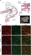
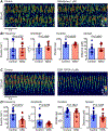
Fig. 1:. Mouse LES morphology and ICC within LES and esophagus.
A Anatomy of the mouse upper gastrointestinal tract. Sagittal section of the mouse upper gastrointestinal tract (from esophagus to duodenum) stained with a trichrome stain to identify SMCs (pink) and connective tissue (blue) (a). Additional structures staining pink include the mucosa. Inset (right) shows a higher magnification image of the LES clasp muscle. Muscular tissues in the mouse esophageal body are composed of skeletal muscle fibers whereas the muscular tissues of the LES, stomach, pyloric sphincter (PS) and duodenum are smooth muscles. The LES clasp is indicated by the dashed green line and is divided into bundles separated by connective tissue septa. An image of a mouse LES dissection indicating the portion used for functional experiments (b). The skeletal muscle of the esophagus was removed (indicated by dashed red line and scissors symbol) and the LES clasp muscles were used for contractile, microelectrode, calcium imaging and immunohistochemical experiments performed in this study. B Immunohistochemical labelling of ICC using a c-Kit antibody (Red: ICC biomarker) and an ANO1 antibody (Green) in mouse (a) and monkey LES clasp (b) and the body of the monkey esophagus (c). Spindle-shaped ICC-IM were distributed through the LES of both animals and these cells appeared to be shorter and of a lower density in the monkey esophagus. c-Kit and ANO1 co-localized in ICC in the LES and esophagus (Images of c-Kit and ANO1 merged), demonstrating that ICC-IM express ANO1 protein in esophageal tissues.
Fig. 2:
Fig. 2:. Cell identification and Ca2+ handling gene expression in LES ICC-IM.
A qPCR data of relative expression of gene transcripts for Ano1, Kit, Myh11, Pdgfra, Gja1 and Gja7 in FACS sorted GFP+ populations of LES-ICC (from Kit+/copGFP mice) and LES-SMC (from SmHC+ /eGFP mice) vs. sorted GFP cells from Kit+/copGFP mice and GFP cells from SmHC+ /eGFP mice (expression relative to Gapdh), n=3. B qPCR data of relative expression of gene transcripts for Itpr1, Itpr2, Itpr3, Ryr1, Ryr2, Ryr3, Orai1, Orai2, Orai3, Stim1 and Stim2 in FACS sorted GFP+ populations of LES-ICC (from Kit+/copGFP mice) and LES-SMC (from SmHC+ /eGFP mice) vs. sorted GFP cells from Kit+/copGFP mice and GFP cells from SmHC+ /eGFP mice (expression relative to Gapdh), n=3.
Fig. 3:
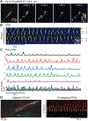
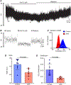
Fig. 3:. LES ICC-IM exhibit dynamic intracellular Ca2+ signaling.
A Time-lapse images of an ICC-IM within an LES muscle strip from a Kit-Cre-GCaMP6f mouse (60x). Left most panel shows the raw GCaMP6f signal. All other panels show colour-coded Ca2+ transients occurring along the length of ICC-IM from multiple intracellular sites (arrows). B STM of Ca2+ transients in a single LES-ICC recorded in situ. Five different firing sites were observed (as indicated) and their activities are plotted as traces in Panel C. Bottom trace in C shows merged traces from the five firing sites. No correlation or entrainment between firing sites was observed. D Leftmost panel shows an image of two adjacent LES ICC-IM from a Kit-KI-GCaMP6f mouse. Merged STMs of the Ca2+ transient activity of the two cells highlighted are shown in the rightmost panel. Ca2+ activity transients were thresholded and shown in a combined STM in uniform red (cell 1) or green (cell 2) colours.
Fig. 4:
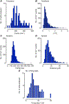
Fig. 4:. Quantification of ICC-IM Ca2+ signaling in the LES.
A-E Histograms show values for Ca2+ transient frequency (A), amplitude (B), duration (C), spatial spread (D), number of firing sites per ICC-IM (E), 1247 events, c=79, N=25.
Fig. 5:
Fig. 5:. Ca2+ release pathways mediate Ca2+ transients in ICC-IM of the LES.
A STMs showing the effects of the SERCA pump inhibitor, CPA (10 μM), on Ca2+ transients. B STMs showing the effects of caffeine (10 mM) on Ca2+ transients. C Summary data of the effects of CPA on Ca2+ transient frequency in ICC-IM (c=12, N=4). D Summary data of the effects of caffeine on Ca2+ transient frequency in ICC-IM (c=10, N=4). This concentration of CPA was chosen because it has shown efficacy in blocking ER mediated Ca2+ release in ICC other GI organs (Baker et al., 2016; Drumm et al., 2019b; Drumm et al., 2020b; Hannigan et al., 2020). This concentration of caffeine was chosen as it has shown efficacy in blocking IP3R1 mediated Ca2+ release in model systems (Saleem et al., 2014) as well as endothelial cells that also rely on IP3R mediated Ca2+ release (Heathcote et al., 2019).
Fig. 6:
Fig. 6:. Ca2+ transients in LES ICC-IM do not rely on RyRs.
A STMs showing the effects of the RyR inhibitor, tetracaine (50 μM), on Ca2+ transients. B Summary data of the effects of tetracaine on ICC Ca2+ transient frequency, amplitude, duration and spatial spread (c=7, N=4). This concentration of tetracaine was chosen as it has shown efficacy in blocking RyR mediated Ca2+ release in several smooth muscle cells and tissues (Bai et al., 2009; MacQuaide et al., 2009; Hilliard et al., 2010; Laver & van Helden, 2011; Pritchard et al., 2018). Higher concentrations were not tested due to concerns with off-target effects of high tetracaine concentrations on IP3Rs (MacMillan et al., 2005).
Fig. 7:
Fig. 7:. Ca2+ transients in ICC-IM of the LES are depend upon extracellular Ca2+.
A STMs showing the effects removing extracellular Ca2+ (Ca2+ free KRB solution, buffered with 0.5 mM EGTA). The frequency of Ca2+ transients in ICC is greatly reduced after 10 minutes exposure to Ca2+ free conditions. B Summary effects of Ca2+ free solution (0.5 mM EGTA) on ICC Ca2+ transient frequency, amplitude, duration and spatial spread after 2-10 min exposures (c=13, N=5).
Fig. 8:
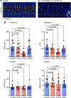
Fig. 8:. Ca2+ transients in ICC-IM of the LES are sustained by Ca2+ influx via Orai channels.
A STMs showing the effects of nifedipine (1 μM) on Ca2+ transients. B Summary data showing that nifedipine has no effect on Ca2+ transient frequency, amplitude, duration and spatial spread (c=10, N=4) in ICC-IM of the LES. C STMs showing the effects of the Orai channel inhibitor GSK 7975A (1 μM) on Ca2+ transients. D Summary data of the effects of GSK 7975A on Ca2+ transient frequency, amplitude, duration and spatial spread (c=13, N=4). Ca2+ transient frequency and amplitude were reduced significantly by GSK 7975A. This concentration of nifedipine was chosen as it has shown efficacy in blocking Ca2+ influx through L-type Ca2+ channels in several smooth muscle cells and tissues (Cobine et al., 2017; Drumm et al., 2020b; Hannigan et al., 2020; Baker et al., 2021b). This concentration of GSK7975A was chosen as it has shown efficacy in blocking Ca2+ influx via Orai channels in several SMCs and tissues (Boie et al., 2017; Chen & Sanderson, 2017; Drumm et al., 2018a; Drumm et al., 2018b; Zheng et al., 2018; Drumm et al., 2020a).
Fig. 9:
Fig. 9:. The effect of Ani9 on membrane potentials in mouse LES clasp muscles.
A Representative trace showing the membrane potential oscillations (MPOs) obtained upon impalment of cells in mouse LES clasp muscle. Ani9 (3 μM) caused hyperpolarization and reduced the amplitudes of MPOs in the presence of nicardipine (100 nM). Activity returned to control after washout of the drug. B Expanded time scale of portions of the recording in A (a=control, b=Ani9, c=washout). C Amplitude histograms from 1 min recordings generated by Clampfit 10.3 (Methods) summarizing RMPs and showing the net hyperpolarizations induced by Ani9 (n=8, N=8). Median values for RMP and MPOs were calculated from Gaussian fits of the data. This concentration of Ani9 was chosen, as there were no effects on contractile responses to high K+ solutions that might suggest off-target effects of the drug (see Fig. 11). D Summary of the effects of Ani9 on RMP. E. Summary of the effects of Ani9 on MPOs (plotted as standard error of the variance in RMP (distribution).
Fig. 10:
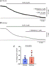
Fig. 10:. ANO1 channel antagonist reduces LES tone.
A Representative contractile trace showing the effect of the ANO1 channel antagonist, Ani9 (3 μM), on tone in mouse LES (n=6, N=6). B Representative contractile trace showing the effect of Ani9 (3 μM) on tone in monkey LES (n=6, N=2). C Summary data for the effects of Ani9 on tone in mouse and monkey LES. This concentration of Ani9 was chosen because it had no effect on contractile responses to high K+ solutions (see Fig. 11). This concentration of sodium nitroprusside (SNP) was chosen as it has shown efficacy in producing maximum relaxation in other GI muscles (Cobine et al., 2017; Cobine et al., 2018).
Fig. 11:
Fig. 11:. Tests to confirm concentrations of ANO1 channel antagonist did not have off-target effects on KCl-induced contractions of LES.
A Representative contractile trace showing the effects of the ANO1 channel antagonist, Ani9 (1 and 3 μM) and nifedipine (1 μM) on contraction of mouse LES induced by 60 mM KCl solution. B Representative contractile trace showing the effects of a range of Ani9 concentrations (300 nM – 10 μM) and nifedipine (1 μM) on contraction of monkey LES induced by 60 mM KCl solution. Ani9 at the concentrations used elsewhere in this study did not block high K+ contractions but the contractions were completely blocked by nifedipine. C Summary data for the effects of Ani9 (3μM) on KCl-induced contractions in mouse (n=6, N=6) and monkey LES (n=4, N=4).
Fig. 12:
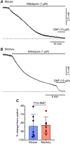
Fig. 12:. Cav1.2 channel antagonist reduces LES tone.
A Representative contractile trace showing the effect of the Cav1.2 channel antagonist, nifedipine (1 μM), on tone in mouse LES (n=7, N=7). B Representative contractile trace showing the effects of nifedipine (1 μM) on tone in the monkey LES (n=6, N=3). SNP (10 μM) was added at the end of the traces in A&B to determine if there was any remaining tone after nifedipine. C Summary data for the effects of nifedipine on mouse and monkey LES tone.

Comment in

  • LEtS set the tone.
    Joshi V, Beyder A. Joshi V, et al. J Physiol. 2022 Jun;600(11):2541-2542. doi: 10.1113/JP283011. Epub 2022 Apr 30. J Physiol. 2022. PMID: 35445403 Free PMC article. No abstract available.

References

    1. Bai Y, Edelmann M & Sanderson MJ. (2009). The contribution of inositol 1,4,5-trisphosphate and ryanodine receptors to agonist-induced Ca2+ signaling of airway smooth muscle cells. American Journal of Physiology - Lung Cellular and Molecular Physiology 297, 347–361. - PMC - PubMed
    1. Baker SA, Drumm BT, Saur D, Hennig GW, Ward SM & Sanders KM. (2016). Spontaneous Ca2+ transients in interstitial cells of Cajal located within the deep muscular plexus of the murine small intestine. The Journal of Physiology 594, 3317–3338. - PMC - PubMed
    1. Baker SA, Hwang SJ, Blair PJ, Sireika C, Wei L, Ro S, Ward SM & Sanders KM. (2021a). Ca(2+) transients in ICC-MY define the basis for the dominance of the corpus in gastric pacemaking. Cell Calcium 99, 102472. - PMC - PubMed
    1. Baker SA, Leigh WA, Del Valle G, De Yturriaga IF, Ward SM, Cobine CA, Drumm BT & Sanders KM. (2021b). Ca(2+) signaling driving pacemaker activity in submucosal interstitial cells of Cajal in the murine colon. Elife 10. - PMC - PubMed
    1. Beckett EAH, Bayguinov YR, Sanders KM, Ward SM & Hirst GDS. (2004). Properties of unitary potentials generated by intramuscular interstitial cells of Cajal in the murine and guinea-pig gastric fundus. Journal of Physiology 559, 259–269. - PMC - PubMed

Publication types