Review
. 2022 Apr;26(7):2049-2062.
doi: 10.1111/jcmm.17228. Epub 2022 Mar 1.

JAK-STAT core cancer pathway: An integrative cancer interactome analysis

Affiliations
Review

JAK-STAT core cancer pathway: An integrative cancer interactome analysis

Fettah Erdogan et al. J Cell Mol Med. 2022 Apr.

Abstract

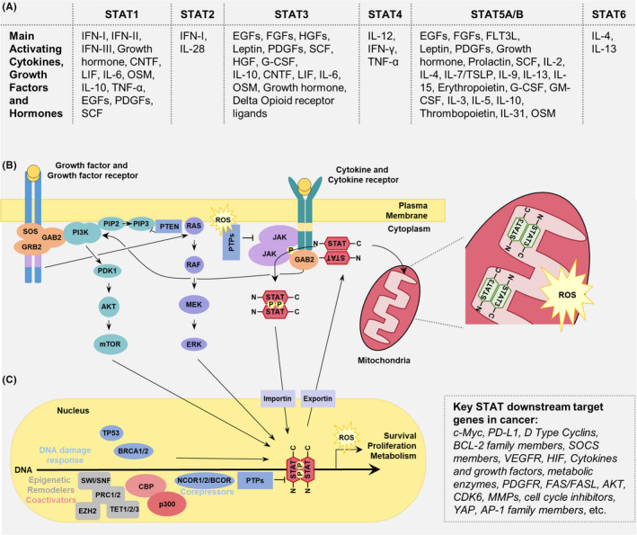
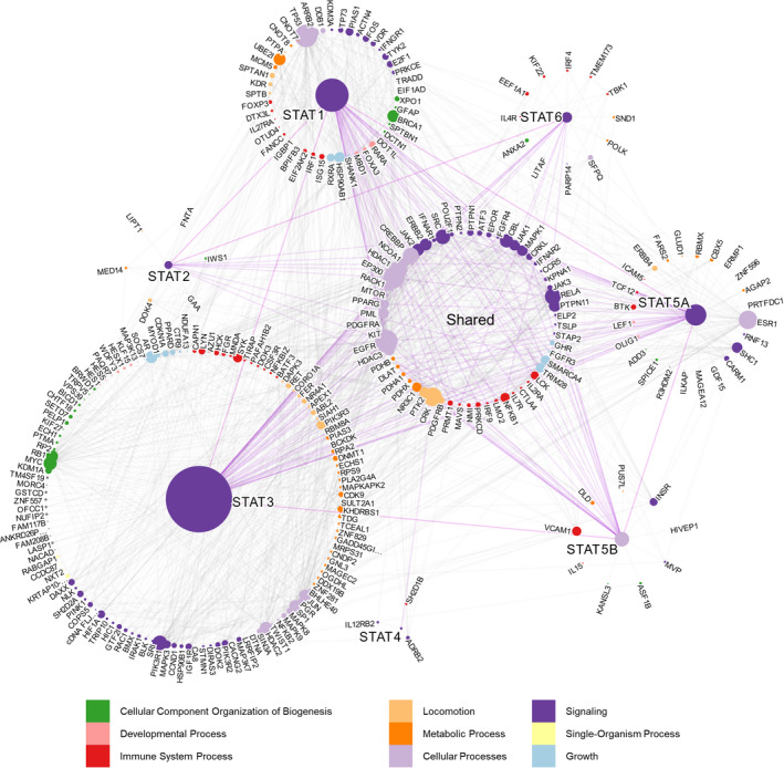
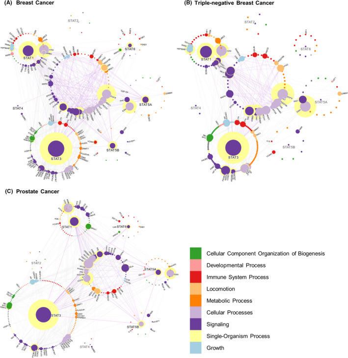
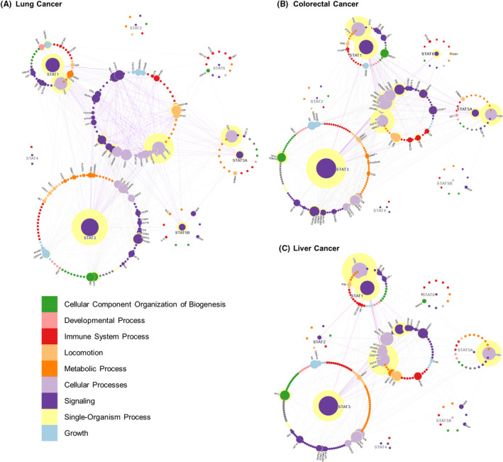
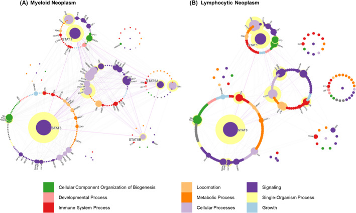
Through a comprehensive review and in silico analysis of reported data on STAT-linked diseases, we analysed the communication pathways and interactome of the seven STATs in major cancer categories and proposed rational targeting approaches for therapeutic intervention to disrupt critical pathways and addictions to hyperactive JAK/STAT in neoplastic states. Although all STATs follow a similar molecular activation pathway, STAT1, STAT2, STAT4 and STAT6 exert specific biological profiles associated with a more restricted pattern of activation by cytokines. STAT3 and STAT5A as well as STAT5B have pleiotropic roles in the body and can act as critical oncogenes that promote many processes involved in cancer development. STAT1, STAT3 and STAT5 also possess tumour suppressive action in certain mutational and cancer type context. Here, we demonstrated member-specific STAT activity in major cancer types. Through systems biology approaches, we found surprising roles for EGFR family members, sex steroid hormone receptor ESR1 interplay with oncogenic STAT function and proposed new drug targeting approaches of oncogenic STAT pathway addiction.

Keywords: JAK/STAT pathway in cancers; blood cancer; breast cancer; colorectal cancers; liver cancers; lung cancer; protein-protein interactions; systems medicine.


Conflict of interest statement

The authors declare no competing interests.

Figures

FIGURE 1
FIGURE 1
Common STAT cellular activation‐inactivation cycle pathways. (A) Variety of cytokines, hormones and growth factors can activate STAT family members. (B) Binding of the ligand to either the cytokine or growth factor (GF) receptor induces series of the activation cascades. Whereas JAK kinase activation is generally exclusive to cytokine receptor signaling, STAT action is triggered by both receptor types. Typically, auto‐phosphorylated receptor‐associated Janus kinases (JAKs) phospho‐activate STATs which form STAT parallel dimer and are transported across nuclear membrane by Importin α’s (KPNA1) and involve the action of Nucleophosmin I decamer (not shown). Binding of activated STATs to DNA is coupled to interactions with various transcriptional regulators discussed in the main text. Phosphatase‐mediated inactivation of the DNA‐bound STAT complex disengages the dimer from the DNA, breaks them into monomers and leads to their export via nuclear exportins (XPO1). Cytokine and growth factor receptors are both inducers of the RAS‐RAF‐MAPK and the PI3K‐AKT‐mTOR pathways. For simplicity, other signaling pathways such as activation of PKC or PLC are excluded. STAT3 is also capable of localizing into the mitochondria inner matrix and influencing ROS production. (C) STATs are also reported to be involved in the DNA damage response (e.g., TP53 and BRCA1/2) and interact with epigenetic modifiers (e.g., EZH2, TET1/2/3, SWI/SNF, PRC1/2, CBP‐p300 and NCOA‐1). CNTF, ciliary neurotrophic factor; EGF, Epidermal growth factor; FGF, fibroblast growth factor; FLT3L, FMS‐like tyrosine kinase 3 ligand; G‐CSF, granulocyte colony‐stimulating factor; GM‐CSF, granulocyte‐macrophage colony‐stimulating factor; HGF, hepatocyte growth factor; IFN, interferon; IL, interleukin; LIF, leukemia inhibitory factor; OSM, oncostatin M; PDGF, platelet‐derived growth factor; SCF, stem cell factor; TGF, transforming growth factor; TNF, tumor necrosis factor; TSLP, thymic stromal lymphopoietin
FIGURE 2
FIGURE 2
STAT interactome. Global STAT interactome presenting all literature‐based STAT PPIs. Each node (dot) is a protein, and each edge (line) is an interaction between two proteins. The size of the nodes is proportional to their degree, and the color of each node is representative of the biological process of the protein as described in the legend. The length of the edges connecting each node is arbitrary. Proteins which were found to interact with multiple STATs are grouped in the middle, and those exclusive to one STAT are grouped around that corresponding STAT member. A summary table of all interactions can be found in Table S1
FIGURE 3
FIGURE 3
STAT interactions in breast cancer, TNBC and prostate cancer. STAT interactions in two of the five most common death‐causing cancers listed by the WHO; breast cancer and prostate cancer, as well as the breast cancer subtype TNBC. Any interaction shared between two or more STATs is grouped in the middle, and the edges connecting those interactions are highlighted. Proteins for which a label is shown are those which are reported to be over‐expressed in that disease. Although not all proteins directly interact with each other, they may communicate indirectly through hub proteins. The proteins important for indirect communication are identified as ‘central’ to the networks and have a yellow halo in the network maps. Centrality measures how important the protein is for communication between the STAT interactome. It is measured using an undirected all‐pairs shortest path algorithm which measures how many of the shortest paths between proteins pass through a node. Therefore, proteins which are more central act as hubs through which signals are efficiently transmitted from one end of the interactome to the other. The centrality of a protein is proportional to the size of the yellow halo
FIGURE 4
FIGURE 4
STAT interactions in lung, colorectal and liver cancers. STAT interactions in the three of the five commonly death‐causing cancers listed by the WHO lung cancer, colorectal cancer and liver cancer. For a descriptor of the interactome labels, see Figure 3
FIGURE 5
FIGURE 5
STAT interactions in hematopoietic cancers. STAT interactions in the categories of myeloid and lymphoid neoplasms. For a descriptor of the interactome labels, see Figure 4

References

    1. Yu H, Pardoll D, Jove R. STATs in cancer inflammation and immunity: a leading role for STAT3. Nat Rev Cancer. 2009;9:798‐809. - PMC - PubMed
    1. Wang Z, Zhu S, Shen M, et al. STAT3 is involved in esophageal carcinogenesis through regulation of Oct‐1. Carcinogenesis. 2013;34:678‐688. - PubMed
    1. Schindler C, Darnell JE Jr Transcriptional responses to polypeptide ligands: the JAK‐STAT pathway. Annu Rev Biochem. 1995;64(1):621‐652. doi:10.1146/annurev.bi.64.070195.003201 - DOI - PubMed
    1. Shuai K, Horvath CM, Huang LH, Qureshi SA, Cowburn D, Darnell JE Jr. Interferon activation of the transcription factor Stat91 involves dimerization through SH2‐phosphotyrosyl peptide interactions. Cell. 1994;76(5):821‐828. doi:10.1016/0092-8674(94)90357-3 - DOI - PubMed
    1. Darnell JE, Kerr LM, Stark GR. Jak‐STAT pathways and transcriptional activation in response to IFNs and other extracellular signaling proteins. Science. 1994;264(5164):1415‐1421. doi:10.1126/science.8197455 - DOI - PubMed

Publication types

MeSH terms