. 2022 Mar 1;14(5):2081-2100.
doi: 10.18632/aging.203924. Epub 2022 Mar 1.

Modulation of RNA splicing associated with Wnt signaling pathway using FD-895 and pladienolide B

Affiliations

Modulation of RNA splicing associated with Wnt signaling pathway using FD-895 and pladienolide B

Deepak Kumar et al. Aging (Albany NY). .

Abstract

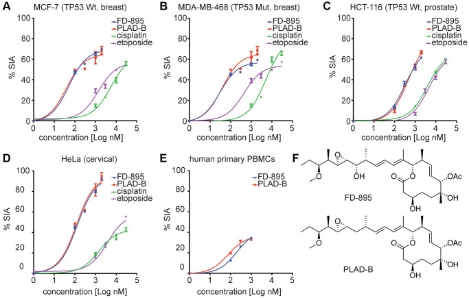
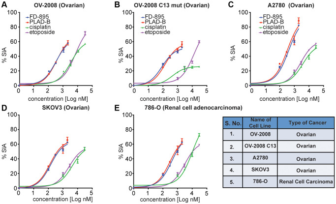
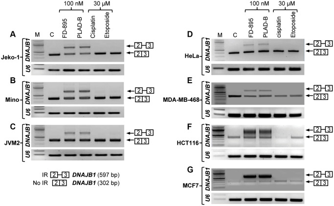
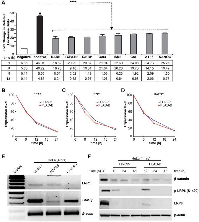
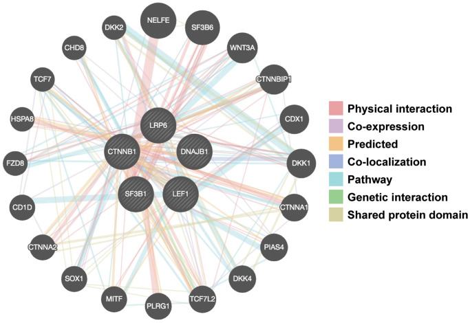
Alterations in RNA splicing are associated with different malignancies, including leukemia, lymphoma, and solid tumors. The RNA splicing modulators such as FD-895 and pladienolide B have been investigated in different malignancies to target/modulate spliceosome for therapeutic purpose. Different cell lines were screened using an RNA splicing modulator to test in vitro cytotoxicity and the ability to modulate RNA splicing capability via induction of intron retention (using RT-PCR and qPCR). The Cignal Finder Reporter Array evaluated [pathways affected by the splice modulators in HeLa cells. Further, the candidates associated with the pathways were validated at protein level using western blot assay, and gene-gene interaction studies were carried out using GeneMANIA. We show that FD-895 and pladienolide B induces higher apoptosis levels than conventional chemotherapy in different solid tumors. In addition, both agents modulate Wnt signaling pathways and mRNA splicing. Specifically, FD-895 and pladienolide B significantly downregulates Wnt signaling pathway-associated transcripts (GSK3β and LRP5) and both transcript and proteins including LEF1, CCND1, LRP6, and pLRP6 at the transcript, total protein, and protein phosphorylation's levels. These results indicate FD-895 and pladienolide B inhibit Wnt signaling by decreasing LRP6 phosphorylation and modulating mRNA splicing through induction of intron retention in solid tumors.

Keywords: FD-895; Wnt signaling; intron retention; pladienolide B; splice modulation; spliceosome; splicing.


Conflict of interest statement

CONFLICTS OF INTEREST: The authors declare no conflicts of interest related to this study.

Figures

Figure 1
Figure 1
In vitro cytotoxicity induced by FD-895 and pladienolide B in different cancer cell lines, and normal human primary PBMCs. Cancer Cells were exposed to FD-895 (100 nM to 2 uM), pladienolide B (100 nM to 2 uM), etoposide (1 μM to 30 μM), and cisplatin (1 μM to 30 μM) for 48 h. Apoptosis were measured in MCF-7 (A), MDA-MB-468 (B), HCT-116 (C) and HeLa (D) cells using MTS assay. The absorbance of the control (cells without treatment) was subtracted from the treated cells of each cell line. (E) Normal PBMC cells were exposed to FD-895, and pladienolide B. Cells were stained with propidium iodide and DiOC6 to differentiate dead and viable cells by using flow cytometer. Data presented in form of % specific induced apoptosis (% SIA). To assess the compound specific induced apoptosis vs. background spontaneous cell death from in vitro culture conditions, we calculated the percentage of SIA using the following formula: % SIA = [(compound induced apoptosis – media only spontaneous apoptosis)/(100- media only spontaneous apoptosis)] × 100. The data shows the results of samples analyzed in duplicate with the mean and its respective SD. (F) Structures of pladienolide-B and FD-895.
Figure 2
Figure 2
FD-895 or by pladienolide B induced apoptosis in ovarian and renal cancer cells. Ovarian cancer cells (A) OV-2008, cisplatin sensitive (B) OV-2008 C13 mut, cisplatin resistant, (C) A2780, (D) SKOV3 or (E) renal cancer cells (786-O) were incubated with FD-895 (100 nM to 2 μM), pladienolide B (100 nM to 2 μM), etoposide (1 μM to 30 μM), or of cisplatin (1 μM to 30 μM) for 48 h. Cells viability were measured as using MTS assay. This experiment was repeated in triplicate independently. Data presented in form of % SIA. The data shows the results of samples analyzed in duplicate with the mean and its respective SD.
Figure 3
Figure 3
Intron retention of DNAJB1 gene in different cancer cell lines. Tumor cell lines were treated with 100 nM of FD-895, 100 nM pladienolide B, 30 μM cisplatin or 30 μM etoposide for 4 h. Analysis of IR of DNAJB1 mRNAs was evaluated by RT-PCR in (AC) Mantle cell lymphoma cells, (Jeko-1, Mino and JMV-2), (D) HeLa, (E) MDA-MB-468 (F) HCT116 and (G) MCF-7 cells. RNU6A, an intronless gene was used as RNA quality and loading control.
Figure 4
Figure 4
Effect of FD-895 on different pathways and effect on Wnt signaling in HeLa cells. (A) HeLa cells were use for introducing pathway reporters into cells via reverse transfection. Post-transfection, the cells were treated with vehicle or 100 nM FD-895 for 3 h. Luciferase and renilla expression was evaluated. HeLa cells were exposed to 100 nM of FD-895 or 100 nM pladienolide B for 6 h, 12 h or 24 h and expression of (B) LEF1, (C) FN1, and (D) CCND1 were determined by qRT-PCR. (E) HeLa cells were treated with 100 nM of FD-895 or 30 μM cisplatin for 4 h. Analysis of IR for GSK3β and LRP5 mRNAs was evaluated by using RT-PCR. (F) HeLa cells were treated with 100 nM of FD-895 or 100 nM pladienolide B for 6 h, 12 h or 24 h. Protein extracts were immunoblotted for β-catenin, phohspho-LRP6, LRP6, and β-actin.
Figure 5
Figure 5
Gene-gene interaction networks among selected genes constructed by GeneMANIA. A gene-gene network was constructed with the search tool for the retrieval of interacting genes available in GeneMANIA annotation information for selected 5 genes, including physical interaction, genetic interaction, co-expression and shared pathways and protein structure domain. The central black nodes denote 5 selected genes used as an “INPUT”, and peripheral nodes denote gene interactions with the black nodes. The network also contains 20 normal human genes. The size of the circles indicates the degree of interaction.

Similar articles

Cited by

References

    1. Wang BD, Lee NH. Aberrant RNA Splicing in Cancer and Drug Resistance. Cancers (Basel). 2018; 10:458. 10.3390/cancers10110458 - DOI - PMC - PubMed
    1. Tripathi G, Tripathi A, Johnson J, Kashyap MK. Role of RNA Splicing in Regulation of Cancer Stem Cell. Curr Stem Cell Res Ther. 2021. [Epub ahead of print]. 10.2174/1574888X16666211207103628 - DOI - PubMed
    1. León B, Kashyap MK, Chan WC, Krug KA, Castro JE, La Clair JJ, Burkart MD. A Challenging Pie to Splice: Drugging the Spliceosome. Angew Chem Int Ed Engl. 2017; 56:12052–63. 10.1002/anie.201701065 - DOI - PMC - PubMed
    1. Jutzi D, Akinyi MV, Mechtersheimer J, Frilander MJ, Ruepp MD. The emerging role of minor intron splicing in neurological disorders. Cell Stress. 2018; 2:40–54. 10.15698/cst2018.03.126 - DOI - PMC - PubMed
    1. Chen W, Moore MJ. The spliceosome: disorder and dynamics defined. Curr Opin Struct Biol. 2014; 24:141–9. 10.1016/j.sbi.2014.01.009 - DOI - PMC - PubMed

Publication types