Review
. 2022 May:84:100889.
doi: 10.1016/j.jbior.2022.100889. Epub 2022 Feb 23.

Phosphorylation-mediated regulation of the Nem1-Spo7/Pah1 phosphatase cascade in yeast lipid synthesis

Affiliations
Review

Phosphorylation-mediated regulation of the Nem1-Spo7/Pah1 phosphatase cascade in yeast lipid synthesis

Shoily Khondker et al. Adv Biol Regul. 2022 May.

Abstract

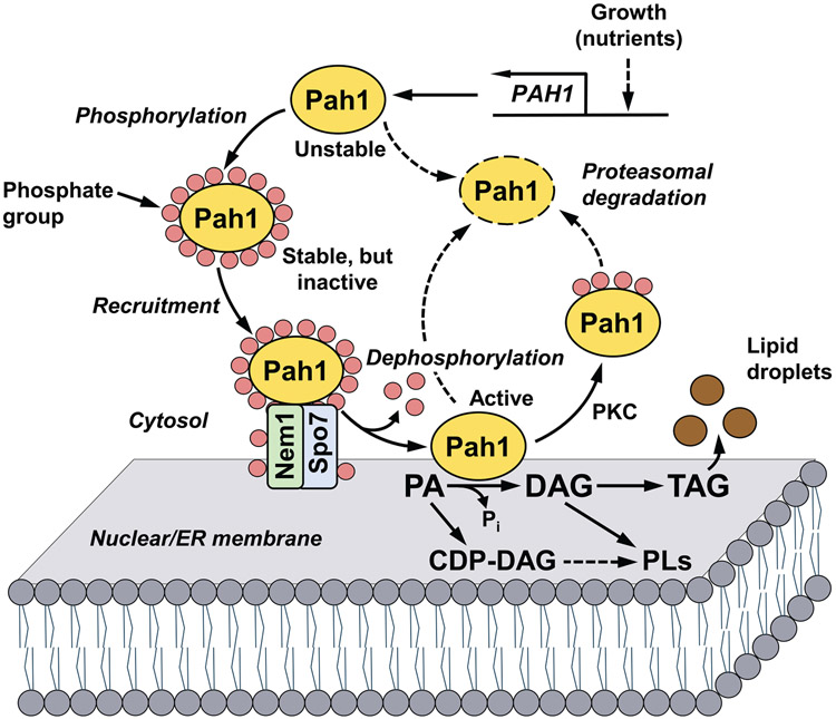
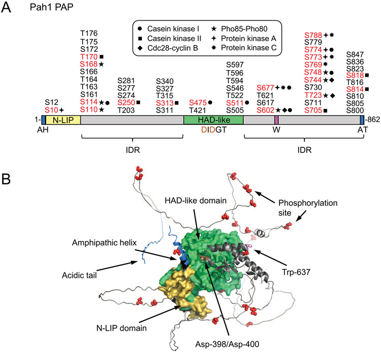
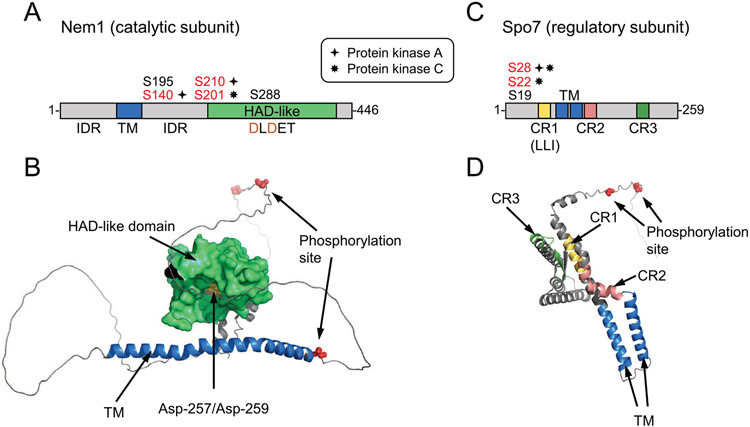
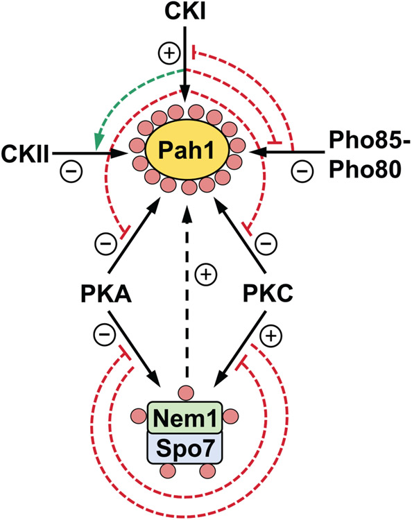
The PAH1-encoded phosphatidate phosphatase, which catalyzes the dephosphorylation of phosphatidate to produce diacylglycerol, controls the divergence of phosphatidate into triacylglycerol synthesis and phospholipid synthesis. Pah1 is inactive in the cytosol as a phosphorylated form and becomes active on the nuclear/endoplasmic reticulum membrane as a dephosphorylated form by the Nem1-Spo7 protein phosphatase complex. The phosphorylation of Pah1 by protein kinases, which include casein kinases I and II, Pho85-Pho80, Cdc28-cyclin B, and protein kinases A and C, controls its cellular location, catalytic activity, and susceptibility to proteasomal degradation. Nem1 (catalytic subunit) and Spo7 (regulatory subunit), which form a protein phosphatase complex catalyzing the dephosphorylation of Pah1 for its activation, are phosphorylated by protein kinases A and C. In this review, we discuss the functions and interrelationships of the protein kinases in the control of the Nem1-Spo7/Pah1 phosphatase cascade and lipid synthesis.

Keywords: Diacylglycerol; Nem1-Spo7 protein phosphatase; Pah1 PA phosphatase; Phosphatidic acid; Phospholipid; Protein kinase; Triacylglycerol; Yeast.


Conflict of interest statement

Conflicts of interest

None.

Figures

Fig. 1.
Fig. 1.
Model for the phosphorylation/dephosphorylation-mediated regulation of Pah1 PAP in lipid synthesis. Expression of the PAP-encoding gene PAH1 is regulated during growth by nutrient status. After expression, its product Pah1 in the cytosol is phosphorylated by multiple protein kinases. Phosphorylated Pah1 translocates to the nuclear/ER membrane through its recruitment and dephosphorylation by the Nem1-Spo7 protein phosphatase complex, which itself is subject to phosphorylation. Dephosphorylated Pah1 that is associated with the membrane catalyzes the conversion of PA to DAG, which is then acylated to form TAG that is stored in lipid droplets. Dephosphorylated Pah1 or PKC-phosphorylated Pah1 that is not phosphorylated at the target sites for Pho85-Pho80/Cdc28-cyclin B is degraded by the proteasome (indicated by the dashed line arrows and ellipse). The PAP substrate PA may also be converted to CDP-DAG, which is then used for the synthesis of the membrane phospholipids phosphatidylserine, phosphatidylethanolamine, phosphatidylcholine, phosphatidylinositol, phosphatidylglycerol, and cardiolipin. The PAP product DAG may also be converted to phosphatidylcholine and phosphatidylethanolamine via the Kennedy pathway. Greater details of the yeast phospholipid synthetic pathways may be found elsewhere (Carman and Han, 2011; Kwiatek et al., 2020).
Fig. 2.
Fig. 2.
Domains/regions and phosphorylation sites in Pah1. A, the diagram shows the positions of the N-terminal amphipathic helix (AH), the conserved N-LIP and HAD-like catalytic domains, the conserved tryptophan (W) residue, the C-terminal acidic tail (AT), and the intrinsically disordered regions (IDR). The serine (S) and threonine (T) residues known to be phosphorylated are grouped at their approximate positions, and marked in red color for the known responsible protein kinases. B, the AlphaFold (Jumper et al., 2021) structure prediction of Pah1 was visualized with the PyMol program.
Fig. 3.
Fig. 3.
Domains/regions and phosphorylation sites in Nem1 and Spo7. A, the diagram denotes the catalytic HAD-like domain, transmembrane (TM) region, and intrinsically disordered regions (IDR) in Nem1. C, the diagram shows the conserved regions (CR) 1, 2, and 3 and transmembrane regions of Spo7. The serine residues phosphorylated (red color) by PKA or PKC in Nem1 and Spo7 are also indicated. B and D, the AlphaFold (Jumper et al., 2021) structure predictions of Nem1 and Spo7 were visualized with the PyMol program.
Fig. 4.
Fig. 4.
Interrelationships between the phosphorylations of Pah1, Nem1, and Spo7. Pah1 is phosphorylated by CKI, CKII, Pho85-Pho80, PKA, and PKC (solid arrows, upper diagram). The CKI phosphorylation of Pah1 stimulates its subsequent phosphorylation by CKII (dashed green arrow), but inhibits its subsequent phosphorylations by Pho85-Pho80, PKA, and PKC (dashed blunted red line). The Pho85-Pho80 phosphorylation of Pah1 inhibits its subsequent phosphorylation by CKI (dashed blunted red line). Nem1 and Spo7 are phosphorylated by PKA and PKC (solid arrows, lower diagram). The phosphorylation of the Nem1-Spo7 complex by PKA inhibits phosphorylation of Spo7 by PKC, whereas the phosphorylation of the complex by PKC inhibits phosphorylation of Nem1 by PKA (dashed blunted red lines). Phosphorylated Pah1, Nem1, and Spo7 are indicated by the small pink circles, and the effects of the phosphorylations by the various protein kinases are indicated by + and − symbols.

References

    1. Adeyo O, Horn PJ, Lee S, Binns DD, Chandrahas A, Chapman KD, Goodman JM, 2011. The yeast lipin orthologue Pah1p is important for biogenesis of lipid droplets. J. Cell Biol 192, 1043–1055. - PMC - PubMed
    1. Albuquerque CP, Smolka MB, Payne SH, Bafna V, Eng J, Zhou H, 2008. A multidimensional chromatography technology for in-depth phosphoproteome analysis. Mol. Cell Proteomics 7, 1389–1396. - PMC - PubMed
    1. Babu P, Bryan JD, Panek HR, Jordan SL, Forbrich BM, Kelley SC, Colvin RT, Robinson LC, 2002. Plasma membrane localization of the Yck2p yeast casein kinase 1 isoform requires the C-terminal extension and secretory pathway function. J. Cell Sci 115, 4957–4968. - PubMed
    1. Barbosa AD, Sembongi H, Su W-M, Abreu S, Reggiori F, Carman GM, Siniossoglou S, 2015. Lipid partitioning at the nuclear envelope controls membrane biogenesis. Mol. Biol. Cell 26, 3641–3657. - PMC - PubMed
    1. Bodenmiller B, Wanka S, Kraft C, Urban J, Campbell D, Pedrioli PG, Gerrits B, Picotti P, Lam H, Vitek O, Brusniak MY, Roschitzki B, Zhang C, Shokat KM, Schlapbach R, Colman-Lerner A, Nolan GP, Nesvizhskii AI, Peter M, Loewith R, von MC, Aebersold R, 2010. Phosphoproteomic analysis reveals interconnected system-wide responses to perturbations of kinases and phosphatases in yeast. Sci. Signal 3, rs4. - PMC - PubMed

Publication types

MeSH terms

LinkOut - more resources