

The emerging roles of extracellular vesicles as intercellular messengers in liver physiology and pathology
- PMID: 35232008
- PMCID: PMC9597227
- DOI: 10.3350/cmh.2021.0390
The emerging roles of extracellular vesicles as intercellular messengers in liver physiology and pathology
Abstract
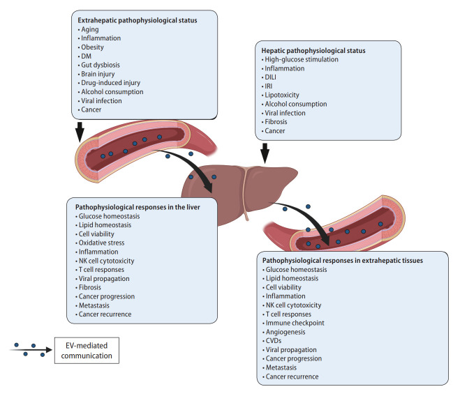
Extracellular vesicles (EVs) are membrane-enclosed particles released from almost all cell types. EVs mediate intercellular communication by delivering their surface and luminal cargoes, including nucleic acids, proteins, and lipids, which reflect the pathophysiological conditions of their cellular origins. Hepatocytes and hepatic non-parenchymal cells utilize EVs to regulate a wide spectrum of biological events inside the liver and transfer them to distant organs through systemic circulation. The liver also receives EVs from multiple organs and integrates these extrahepatic signals that participate in pathophysiological processes. EVs have recently attracted growing attention for their crucial roles in maintaining and regulating hepatic homeostasis. This review summarizes the roles of EVs in intrahepatic and interorgan communications under different pathophysiological conditions of the liver, with a focus on chronic liver diseases including nonalcoholic steatohepatitis, alcoholic hepatitis, viral hepatitis, liver fibrosis, and hepatocellular carcinoma. This review also discusses recent progress for potential therapeutic applications of EVs by targeting or enhancing EV-mediated cellular communication for the treatment of liver diseases.
Keywords: Communication; Extracellular vesicles; Liver; Pathology; Physiology.
Conflict of interest statement
The authors have no conflicts to disclose.
Figures
Similar articles
-
Extracellular Vesicles in NAFLD/ALD: From Pathobiology to Therapy.Cells. 2020 Mar 27;9(4):817. doi: 10.3390/cells9040817. Cells. 2020. PMID: 32231001 Free PMC article. Review.
-
Extracellular vesicles in liver disease and beyond.World J Gastroenterol. 2018 Oct 28;24(40):4519-4526. doi: 10.3748/wjg.v24.i40.4519. World J Gastroenterol. 2018. PMID: 30386101 Free PMC article.
-
The promise of small particles: extracellular vesicles as biomarkers in liver pathology.J Physiol. 2023 Nov;601(22):4953-4971. doi: 10.1113/JP283074. Epub 2022 Jul 1. J Physiol. 2023. PMID: 35708653 Review.
-
Extracellular vesicles in liver pathobiology: Small particles with big impact.Hepatology. 2016 Dec;64(6):2219-2233. doi: 10.1002/hep.28814. Epub 2016 Oct 20. Hepatology. 2016. PMID: 27628960 Free PMC article. Review.
-
The role of hepatocyte-derived extracellular vesicles in liver and extrahepatic diseases.Biomed Pharmacother. 2024 Nov;180:117502. doi: 10.1016/j.biopha.2024.117502. Epub 2024 Oct 1. Biomed Pharmacother. 2024. PMID: 39357327 Review.
Cited by
-
The Molecular Dynamics of Extracellular Vesicles and their Protein Corona in the Secretomes of Stem Cells.Stem Cell Rev Rep. 2025 Aug 23. doi: 10.1007/s12015-025-10961-1. Online ahead of print. Stem Cell Rev Rep. 2025. PMID: 40848102 Review.
-
Current updates on the molecular and genetic signals as diagnostic and therapeutic targets for hepatitis B virus-associated hepatic malignancy.Heliyon. 2024 Jul 8;10(14):e34288. doi: 10.1016/j.heliyon.2024.e34288. eCollection 2024 Jul 30. Heliyon. 2024. PMID: 39100497 Free PMC article. Review.
-
A century journey of organelles research in the plant endomembrane system.Plant Cell. 2024 May 1;36(5):1312-1333. doi: 10.1093/plcell/koae004. Plant Cell. 2024. PMID: 38226685 Free PMC article. Review.
-
Plasma Pattern of Extracellular Vesicles Isolated from Hepatitis C Virus Patients and Their Effects on Human Vascular Endothelial Cells.Int J Mol Sci. 2023 Jun 15;24(12):10197. doi: 10.3390/ijms241210197. Int J Mol Sci. 2023. PMID: 37373343 Free PMC article.
-
Cell-to-cell and organ-to-organ crosstalk in the pathogenesis of alcohol-associated liver disease.eGastroenterology. 2024 Oct;2(4):e100104. doi: 10.1136/egastro-2024-100104. Epub 2024 Dec 9. eGastroenterology. 2024. PMID: 39735421 Free PMC article.
References
-
- Théry C, Witwer KW, Aikawa E, Alcaraz MJ, Anderson JD, Andriantsitohaina R, et al. Minimal information for studies of extracellular vesicles 2018 (MISEV2018): a position statement of the international society for extracellular vesicles and update of the MISEV2014 guidelines. J Extracell Vesicles. 2018;7:1535750. - PMC - PubMed
-
- Vlachakis D, Mitsis Τ, Nicolaides N, Efthimiadou A, Giannakakis A, Bacopoulou F, et al. Functions, pathophysiology and current insights of exosomal endocrinology (review) Mol Med Rep. 2021;23:26. - PubMed
Publication types
MeSH terms
Substances
Grants and funding
LinkOut - more resources
Full Text Sources
Other Literature Sources
Medical
