. 2022 Mar;12(3):210271.
doi: 10.1098/rsob.210271. Epub 2022 Mar 2.

Crystals of TELSAM-target protein fusions that exhibit minimal crystal contacts and lack direct inter-TELSAM contacts

Affiliations

Crystals of TELSAM-target protein fusions that exhibit minimal crystal contacts and lack direct inter-TELSAM contacts

Supeshala Nawarathnage et al. Open Biol. 2022 Mar.

Abstract

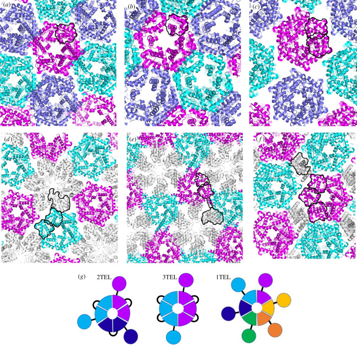
While conducting pilot studies into the usefulness of fusion to TELSAM polymers as a potential protein crystallization strategy, we observed novel properties in crystals of two TELSAM-target protein fusions, as follows. (i) A TELSAM-target protein fusion can crystallize more rapidly and with greater propensity than the same target protein alone. (ii) TELSAM-target protein fusions can be crystallized at low protein concentrations. This unprecedented observation suggests a route to crystallize proteins that can only be produced in microgram amounts. (iii) The TELSAM polymers themselves need not directly contact one another in the crystal lattice in order to form well-diffracting crystals. This novel observation is important because it suggests that TELSAM may be able to crystallize target proteins too large to allow direct inter-polymer contacts. (iv) Flexible TELSAM-target protein linkers can allow target proteins to find productive binding modes against the TELSAM polymer. (v) TELSAM polymers can adjust their helical rise to allow fused target proteins to make productive crystal contacts. (vi). Fusion to TELSAM polymers can stabilize weak inter-target protein crystal contacts. We report features of these TELSAM-target protein crystal structures and outline future work needed to validate TELSAM as a crystallization chaperone and determine best practices for its use.

Keywords: TELSAM; X-ray crystallography; covalent protein crystallization chaperone; polymer-forming protein crystallization chaperone; protein crystallization method; protein polymer.


Conflict of interest statement

We declare we have no competing interests

Figures

Figure 1.
Figure 1.
All currently reported structures involving TELSAM polymers feature direct inter-polymer crystal contacts. TELSAM polymers are shown in cartoon representation and coloured in magenta, cyan and violet. Fused target proteins are coloured grey. In each image, a single polypeptide has been indicated with black outlines around each of its constituent sub-domains. (a) PDB ID 1JI7: 1TEL alone [5], (b) PDB ID 1LKY: 1TEL E222R mutant [8], (c) PDB ID 2QB1: 2TEL alone [6], (d) PDB ID 2QB0: 2TEL–lysozyme fusion [6], (e) PDB ID 2QAR: 2TEL–helix–lysozyme fusion [6], (f) PDB ID 5L0P: 3TEL–ferric uptake regulator fusion [7], (g) schematic of 2TEL, 3TEL and 1TEL. Individual polypeptides are offset with unique colours. Circles denote target proteins, wedges denote TELSAM subunits and black lines denote linkers.
Figure 2.
Figure 2.
TELSAM accelerates the crystallization rate of a genetically fused vWa domain. (a) SEC trace of vWa alone. (b) A post-SEC polyacrylamide gel electrophoresis (PAGE) gel of purified vWa alone. (c) Representative crystals of vWa alone. Scale bar is 100 μm. (d) Design model of a 1TEL-flex-vWa fusion in cartoon representation, with TELSAM in magenta and the vWa in cyan. Other subunits of the TELSAM polymer are shown in white. The linker is coloured yellow and indicated with an arrow. (e) SEC trace of 1TEL-flex-vWa. (f) A post-SEC PAGE gel of purified 1TEL-flex-vWa. (g) Crystals of 1TEL-flex-vWa. Scale bar is 100 μm. (h) Representative diffraction pattern from a crystal of 1TEL-flex-vWa.
Figure 3.
Figure 3.
Detail of the 1TEL-flex-vWa crystal structure and lattice. (a) Crystal lattice of 1TEL-flex-vWa, in cartoon representation with TELSAM in magenta and the vWa in cyan. A black outline denotes each sub-domain of a single polypeptide. (b) Side view of the 1TEL-flex-vWa crystal lattice, showing two TELSAM polymers (magenta) and selected vWA domains (cyan and purple). (c) Superposition of the vWa domain from 1TEL-flex-vWa (cyan) onto previously published vWa structures (grey, PDB IDs: 1SHU, 1SHT [11], 1T6B [24] and 1TZN [25]). Significant differences from previous structures are indicated with arrows. (d) Comparison of the design model (white) and crystal structure (magenta and cyan) of 1TEL-flex-vWa (the region of the linker that becomes α-helical is indicated with an arrow). Other copies of the vWa domain have been omitted for clarity. (e) Detail of the cis interface between 1TEL (magenta) and vWa (cyan). Hydrogen bonds are shown as black dashes. The single alanine linker is shown in yellow. (f) Detail of the trans interface between two vWa units, coloured cyan and grey. (g) Comparison of the helical rise from previously published TELSAM crystal structures [–8]. The relative rise of a single turn of each helix is denoted with a black bar. Fused target proteins have been omitted. (h) Detail of T4 lysozyme (cyan) intercalation into the TELSAM polymer helix (magenta) of PDB ID: 2QAR [6].
Figure 4.
Figure 4.
Production and detail of the 3TEL-rigid-DARPin crystal structure. (a) Design model of 3TEL-rigid-DARPin, with successive 1TEL domains shown in purple, orange and magenta and the DARPin in cyan. Linkers are coloured yellow and indicated with arrows. (b) SEC trace of 3TEL-rigid-DARPin. (c) A post-SEC PAGE gel of 3TEL-rigid-DARPin. (d) Representative crystals of 3TEL-rigid-DARPin. Scale bar is 100 μm. (e) Representative diffraction pattern of 3TEL-rigid-DARPin. (f) Comparison of the design model (magenta) with the crystal structure (cyan and magenta) of 3TEL-rigid-DARPin, with an arrow to indicate the shift of the DARPin and connecting α-helix. (g) Superposition of the DARPin domains from the 3TEL-rigid-DARPin crystal structure (cyan) and from the published structure of this same DARPin (white, PDB ID: 4J7W) [12]. Significant differences from the previous structure are indicated with arrows. (h) Detail of the crystal packing from the previous crystal structure of the DARPin domain. One of the DARPin domains has been coloured according to the crystallographic B-factor. (i) Detail of the crystal contacts from the crystal structure of 3TEL-rigid-DARPin. One of the 3TEL-rigid-DARPin units has been coloured according to the crystallographic B-factor. The view angle is the same as in (h).
Figure 5.
Figure 5.
Additional features of the 3TEL-rigid-DARPin crystal structure. (a) Crystal lattice of 3TEL-rigid-DARPin. Black outlines highlight a single polymer as well as the domains of a single polypeptide subunit within that polymer. (b) Side view of the 3TEL-rigid-DARPin crystal lattice, showing two TELSAM polymers (light blue and light pink) and selected DARPin domains (cyan and magenta). (c–e). Diagram relating the 3TEL-rigid-DARPin crystal packing to the crystallographic unit cell, shown from three view angles. Two polymer layers are shown, with three polymers in each layer and 6 × 3TEL-rigid-DARPin subunits in each polymer (three turns of each polymer). The unit cell axes are denoted with labelled arrows and approximately correspond to the reciprocal space axes and the axes of an ellipsoid approximating the anisotropic diffraction limits. In each view angle, the origin lies in the plane of the page while the unseen unit cell axis projects out of the page towards the reader. These anisotropic diffraction limits approximately correspond to each of the unit cell vectors and appear in coloured type. Schematics are given below each view angle to indicate the orientation of both the TELSAM polymers (pink and blue) and the crystal as a whole (white prismatic disc) in each view. The predicted vertical displacement of the two polymer sheets relative to one another is indicated with thick white arrows in (e). (f) Schematic of crystal contacts made by a single DARPin molecule, highlighted with a black line. Contacts to its own 3TEL polymer layer are indicated with red arrows, while contacts to DARPins from an adjacent polymer layer are indicated with black arrows. (g) Interface between a DARPin (cyan) and another DARPin (magenta) from an apposed 3TEL polymer layer, with selected amino acid side chains shown as sticks and transparent spheres and a salt bridge shown as a black dash. This is the same interface indicated with black arrows in (f).

Similar articles

Cited by

References

    1. Shi D, Nannenga BL, Iadanza MG, Gonen T. 2013. Three-dimensional electron crystallography of protein microcrystals. Elife 2, e01345. (10.7554/eLife.01345) - DOI - PMC - PubMed
    1. Tenboer J, et al. 2014. Time-resolved serial crystallography captures high-resolution intermediates of photoactive yellow protein. Science 346, 1242–6. (10.1126/science.1259357) - DOI - PMC - PubMed
    1. Chen L, Oughtred R, Berman HM, Westbrook J. 2004. TargetDB: a target registration database for structural genomics projects. Bioinformatics 20, 2860-2862. (10.1093/bioinformatics/bth300) - DOI - PubMed
    1. Dale GE, Oefner C, D'Arcy A. 2003. The protein as a variable in protein crystallization. J. Struct. Biol. 142, 88-97. (10.1016/s1047-8477(03)00041-8) - DOI - PubMed
    1. Kim CA, Phillips ML, Kim W, Gingery M, Tran HH, Robinson MA, Faham S, Bowie JU. 2001. Polymerization of the SAM-domain of TEL in leukemogenesis and transcriptional repression. EMBO J. 20, 4173-4182. (10.1093/emboj/20.15.4173) - DOI - PMC - PubMed

Publication types

LinkOut - more resources