. 2022 Nov;71(11):2253-2265.
doi: 10.1136/gutjnl-2021-325851. Epub 2022 Mar 1.

Squalene epoxidase drives cancer cell proliferation and promotes gut dysbiosis to accelerate colorectal carcinogenesis

Affiliations

Squalene epoxidase drives cancer cell proliferation and promotes gut dysbiosis to accelerate colorectal carcinogenesis

Chuangen Li et al. Gut. 2022 Nov.

Abstract

Objective: Aberrant lipid metabolism is a hallmark of colorectal cancer (CRC). Squalene epoxidase (SQLE), a rate-limiting enzyme in cholesterol biosynthesis, is upregulated in CRC. Here, we aim to determine oncogenic function of SQLE and its interplay with gut microbiota in promoting colorectal tumourigenesis.

Design: Paired adjacent normal tissues and CRC from two cohorts were analysed (n=202). Colon-specific Sqle transgenic (Sqle tg) mice were generated by crossing Rosa26-lsl-Sqle mice to Cdx2-Cre mice. Stools were collected for metagenomic and metabolomic analyses.

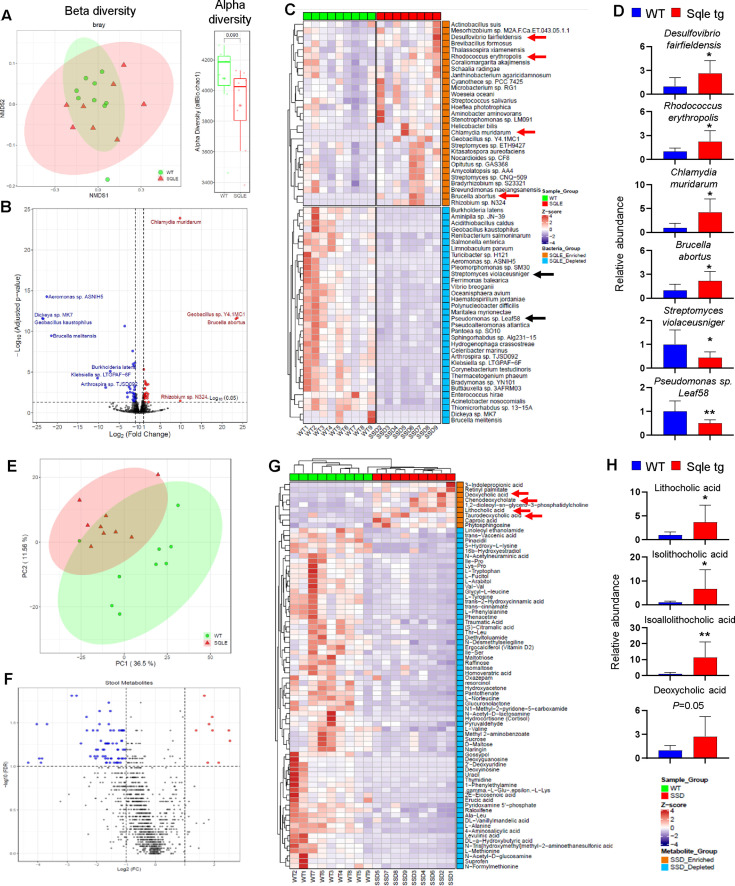
Results: SQLE messenger RNA and protein expression was upregulated in CRC (p<0.01) and predict poor survival of patients with CRC. SQLE promoted CRC cell proliferation by inducing cell cycle progression and suppressing apoptosis. In azoxymethane-induced CRC model, Sqle tg mice showed increased tumourigenesis compared with wild-type mice (p<0.01). Integrative metagenomic and metabolomic analyses unveiled gut dysbiosis in Sqle tg mice with enriched pathogenic bacteria, which was correlated to increased secondary bile acids. Consistent with detrimental effect of secondary bile acids, gut barrier function was impaired in Sqle tg mice, with reduced tight junction proteins Jam-c and occludin. Transplantation of Sqle tg mice stool to germ-free mice impaired gut barrier function and stimulated cell proliferation compared with control mice stool. Finally, we demonstrated that terbinafine, a SQLE inhibitor, could be repurposed for CRC by synergising with oxaliplatin and 5-fluorouracil to inhibit CRC growth.

Conclusion: This study demonstrates that SQLE mediates oncogenesis via cell intrinsic effects and modulation of gut microbiota-metabolite axis. SQLE represents a therapeutic target and prognostic marker in CRC.

Keywords: colonic microflora; colorectal cancer; colorectal neoplasm.


Conflict of interest statement

Competing interests: None declared.

Figures

Figure 1
Figure 1
Squalene epoxidase (SQLE) is upregulated in colorectal cancer (CRC) and its expression predicts poor survival. (A) SQLE messenger RNA (mRNA) expression was determined in paired tumour and adjacent normal tissues from Hong Kong (HK) cohort (Beijing, n=144), The Cancer Genome Atlas (TCGA) cohort (n=50) and the GDS4382 cohort (n=17). Paired two-tailed Student’s t-test was used to calculate p value. (B) SQLE protein expression was determined in paired tumour and adjacent normal tissues (n=10) by immunohistochemistry (IHC). Paired two-tailed Student’s t-test was used to calculate p value. Scale bars, 50 µm. (C) Kaplan-Meier curve analysis revealed that patients with CRC with high SQLE mRNA has significantly lower 5-year survival as compared with patients with low SQLE mRNA in HK cohort (left). Cox regression analysis (right). (D) SQLE mRNA expression is associated with poor survival by Kaplan-Meier curve (left) and Cox regression analysis (right) in GSE17538 cohort. (E) Tissue microarray (TMA) analysis demonstrated that patients with CRC with high SQLE protein expression have significantly lower 5-year survival, as evidenced by Kaplan-Meier curve (left) and Cox regression analysis (right). Nuclear staining of 10% was used as the cut-off value. *P<0.05; **p<0.01; ***p<0.001. AJCC, The American Joint Committee on Cancer; TNM, tumour, node, metastases.
Figure 2
Figure 2
Squalene epoxidase (SQLE) promotes colorectal cancer (CRC) cell proliferation. (A) SQLE overexpression in LOVO and SW1116 cells promoted cell viability and (B) colony formation. (C) Knockdown of SQLE in HT29 and DLD1 cells significantly suppressed cell viability and (D) colony formation. (E) SQLE promotes tumour growth in vivo. SW1116 and LOVO cells (5×10⁶ cells/tumour) transfected with SQLE or control vector were injected to nude mice aged 4 weeks. Tumour growth was significantly faster in SQLE overexpressing cells as compared with controls. (F) SQLE is overexpressed in two primary CRC organoids, and knockout of SQLE suppressed their growth. The significance of the difference between growth curves was determined by repeated-measures analysis of variance. Difference in colony formation was determined by two-tailed Student’s t-test. *P<0.05; **p<0.01; ***p<0.001; ****p<0.0001. All experiments were conducted 3 times in triplicate.
Figure 3
Figure 3
Squalene epoxidase (SQLE) suppresses cell apoptosis and promotes cell cycle progression in colorectal cancer (CRC) cells. (A) SQLE overexpression suppressed apoptosis in LOVO and SW1116 cells, as determined by annexin V/7-aminoactinomycin D (7-AAD) staining and flow cytometry analysis. (B) Western blot analysis indicated that SQLE overexpression suppressed the expression of cleaved PARP, caspase-3 and caspase-7. (C) Knockdown of SQLE in HT29 and DLD1 cells induced apoptosis by the annexin V/7-AAD assay. (D) SQLE knockdown increased cleaved PARP, caspase-3 and caspase-7 expression. (E) SQLE overexpression in LOVO and SW1116 cells led to a decrease of G0/G1 phase cell population concomitant with an increase of S phase cell population, as assessed by propidium iodide (PI) staining and flow cytometry. (F) SQLE overexpression enhanced the expression of PCNA, cyclin D1 and CDK4 that promotes G1 phase progression, while suppressing the expression of G1 gatekeepers p21Cip1, p27Kip1 and p53. (G) SQLE knockdown in HT29 and DLD1 cells led to accumulation of cells in G0/G1 phase with decreased S phase cell population. (H) SQLE silencing inhibited PCNA, cyclin D1 and CDK4, while inducing p21Cip1, p27Kip1 and p53 expression. The difference between two groups was determined by two-tailed Student’s t-test. *P<0.05; **p<0.01. All experiments were conducted 3 times in triplicate.
Figure 4
Figure 4
Colon-specific squalene epoxidase (Sqle) transgenic (tg) expression promotes colorectal tumourigenesis in mice. (A) The construction of colon-specific, conditional Sqle tg mice. Sqle transgene was introduced into the Rosa26 locus with Loxp sites flanking a STOP codon at 5’-end. These mice were then crossed to Cdx2-CreERT2 mice to obtain colon-specific Sqle tg mice. At 6 weeks of age, tamoxifen injection was given (100 mg/kg, intraperitoneally) for 4 times (one dose/day). Western blot analysis confirmed SQLE overexpression in colon tissues. (B) SQLE promotes chemically induced colorectal cancer (CRC). Sqle tg mice aged 6 weeks and wild-type (WT) littermates (n=10 per group) were injected with tamoxifen (100 mg/kg, intraperitoneally, 4 times), followed by azoxymethane (AOM) injection (10 mg/kg, intraperitoneally) for 6 times (once per week). Mice were sacrificed at week 24. (C) Colon-specific Sqle overexpression significantly increased the tumour number and burden in the colon (n=10 in each group). (D) Histological examination performed by a pathologist blinded to the nature of the samples confirmed that SQLE promoted tumour development, as the ratio of high grade of dysplasia in Sqle tg mice was 70%, while the corresponding ratio in WT mice was 20%. (E) Ki-67 staining showed that colon tissues of Sqle tg mice had higher cell proliferation as compared with WT mice. (F) TUNEL staining revealed that Sqle tg mice colon had reduced apoptosis as compared with WT mice. (G) Cancer Pathway PCR array analysis of colon tissues from Sqle tg and WT mice, which were first injected with tamoxifen (100 mg/kg, intraperitoneally, 4 times) and then sacrificed after 3 months. (H) The construction of CRISPR/Cas9-induced whole body Sqle knockout (KO) mice. Downregulation of SQLE in heterozygous Sqle KO mice colon was confirmed by western blot analysis. (I) Sqle KO mice and WT littermates were subjected to AOM-DSS regimen to induce CRC. Sqle KO mice developed significantly less colon tumours as compared with WT mice (n=5 in each group). The difference between two groups was determined by two-tailed Student’s t-test.
Figure 5
Figure 5
Colon-specific squalene epoxidase (Sqle) transgenic (tg) expression in mice dysregulate gut microbiota and metabolites. (A) Metagenomic sequencing was performed on stool obtained from Sqle tg mice and wild-type (WT) littermates at 3 months after tamoxifen injection. Principal component analysis (PCA) of bacteria composition between Sqle tg (n=8) and WT (n=9) mice (left). Alpha-diversity showed a decreasing trend in Sqle tg mice (right). (B and C) Volcano plot and heat map analysis revealed 29 enriched bacteria, including pathogenic bacteria, Desulfovibrio fairfieldensis, Rhodococcus erythropolis, Brucella abortus and Chlamydia muridarum, and 34 depleted bacteria such as Streptomyces violaceoruber, Pseudomonas sp Leaf58 in Sqle tg mice. (D) Quantitative PCR (qPCR) validation of key bacterial species altered in Sqle tg mice. (E) Global metabolomics profiling was performed on stool samples from Sqle tg mice and WT littermates at 3 months of age. PCA analysis of metabolite profiles (n=9 for each group). (F and G) Volcano plot and heat map analysis revealed nine enriched metabolites such as deoxycholic acid (DCA), lithocholic acid (LCA), taurodeoxycholic acid (TDCA) and chenodeoxycholic acid (CDCA); plus 64 depleted metabolites in Sqle tg mice as compared with WT littermates. (H) Targeted metabolomic analysis of secondary bile acids by liquid chromatography-mass spectrometry. *P<0.05; **p<0.01.
Figure 6
Figure 6
Gut microbiota-metabolite axis contributes to gut barrier dysfunction in squalene epoxidase (Sqle) transgenic (tg) mice and in germ-free mice receiving faecal microbiota transplantation (FMT) from Sqle tg mice. (A) Correlation analysis revealed that pathogenic bacteria are positively correlated with altered metabolites, including secondary bile acids. (B) Fluorescein isothiocyanate (FITC)-dextran intestinal permeability test was performed on Sqle tg mice and wild-type (WT) littermates at 3 months after tamoxifen injection, revealing that the gut barrier was impaired in Sqle tg mice. (C) Electron microscopy confirmed impaired intestinal tight junction in Sqle tg mice. (D) Western blot analysis showed that tight junction proteins Jam-c and occludin were downregulated in Sqle tg mice. (E) Muc2 messenger RNA (mRNA) was downregulated in Sqle tg mice. (F) Stool collected from Sqle tg and WT littermates was gavaged to germ-free mice aged 6 weeks. One month later, FITC-dextran was gavaged 4 hours before sacrifice to perform permeability assay. Intestinal permeability was significantly induced in germ-free mice gavaged with stool from Sqle tg mice. (G) Electron microscopy confirmed the impaired intestinal tight junction germ-free mice fed with stool from Sqle tg mice. (H) Western blot analysis showed that tight junction proteins Jam-c and occludin were downregulated in germ-free mice gavaged with stool from Sqle tg mice. (I) Germ-free mice transplanted with Sqle tg mice stool showed increased Ki-67 staining in the colon epithelium as compared with those gavaged with WT mice stool. Two-tailed Student’s t-test was used for the difference determination between two groups.
Figure 7
Figure 7
Squalene epoxidase (SQLE) is a therapeutic target in colorectal cancer (CRC). (A) Terbinafine, a Food and Drug Administration-approved drug targeting SQLE, dose-dependently suppressed cell viability of HT29 and DLD1 cells, and primary CRC organoids. (B) Terbinafine co-treatment sensitised HT29 and DLD1 cells to the growth inhibitory effect of 5-fluorouracil (5-FU) or oxaliplatin. (C) Combination index (CI) indicated that terbinafine+5-FU or oxaliplatin exerted synergistic effects for the treatment of CRC cells, with CI <1. (D) Apoptosis assay showed that terbinafine+5-FU or oxaliplatin (48 hours) synergistically induced apoptosis in CRC cells. (E) Nude mice were injected subcutaneously with HT29 cells, randomised and then treated with (1) vehicle, (2) terbinafine (80 mg/kg, oral daily), (3) 5-FU (30 mg/mg, intraperitoneally, once per week), (4) oxaliplatin (10 mg/kg, intraperitoneally, twice per week), (5) terbinafine+5-FU and (6) terbinafine+oxaliplatin. Terbinafine treatment alone suppressed tumour growth significantly by ~50%. On the other hand, terbinafine combined with 5-FU or oxaliplatin showed synergistic effects in suppressing tumour size in nude mice as compared with 5-FU or oxaliplatin single treatments. The significance of the difference between tumour growth rate was determined by repeated-measures analysis of variance. **p<0.01; ***p<0.001; ****p<0.0001. All in vitro experiments were conducted 3 times in triplicate.

References

    1. Liu P-H, Wu K, Ng K, et al. Association of obesity with risk of early-onset colorectal cancer among women. JAMA Oncol 2019;5:37–44. 10.1001/jamaoncol.2018.4280 - DOI - PMC - PubMed
    1. Yang MH, Rampal S, Sung J, et al. The association of serum lipids with colorectal adenomas. Am J Gastroenterol 2013;108:833–41. 10.1038/ajg.2013.64 - DOI - PubMed
    1. Yang J, Yu J. The association of diet, gut microbiota and colorectal cancer: what we eat may imply what we get. Protein Cell 2018;9:474–87. 10.1007/s13238-018-0543-6 - DOI - PMC - PubMed
    1. Beyaz S, Mana MD, Roper J, et al. High-Fat diet enhances stemness and tumorigenicity of intestinal progenitors. Nature 2016;531:53–8. 10.1038/nature17173 - DOI - PMC - PubMed
    1. Ecker J, Benedetti E, Kindt ASD, et al. The colorectal cancer lipidome: identification of a robust tumor-specific lipid species signature. Gastroenterology. In Press 2021;161:910–23. 10.1053/j.gastro.2021.05.009 - DOI - PubMed

Publication types