Review
. 2022 Mar 1;14(3):a041012.
doi: 10.1101/cshperspect.a041012.

Caspases and Their Substrates

Affiliations
Review

Caspases and Their Substrates

Douglas R Green. Cold Spring Harb Perspect Biol. .
No abstract available


Figures

Figure 1.
Figure 1.
The acylation step of caspase cleavage.
Figure 2.
Figure 2.
The deacylation step of caspase cleavage. The mechanism is based on the function of other cysteine proteases and has not been confirmed in caspases.
Figure 3.
Figure 3.
Schematics of several human caspases. Caspases-2 and -10 are grouped with the initiator caspases, although their classification is problematic. The representation aligns related sequences and should not be taken to indicate the actual structures of the proteins.
Figure 4.
Figure 4.
The linear organization of CED-3. CARD, caspase recruitment domain. The arrows are auto-cleavage sites.
Figure 5.
Figure 5.
Caspases in Drosophila.
Figure 6.
Figure 6.
Numbering scheme for amino acids around a site in a substrate protein that can be cut by caspases.
Figure 7.
Figure 7.
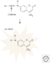
Caspase activity detected using a peptide substrate that generates a fluorescent signal after cleavage.
Figure 8.
Figure 8.
Representation of amino acid preferences for caspase-3. Amino acids are shown in single-letter code, and the size of each letter corresponds to its frequency in cleaved peptides. Cleavage occurs between P1 and P1′. (Reprinted from Mahrus et al. 2008. ©2008 with permission from Elsevier.)
Figure 9.
Figure 9.
Caspase preferences, based on peptides. Sequences shown are optimally cleaved, although other peptide sequences can be cleaved nearly as well.
Figure 10.
Figure 10.
Representation of caspase-3 cleavage sites in known substrates. (Reprinted from Mahrus et al. 2008. ©2008 with permission from Elsevier.)
Figure 11.
Figure 11.
Representation of protein cleavage events in cells undergoing apoptosis. (Reprinted from Mahrus et al. 2008. ©2008 with permission from Elsevier.)
Figure 12.
Figure 12.
Cleavage of the iCAD–CAD complex by caspases is responsible for DNA fragmentation during apoptosis. Caspase-3 (right) cleaves iCAD (center), releasing the active CAD, which randomly cuts the chromatin at accessible sites between the nucleosomes (top left). When run on an agarose gel, the DNA forms a ladder (far left) composed of multiples of nucleosome-sized lengths. (Image courtesy of Dr. Yufang Shi, Institutes for Translational Medicine, Soochow University, Suzhou, China; structure [inset], PDB 1GQF [Riedl et al. 2001].)
Figure 13.
Figure 13.
Silencing or inhibition of ROCK1 prevents blebbing during apoptosis. Blebbing seen during apoptosis (A), is eliminated when ROCK1 is silenced with small interfering RNA (siRNA) (B) or blocked with an inhibitor (C). (Courtesy of Michael Olson, Beatson Institute.)
Figure 14.
Figure 14.
Caspases induce exposure of phosphatidylserine on the outer leaflet of the plasma membrane by two mechanisms. Caspase cleavage (scissors) disrupts the flippase activity of ATP11C (left) and induces the scramblase activity of Xkr8 (right). Both result in loss of phospholipid asymmetry, resulting in exposure of phosphatidylserine on the cell surface.
Figure 15.
Figure 15.
Extra cells in CED3 mutant nematodes. Normal cell deaths in a wild-type larva (arrows, left) are not seen in a CED3 mutant animal (right). (Reprinted from Ellis and Horvitz 1986. ©1986 with permission from Elsevier.)
Figure 16.
Figure 16.
Extra cells in Dronc-deficient fly embryos. Cell deaths (stained blue) in wild-type embryos (left) are not seen in embryos lacking the caspase Dronc (right). (Reprinted from Quinn et al. 2000. ©2000 with permission from the American Society for Biochemistry and Molecular Biology.)
Figure 17.
Figure 17.
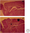
Extra cells (arrow and asterisk) in the brains of caspase-3-deficient mice (−/−; lower) compared with wild-type mice (wt; upper). (Reprinted with permission from Macmillan Publishers Ltd.: Kuida et al. 1996. ©1996.)

References

    1. Green DR. 2022a. Caspase activation and inhibition. Cold Spring Harb Perspect Biol 10.1101/cshperspect.a041020 - DOI - PMC - PubMed
    1. Green DR. 2022b. The burial: clearance and consequences. Cold Spring Harb Perspect Biol 10.1101/cshperspect.a041087 - DOI - PMC - PubMed
    1. Green DR. 2022c. Cell death and cancer. Cold Spring Harb Perspect Biol 10.1101/cshperspect.a041103 - DOI - PubMed
    1. Green DR. 2022d. The mitochondrial pathway of apoptosis, Part 1: MOMP and beyond. Cold Spring Harb Perspect Biol 10.1101/cshperspect.a041038 - DOI - PMC - PubMed
    1. Green DR. 2022e. Cell death in development. Cold Spring Harb Perspect Biol 10.1101/cshperspect.a041095 - DOI - PMC - PubMed

FIGURE CREDITS

    1. Ellis HM, Horvitz HR. 1986. Genetic control of programmed cell death in the nematode C. elegans. Cell 44: 817–829. 10.1016/0092-8674(86)90004-8 - DOI - PubMed
    1. Kuida K, Zhend TS, Na S, Kuan C-Y, Yang D, Karasuyma H, Rakic P, Flavell RA. 1996. Nature 384: 368–372. 10.1038/384368a0 - DOI - PubMed
    1. Mahrus S, Trinidad JC, Barkan DR, Sali A, Burlingame AL, Wells JA. 2008. Global sequencing of proteolytic cleavage sites in apoptosis by specific labeling of protein N termini. Cell 134: 866–876. 10.1016/j.cell.2008.08.012 - DOI - PMC - PubMed
    1. Quinn LM, Dorstyn L, Mills K, Colussi P, Chen P, Coombe M, Abrams J, Kumar S, Richardson H. 2000. An essential role for the caspase Dronc in developmentally programmed cell death in Drosophila. J Biol Chem 275: 40416–40424. 10.1074/jbc.M002935200 - DOI - PubMed
    1. Riedl SJ, Fuentes-Prior P, Renatus M, Kairies N, Krapp S, Huber R, Salvesen GS, Bode W. 2001. Structural basis for the activation of human procaspase-7. Proc Natl Acad Sci 98: 14790–14795. 10.1073/pnas.22150098 - DOI - PMC - PubMed

LinkOut - more resources