. 2022 Mar 1;5(1):160.
doi: 10.1038/s42003-022-03084-7.

Modulation of the monomer-dimer equilibrium and catalytic activity of SARS-CoV-2 main protease by a transition-state analog inhibitor

Affiliations

Modulation of the monomer-dimer equilibrium and catalytic activity of SARS-CoV-2 main protease by a transition-state analog inhibitor

Nashaat T Nashed et al. Commun Biol. .

Abstract

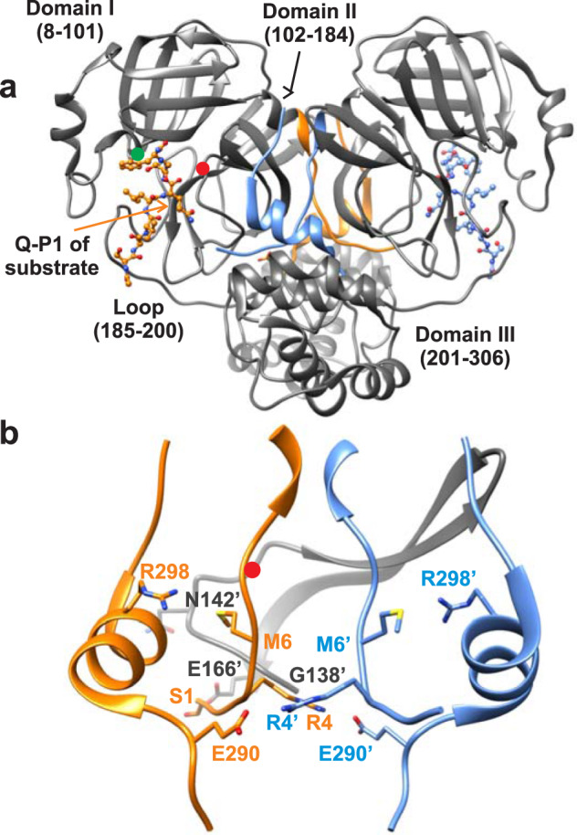
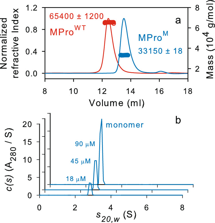
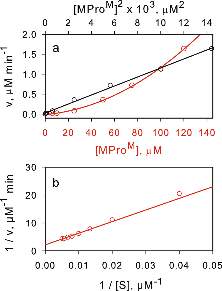
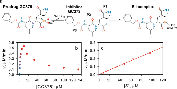
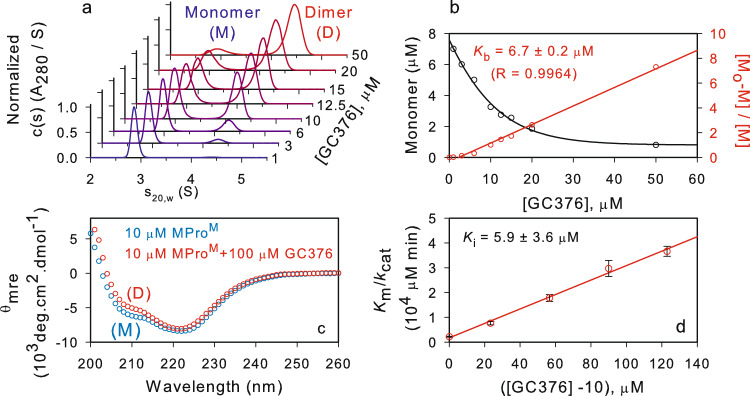
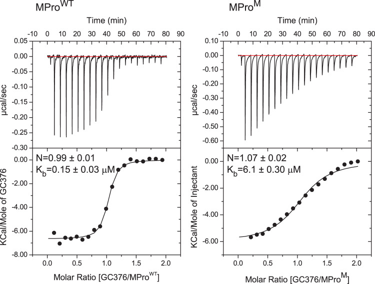
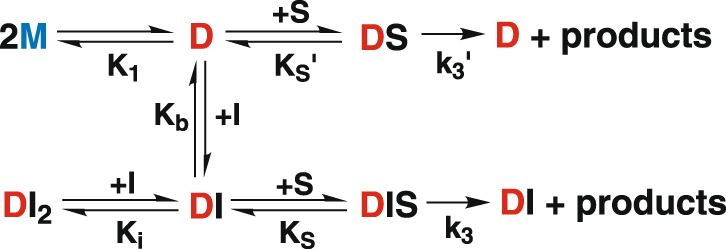
The role of dimer formation for the onset of catalytic activity of SARS-CoV-2 main protease (MProWT) was assessed using a predominantly monomeric mutant (MProM). Rates of MProWT and MProM catalyzed hydrolyses display substrate saturation kinetics and second-order dependency on the protein concentration. The addition of the prodrug GC376, an inhibitor of MProWT, to MProM leads to an increase in the dimer population and catalytic activity with increasing inhibitor concentration. The activity reaches a maximum corresponding to a dimer population in which one active site is occupied by the inhibitor and the other is available for catalytic activity. This phase is followed by a decrease in catalytic activity due to the inhibitor competing with the substrate. Detailed kinetics and equilibrium analyses are presented and a modified Michaelis-Menten equation accounts for the results. These observations provide conclusive evidence that dimer formation is coupled to catalytic activity represented by two equivalent active sites.


Conflict of interest statement

The authors declare no competing interests.

Figures

Fig. 1
Fig. 1. Molecular representation of the SARS-CoV-2 main protease dimer and critical interactions which influence the monomer-dimer (M-D) equilibrium.
a The two subunits of the mature dimer (PDB ID: 7N89) with N- (1–11) and C-terminal (288–306) residues highlighted with subunit A in orange and subunit B in blue. Substrate SAVLQSGF bound to the active site of each subunit is shown with Q-P1 which forms the S1 subsite indicated by the orange arrow. The interface formed by the free N-finger residues 1–7, shown in the middle of the dimer, is critical for dimer stability. The dimer interface is formed by an extensive network of hydrogen bonds and hydrophobic interactions involving N-terminal residues 1′-16′, β-strand residues 118′-125′ and loop residues 137′-142′ as illustrated in reference. Red and green circles denote position of active site C145 and H41 residues, respectively. b Enlargement of the region showing residue positions with the same coloring as in (A) critical for dimer interface stability. Mutations E290A and R298A increase the Kd by ~5000-fold based on our estimate shown in Table 1, and published reports,,. The region (G138 to E166) encompassing the oxyanion loop (S139 to L141) for subunit B are shown in gray. S1 of subunit A interacts with E166 of subunit B. Residues from subunit B are denoted with prime (‘).
Fig. 2
Fig. 2. Molecular mass estimation of MProM.
a Mass estimation by SEC-MALS by injecting 125 µL of MProM and MProWT at ~58 µM and 30 µM, respectively. b SV-AUC absorbance c(s) distributions at concentrations ranging from 18 to 90 µM of MProM. SEC-MALS and SV-AUC were carried out in buffer A at 25 °C.
Fig. 3
Fig. 3. Evaluation of the catalytic efficiency of MProM.
a Non-linear relationship between the rate of catalyzed hydrolysis vs the protein concentration (red line), and the linear relationship between the rate of catalyzed hydrolysis vs the square of the protein concentration (black line). b Lineweaver-Burk plot for the catalyzed hydrolysis at a concentration of 40 µM MProM.
Fig. 4
Fig. 4. Mechanism of catalysis by MProWT and MProM.
M, D and S denote monomer, dimer and substrate, respectively.
Fig. 5
Fig. 5. Modulation of the catalytic activity of MProM by feline coronavirus prodrug GC376.
a Chemical structure of GC376 and steps in its binding to the active site of 3C and 3CL proteases,. b Catalytic activity of 10 µM MProM as a function of increasing GC376 concentration. c A plot of the rate vs substrate concentration at a final concentration of 10 µM MProM and 10 µM GC376.
Fig. 6
Fig. 6. Modulation of the M-D equilibrium of MProM and competitive inhibition by GC376.
a SV-AUC absorbance c(s) distribution of MProM (6–7 µM) in the presence of increasing GC376 concentration ranging from 1-50 µM. b Plot of the monomer amount vs inhibitor concentration (black trace) from data derived from (A). Estimation of the Kb for the binding of GC376 to MProM (red trace). c CD spectra of 10 µM MProM in the absence (blue) and presence (red) of 100 µM inhibitor GC376. d A plot of the Km/kcat vs increasing concentration of GC376 at a final concentration of 10 µM MProM. Error values indicate a standard deviation of data points recorded 4 times in duplicate (Supplementary Table 2).
Fig. 7
Fig. 7. Binding isotherm of GC376 to MProWT and MProM.
Titrations were carried out with 30 µM MProWT and 98 µM MProM (in the cell) vs 300 µM and 1 mM GC376 (in the syringe), respectively, in buffer C at 28 °C. Thermodynamic parameters are listed in Supplementary Table 1.
Fig. 8
Fig. 8. Mechanism of activation and inhibition of MProM by GC376.
M, D, S, I, DS, DI, DI2, DIS denote monomer, dimer, substrate, inhibitor, dimer-substrate complex, dimer-inhibitor complex, dimer bound to 2 inhibitors, dimer bound to 1 inhibitor and 1 substrate, respectively.

References

    1. V’kovski P, Kratzel A, Steiner S, Stalder H, Thiel V. Coronavirus biology and replication: implications for SARS-CoV-2. Nat. Rev. Microbiol. 2020;19:155–170. - PMC - PubMed
    1. Wu F, et al. A new coronavirus associated with human respiratory disease in China. Nature. 2020;579:265–269. - PMC - PubMed
    1. Zhang L, et al. Crystal structure of SARS-CoV-2 main protease provides a basis for design of improved alpha-ketoamide inhibitors. Science. 2020;368:409–412. - PMC - PubMed
    1. Mariano G, Farthing RJ, Lale-Farjat SLM, Bergeron JRC. Structural Characterization of SARS-CoV-2: Where We Are, and Where We Need to Be. Front Mol. Biosci. 2020;7:605236. - PMC - PubMed
    1. Wang C, et al. The establishment of reference sequence for SARS-CoV-2 and variation analysis. J. Med Virol. 2020;92:667–674. - PMC - PubMed

Publication types