Review
. 2022 Oct;43(10):2439-2447.
doi: 10.1038/s41401-022-00879-6. Epub 2022 Mar 1.

Mitochondrial-derived damage-associated molecular patterns amplify neuroinflammation in neurodegenerative diseases

Affiliations
Review

Mitochondrial-derived damage-associated molecular patterns amplify neuroinflammation in neurodegenerative diseases

Miao-Miao Lin et al. Acta Pharmacol Sin. 2022 Oct.

Abstract

Both mitochondrial dysfunction and neuroinflammation are implicated in neurodegeneration and neurodegenerative diseases. Accumulating evidence shows multiple links between mitochondrial dysfunction and neuroinflammation. Mitochondrial-derived damage-associated molecular patterns (DAMPs) are recognized by immune receptors of microglia and aggravate neuroinflammation. On the other hand, inflammatory factors released by activated glial cells trigger an intracellular cascade, which regulates mitochondrial metabolism and function. The crosstalk between mitochondrial dysfunction and neuroinflammatory activation is a complex and dynamic process. There is strong evidence that mitochondrial dysfunction precedes neuroinflammation during the progression of diseases. Thus, an in-depth understanding of the specific molecular mechanisms associated with mitochondrial dysfunction and the progression of neuroinflammation in neurodegenerative diseases may contribute to the identification of new targets for the treatment of diseases. In this review, we describe in detail the DAMPs that induce or aggravate neuroinflammation in neurodegenerative diseases including mtDNA, mitochondrial unfolded protein response (mtUPR), mitochondrial reactive oxygen species (mtROS), adenosine triphosphate (ATP), transcription factor A mitochondria (TFAM), cardiolipin, cytochrome c, mitochondrial Ca2+ and iron.

Keywords: microglia; mitochondrial dysfunction; mitochondrial-derived damage-associated molecular pattern; neurodegenerative diseases; neuroinflammation.


Conflict of interest statement

The authors declare no competing interests.

Figures

Fig. 1
Fig. 1. mtDNA acts as a DAMP to activate neuroinflammation.
Mitochondrial dysfunction causes mtDNA to flow into the cytoplasm and out of glia (the left cell). mtDNA in the cytoplasm is sensed by the cGAS-STING signaling pathway, which induces TBK1-IRF3-IFN signaling and promotes the expression of inflammatory factors. mtDNA in the cytoplasm can also activate the NLRP3 inflammasome and promote the expression of IL-18 and IL-1β by activating caspase-1. Extracellular mtDNA activates TLR9 on the endosomes of other glia (the right cell) to initiate nuclear transcription factor NF-κB signaling.
Fig. 2
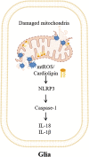
Fig. 2. mtROS and cardiolipin act as DAMPs to activate neuroinflammation.
When damaged, mitochondrial dysfunction of glia leads to excessive production of mtROS and the release of cardiolipin located on the inner mitochondrial membrane into the cytoplasm. mtROS or cardiolipin activates the NLRP3 inflammasome to initiate a caspase-1-dependent neuroinflammatory response and release IL-18 and IL-1β.
Fig. 3
Fig. 3. ATP acts as a DAMP to activate neuroinflammation.
Under stress conditions, ATP from neurons or glia (the left cell) is released outside the cell. ATP activates the caspase-1 signaling cascade mediated by NLRP3 through the P2X7 receptor on the membrane surface of glia (the right cell), leading to neuroinflammation.
Fig. 4
Fig. 4. TFAM acts as a DAMP to activate neuroinflammation.
Under stress conditions, TFAM from neurons or glia (the left cell) is released outside the cell. TFAM activates RAGE or TLR on the membrane surface of glia (the right cell) and initiates the NF-κB-mediated nuclear signaling cascade.
Fig. 5
Fig. 5. Cytochrome c acts as a DAMP to activate neuroinflammation.
Under stress conditions, the mitochondrial membrane of neurons or glia (the left cell) ruptures and cytochrome c is released outside the mitochondria. Cytochrome c in the cytoplasm activates the apoptosis signaling pathway. Extracellular cytochrome c activates the MAPK-JNK signaling cascade through TLR4 on the membrane surface of glia (the right cell), leading to neuroinflammation.
Fig. 6
Fig. 6. Cross-talk between mitochondrial dysfunction and neuroinflammation in neurodegenerative diseases.
After exposure to external stimuli, the function of mitochondria in neurons or glia is impaired, which results in a decrease in mitochondrial membrane potential. Some molecules that originally existed inside the mitochondria, such as mtDNA and cardiolipin, were released into the cytoplasm or even outside the cell. These released molecules, also called damage-associated molecular patterns, interact with pattern recognition receptors on glia to activate inflammatory pathways. Activated glia release proinflammatory factors, which aggravate mitochondrial damage, forming a vicious cycle of mitochondrial dysfunction and neuroinflammation. The interaction between mitochondrial dysfunction and neuroinflammation ultimately results in neurodegenerative diseases. ATP adenosine triphosphate, Cyt c cytochrome c, mtDNA mitochondrial DNA, mtROS mitochondrial reactive oxygen species, TFAM mitochondrial transcription factor A.

Similar articles

Cited by

References

    1. Vaquer-Alicea J, Diamond MI. Propagation of protein aggregation in neurodegenerative diseases. Annu Rev Biochem. 2019;88:785–810. doi: 10.1146/annurev-biochem-061516-045049. - DOI - PubMed
    1. Wang Y, Qin ZH. Molecular and cellular mechanisms of excitotoxic neuronal death. Apoptosis. 2010;15:1382–402. doi: 10.1007/s10495-010-0481-0. - DOI - PubMed
    1. Dong XX, Wang Y, Qin ZH. Molecular mechanisms of excitotoxicity and their relevance to pathogenesis of neurodegenerative diseases. Acta Pharmacol Sin. 2009;30:379–87. doi: 10.1038/aps.2009.24. - DOI - PMC - PubMed
    1. Wang Y, Gu ZL, Cao Y, Liang ZQ, Han R, Bennett MC, et al. Lysosomal enzyme cathepsin B is involved in kainic acid-induced excitotoxicity in rat striatum. Brain Res. 2006;1071:245–9. doi: 10.1016/j.brainres.2005.10.074. - DOI - PubMed
    1. Peruzzotti-Jametti L, Pluchino S. Targeting mitochondrial metabolism in neuroinflammation: towards a therapy for progressive multiple sclerosis. Trends Mol Med. 2018;24:838–55.. doi: 10.1016/j.molmed.2018.07.007. - DOI - PubMed

MeSH terms