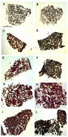
Reinnervation of Vastus lateralis is increased significantly in seniors (70-years old) with a lifelong history of high-level exercise (2013, revisited here in 2022)
- PMID: 35234026
- PMCID: PMC8992670
- DOI: 10.4081/ejtm.2022.10420
Reinnervation of Vastus lateralis is increased significantly in seniors (70-years old) with a lifelong history of high-level exercise (2013, revisited here in 2022)
Abstract
In 2013 we presented results showing that at the histological level lifelong increased physical activity promotes reinnervation of muscle fibers in aging muscles. Indeed, in muscle biopsies from 70-year old men with a lifelong history of high-level physical activity, we observed a considerable increase in fiber-type groupings (F-TG), almost exclusively of the slow type. Slow-type transformation by denervation-reinnervation in senior sportsmen seems to fluctuate from those with scarce fiber-type transformation and groupings to almost fully transformed muscle, going through a process in which isolated fibers co-expressing fast and slow Myosin Heavy Chains (MHCs) seems to fill the gaps. Taken together, our results suggest that, beyond the direct effects of aging on the muscle fibers, changes occurring in skeletal muscle tissue appear to be largely, although not solely, a result of sparse denervation-reinnervation. The lifelong exercise allows the body to adapt to the consequences of the age-related denervation and to preserve muscle structure and function by saving otherwise lost muscle fibers through recruitment to different, mainly slow, motor units. These beneficial effects of high-level life-long exercise on motoneurons, specifically on the slow type motoneurones that are those with higher daily activity, and on muscle fibers, serve to maintain size, structure and function of muscles, delaying the functional decline and loss of independence that are commonly seen in late aging. Several studies of independent reserchers with independent analyses confirmed and cited our 2013 results. Thus, the results we presented in our paper in 2013 seem to have held up rather well.
Conflict of interest statement
We confirm that we have read the Journal’s position on issues involved in ethical publication and affirm that this report is consistent with those guidelines.
It has long been accepted that histological changes seen in aging muscle suggest that denervation significantly contributes to tissue atrophy., Corroborating evidence of a progressive loss of α - motoneurons has been described with aging. Electrophysiological studies have confirmed a decrease in the number of motor units with some increase in their size, suggesting reinnervation events. Further evidence supporting rounds of denervation and reinnervation is based on the observation that in young humans, fiber types appear randomly distributed across the muscle but become increasingly clustered or grouped together with age. Therefore, it has been proposed that apoptosis of motoneurons in the spinal cord (with subsequent incomplete reinnervation of fibers by surviving motoneurons) contributes to the loss of muscle strength and mass that occurs with age. All of these processes are accompanied by a progressive increase in slow muscle fibers, although the literature provides some contradictions (see a recent review). Some of this discrepancy has been dispelled by comparisons of muscle from active and immobile patients: the immobile elderly have a shift toward fast isoform expression, as is common in “unloaded” muscle (e.g., during spaceflight or limb immobilization), whereas muscle wasting is accompanied by a shift toward a fast twitch phenotype. Thus the actual expression pattern of myosin isoforms in the elderly is modulated by complex factors because it depends upon the conflicting influences of both aging and reduced activity tending to shift toward slow and fast isoforms, respectively. To further complicate the situation, conflicting results regarding fast to slow myosin transition arise in endurance training studies using animal models and in clinical trials of humans involving either voluntary exercise or electrical stimulation (directly to muscle or indirectly through nerve stimulation)., Whether these shifts are under neural control or the direct effect of use/disuse on muscle fibers remains to be clarified. In the presents study, we analyzed muscle biopsies harvested from the
Figures

References
-
- Tomlinson BE, Walton JN, Rebeiz JJ. The effects of ageing and of cachexia upon skeletal muscle. A histopathological study. J Neurol Sci 1969;9:321-346. - PubMed
-
- Urbanchek MG, Picken EB, Kalliainen LK, Kuzon WM Jr. Specific force deficit in skeletal muscles of old rats is partially explained by the existence of denervated muscle fibers. J Gerontol A Biol Sci Med Sci 2001;56:B191–B197. - PubMed
-
- Tomlinson BE, Irving D. The numbers of limb motor neurons in the human lumbosacral cord throughout life. J Neurol Sci 1977;34:213-219. - PubMed
-
- Doherty TJ, Vandervoort AA, Taylor AW, Brown WF. Effects of motor unit losses on strength in older men and women. J Appl Physiol 1993;74: 868-874. doi: 10.1063/1.354879. - PubMed
-
- Andersen JL. Muscle fibre type adaptation in the elderly human muscle. Scand J Med Sci Sports 2003;13:40–47. doi: 10.1034/j.1600-0838.2003.00299.x. - PubMed
LinkOut - more resources
Full Text Sources
Medical
Miscellaneous

