. 2022 Jul;87(1):28-39.
doi: 10.1111/cod.14088. Epub 2022 Mar 18.

Obesity aggravates contact hypersensitivity reaction in mice

Affiliations

Obesity aggravates contact hypersensitivity reaction in mice

Monika Majewska-Szczepanik et al. Contact Dermatitis. 2022 Jul.

Abstract

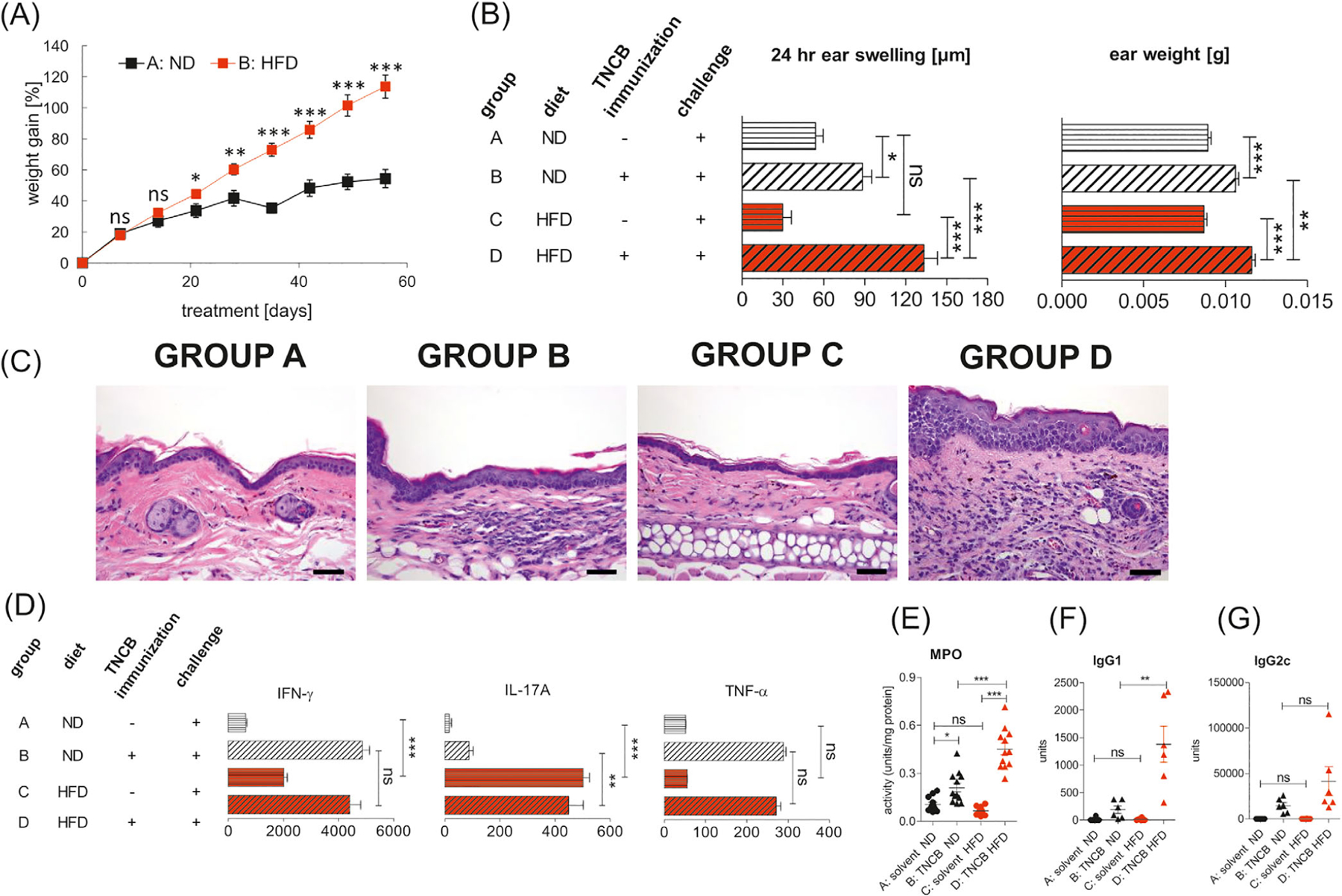
Background: Obesity is associated with chronic, low-grade inflammation in tissues and predisposes to various complications, including inflammatory skin diseases. However, the link between obesity and contact hypersensitivity (CHS) is not fully understood.

Objectives: We sought to determine the influence of obesity on T helper 1 (Th1)-mediated CHS.

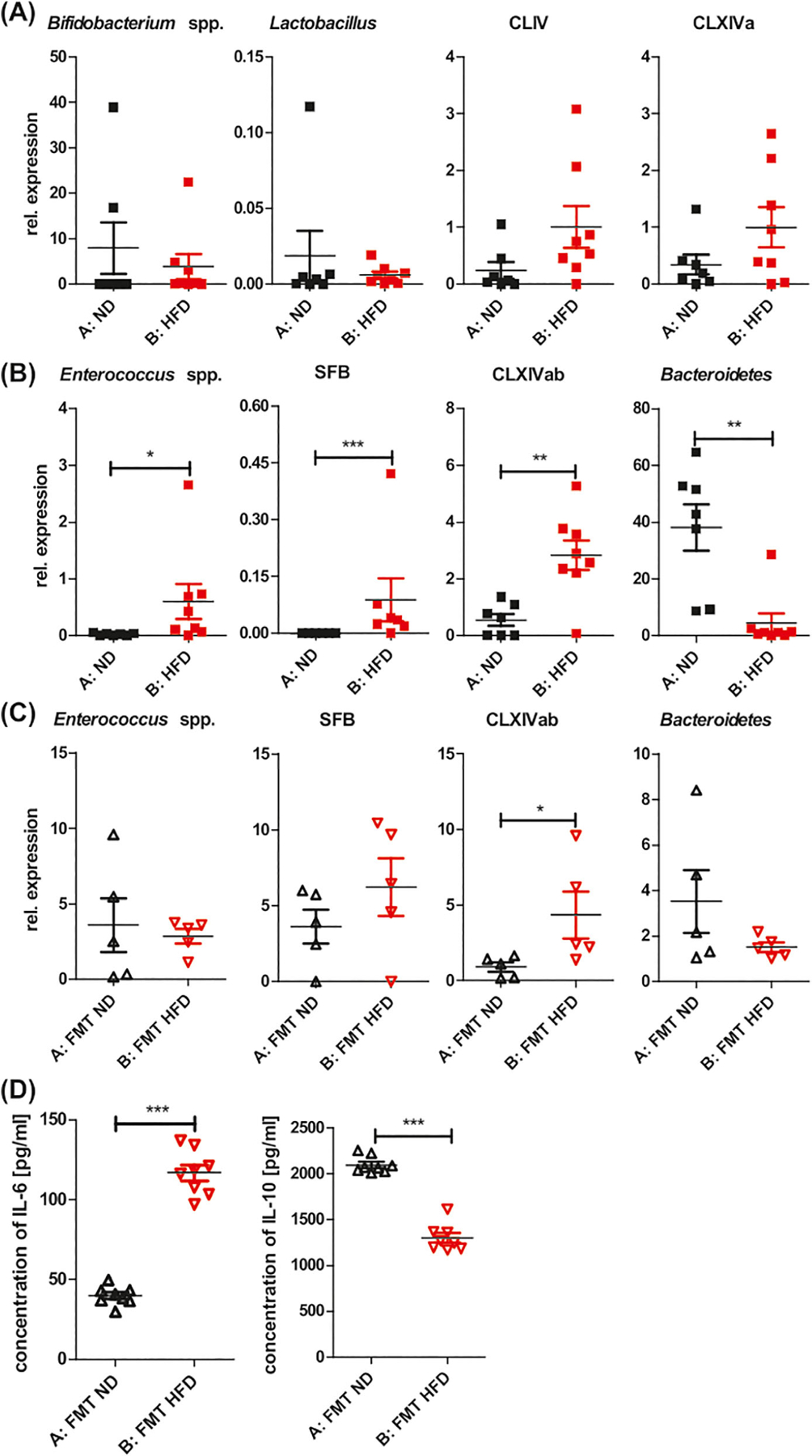
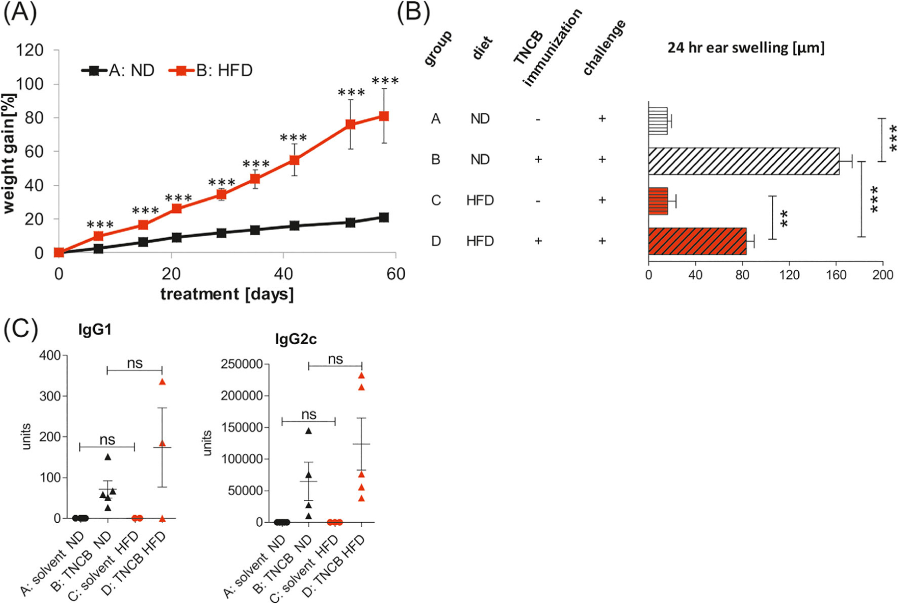
Methods: The activity/phenotype/cytokine profile of the immune cells was tested in vivo and in vitro. Using quantitative polymerase chain reaction (qPCR) and fecal microbiota transplantation (FMT), we tested the role of a high-fat diet (HFD)-induced gut microbiota (GM) dysbiosis in increasing the effects of CHS.

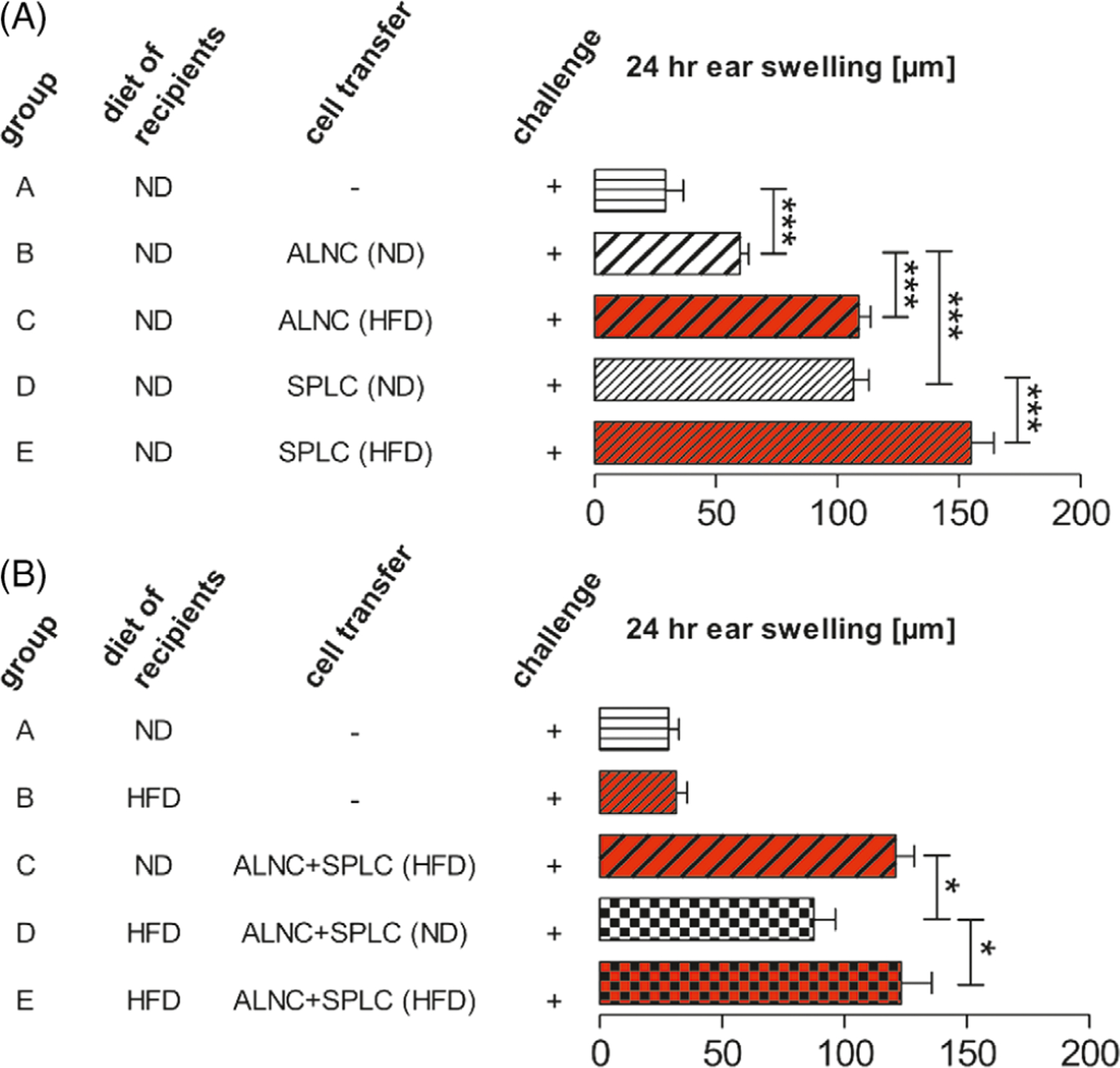
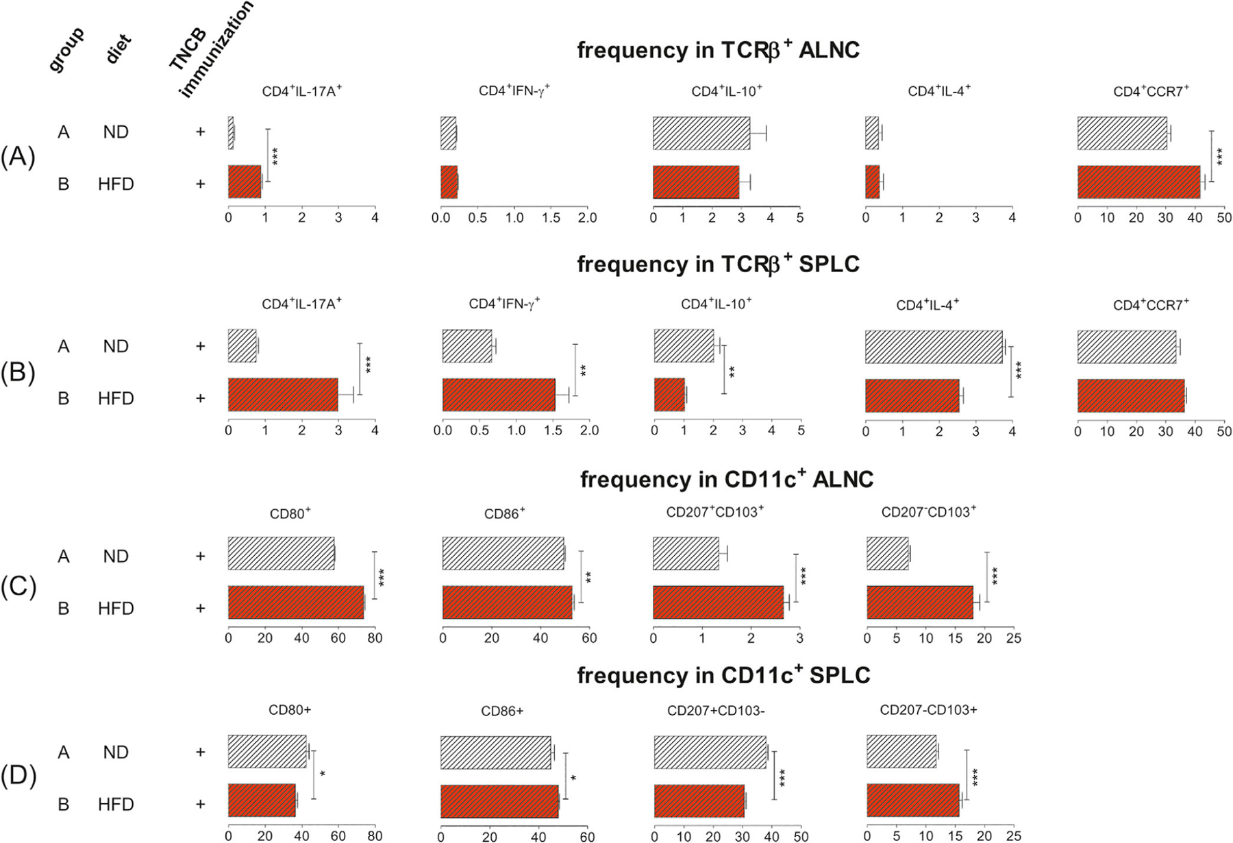
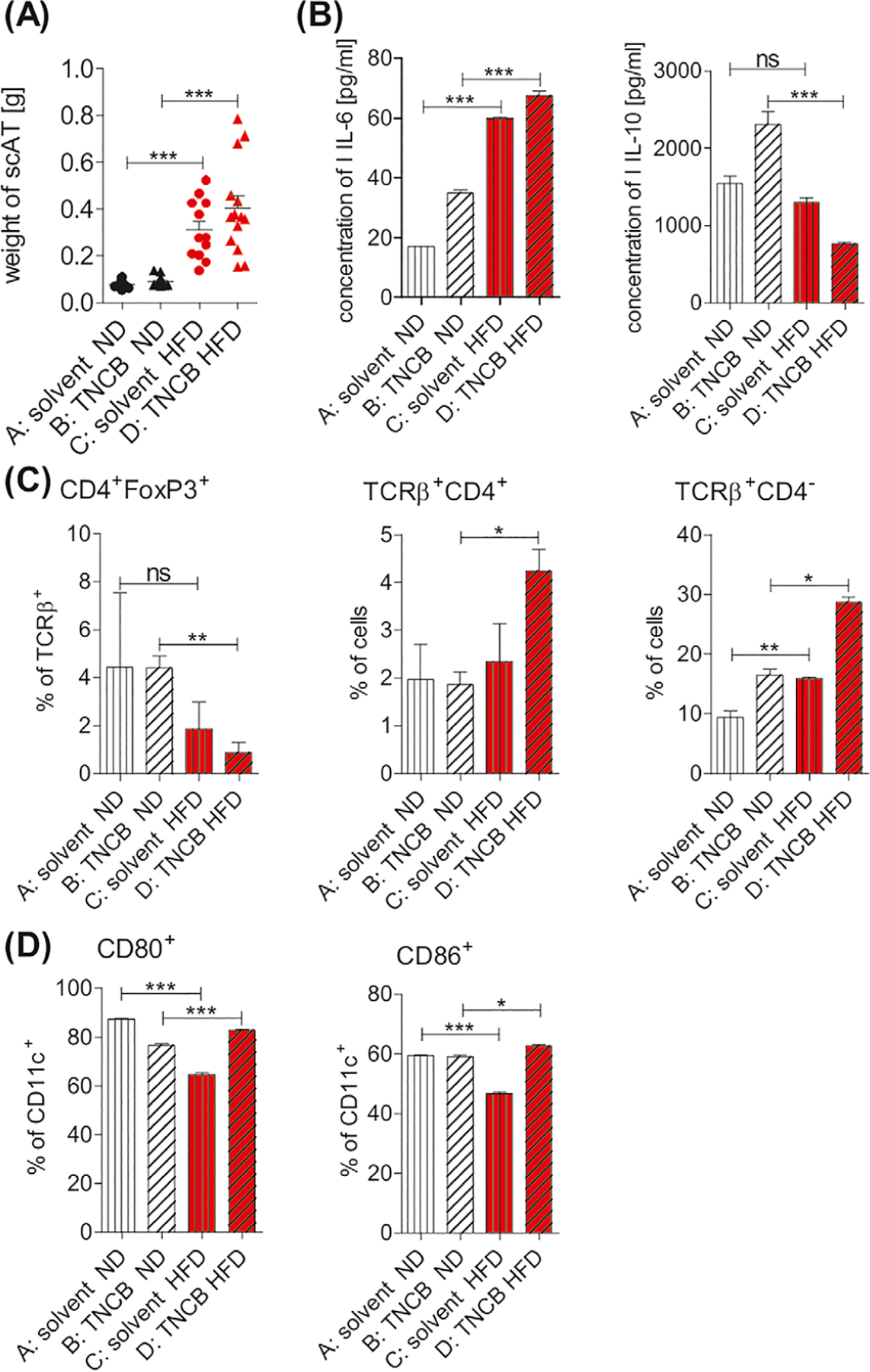
Results: Exacerbated CHS correlates with an increased inflammation-inducing GM in obese mice. We showed a proinflammatory milieu in the subcutaneous adipose tissue of obese mice, accompanied by proinflammatory CD4+ T cells and dendritic cells in skin draining lymph nodes and spleen. Obese interleukin (IL)-17A-/-B6 mice are protected from CHS aggravation, suggesting the importance of IL-17A in CHS aggravation in obesity.

Conclusions: Obesity creates a milieu that induces more potent CHS-effector cells but does not have effects on already activated CHS-effector cells. IL-17A is essential for the pathogenesis of enhanced CHS during obesity. Our study provides novel knowledge about antigen-specific responses in obesity, which may help with the improvement of existing treatment and/or in designing novel treatment for obesity-associated skin disorders.

Keywords: contact hypersensitivity; dendritic cells; high-fat diet-induced obesity; skin inflammation.


Conflict of interest statement

CONFLICT OF INTEREST

The authors declare that they have no conflict of interest.

Figures

FIGURE 1
FIGURE 1
Enhanced CHS in obese C57BL/6 mice. CHS was induced after 8 weeks of feeding with HFD or ND. (A) Weight gain in mice fed with ND (group A) and HFD (group B); n = 12/group. (B) Ear swelling and ear weight in mice fed with ND (group A and B) or HFD (group C and D). Mice were sensitized (day 0) by application of 5% TNCB to the shaved abdomen (group B and D). Unsensitized mice fed with ND (group A) or HFD (group C) were used as negative controls. CHS and ear weight were measured 24 hours after challenge with TNCB (day +4); n = 12/group. (C) Histology of ear tissue after challenge with hapten TNCB in unsensitized mice on the ND (group A) and HFD (group C) and in sensitized mice fed with ND (group B) and HFD (group D). Hematoxylin and eosin staining. Scale bar = 50 μm. Representative images (magnification: ×40). (D) Concentrations of IFN-γ, IL-17A, and TNF-α in ELNC culture supernatants. Mice were fed with ND (group B) or HFD (group D) and subsequently sensitized and challenged with hapten TNCB. Negative control mice were fed with ND (Group A) or HFD (Group C) and subsequently challenged with hapten. ELNCs were isolated 24 hours after challenge and were cultured with antigen for 48 hours. The culture supernatants of ELNCs were collected and tested for cytokine concentration using an ELISA Kit; n = 5–7/group. (E), MPO activity in ear tissue after challenge with hapten TNCB in unsensitized mice (negative) on the ND (group A) and HFD (group C) and in TNCB-sensitized mice (positive) fed with ND (group B) and HFD (group D). MPO colorimetric activity assay was used to determine the activity of MPO in samples; n = 6–12/group. (F,G) Concentration of anti-TNP IgG1 and IgG2c antibodies in serum after challenge with hapten TNCB in unsensitized mice (negative) on the ND (group A) and HFD (group C) and in TNCB-sensitized (positive) mice fed with ND (group B) and HFD (group D). The collected sera were tested for antibodies concentration using an ELISA; n = 6–12/group. *P < .05, **P < .01, ***P < .001. Abbreviations: CHS, contact hypersensitivity reaction; ELNC, auricular lymph node cells; HFD, high fat diet; MPO, myeloperoxidase; ND, normal diet; TNCB, 2,4,6-trinitrochlorobenzene; TNP, 2,4,6-trinitrophenyl
FIGURE 2
FIGURE 2
HFD modifies GM toward bacteria that induce a proinflammatory profile. Relative abundance of bacterial conserved 16S rDNA fragments in gut contents. qPCR was used to evaluate the alteration of gut bacteria. (A) Unchanged relative abundance of Bifidobacterium spp, Lactobacillus, Clostridium cluster IV (CLIV), and Clostridium cluster XIVa (CLXIVa) in mice fed with HFD vs ND for 8 weeks. (B) Increase in relative abundance of Enterococcus spp, SFB, and Clostridium cluster XIVab (CLXIVab) in mice fed with HFD vs ND for 8 weeks. Decrease in relative abundance of Bacteroidetes in mice fed with HFD vs ND for 8 weeks. (C) Relative abundance of Enterococcus spp, SFB, and Clostridium cluster XIVab (CLXIVab), Bacteroidetes in recipients of 2-week FMT HFD vs FMT ND. (D) Concentration of IL-6 and IL-10 in scAT of recipients of 2-week FMT HFD and FMT ND. The culture supernatants of scAT were tested for cytokine concentration using an ELISA; n = 6–8/group. *P < .05, **P < .01, ***P < .001. Abbreviations: FMT, fecal microbiota transplantation; GM, gut microbiota; qPCR, quantitative polymerase chain reaction; SFB, segmented filamentous bacteria
FIGURE 3
FIGURE 3
ALNCs and SPLCs from HFDIO donors transfer elevated CHS into naive syngeneic recipients. (A) Transfer of ALNCs (group B) and SPLCs (group D) from donors fed with ND and TNCB-sensitized. Transfer of ALNCs (group C) and SPLCs (group E) from donors fed with HFD for 8 weeks and TNCB-sensitized. Number of transferred 4-day immune cells: 8 × 10⁶ ALNC/recipient and 16 × 106 SPLC/recipient. Negative control mice did not receive any cells (group A). All mice were challenged with hapten TNCB. CHS was measured 24 hours after challenge with TNCB. (B) HFDIO does not influence T-cell function in the effector phase of CHS. Transfer of ALNCs+SPLCs from 8-week HFD-fed and TNCB-sensitized donors into ND-fed recipients (group C) or 8-week HFD-fed recipients (group E). Transfer of ALNCs+SPLCs from ND-fed and TNCB-sensitized donors into 8-week HFD-fed recipients (group D). Number of transferred 4-day immune cells: 2×10⁷. Negative control mice did not receive any cells (group A and group B). All mice were challenged with TNCB. CHS was measured 24 hours after challenge with TNCB. n = 7–16/group. *P < .05, ***P < .001. Abbreviations: ALNC, axillary and inguinal lymph node cells; HFDIO, HFD-induced obesity/HFD-induced obese; SPLC, splenocytes
FIGURE 4
FIGURE 4
HFDIO promotes pro-inflammatory T cells and DCs in obese (group B) vs lean mice (group A) 4 days after TNCB sensitization. (A) The frequency of CD4+IL-17A+, CD4+IFN-γ+, CD4+IL-10+, CD4+IL-4+, and CD4+CCR7+ T cells in the TCRβ+ population in ALNCs. (B) The frequency of CD4+IL-17A+, CD4+IFN-γ+, CD4+IL-10+, CD4+IL-4+, and CD4+CCR7+ T cells in the TCRβ+ population in SPLCs. (C) The expression of CD80 and CD86, and frequency of CD207+CD103+ and CD207CD103+ cells in the CD11c+ population in ALNCs. (D) The expression of CD80 and CD86, and frequency of CD207+CD103 and CD207CD103+ cells in the CD11c+ population in SPLCs. Samples were analyzed in a flow cytometer. Gating information is presented as Supplementary Information. n = 5/group. *P < .05, **P < .01, ***P < .001. Abbreviation: DCs, dendritic cells
FIGURE 5
FIGURE 5
Mice with HFDIO have a proinflammatory milieu in scAT. Mice were fed with ND (group A and B) or HFD (group C and D). Immunization with hapten TNCB was performed in group B and group D (positive groups). (A) Weight of scAT; n = 10–14/group. (B) Concentration of IL-6 and IL-10 in scAT tissue culture supernatants using an ELISA; n = 2–7/group. (C) Frequency of CD4+FoxP3+, TCRβ+CD4+, and TCRβ+CD4 (expected as TCRβ+CD8+ T cells); n = 2–5/group. (D) Frequency of CD80+ and CD86+ among CD11c+ cells; n = 2–5/group. Samples were analyzed in a flow cytometer. Gating information is presented as Supplementary Information. *P < .05, **P < .01, ***P < .001. Abbreviation: scAT, subcutaneous adipose tissue
FIGURE 6
FIGURE 6
Lack of increased CHS in obese IL-17A−/−B6 mice. CHS was induced after 8 weeks of feeding with HFD or ND. (A) Weight gain in mice fed with ND (group A) and HFD (group B); n = 5–8/group. (B) Ear swelling in mice fed with ND (group A and B) or HFD (group C and D). Mice were sensitized by application of 5% TNCB on the shaved abdomen (group B and D). Unsensitized mice fed with HFD (group C) or ND (group A) were used as negative controls. CHS was measured 24 hours after challenge with TNCB. (C) Concentration of anti-TNP IgG1 and IgG2a antibodies in serum after challenge with hapten TNCB in unsensitized mice (negative) on the ND (group A) and HFD (group C) and in TNCB-sensitized mice (positive) fed with ND (group B) and HFD (group D). The collected sera were tested for antibodies concentration using an ELISA; n = 2–8/group. ***P < .001

References

    1. Honda T, Egawa G, Grabbe S, Kabashima K. Update of immune events in the murine contact hypersensitivity model: toward the understanding of allergic contact dermatitis. J Invest Dermatol 2013;133(2):303–315. doi:10.1038/jid.2012.284 - DOI - PubMed
    1. Kaplan DH, Jenison MC, Saeland S, Shlomchik WD, Shlomchik MJ. Epidermal langerhans cell-deficient mice develop enhanced contact hypersensitivity. Immunity 2005;23(6):611–620. - PubMed
    1. Wang L, Bursch LS, Kissenpfennig A, Malissen B, Jameson SC, Hogquist KA. Langerin expressing cells promote skin immune responses under defined conditions. J Immunol 2008;180(7):4722–4727. - PubMed
    1. Bursch LS, Wang L, Igyarto B, et al. Identification of a novel population of Langerin+ dendritic cells. J Exp Med 2007;204(13):3147–3156. - PMC - PubMed
    1. Wang B, Fujisawa H, Zhuang L, et al. CD4+ Th1 and CD8+ type 1 cytotoxic T cells both play a crucial role in the full development of contact hypersensitivity. J Immunol 2000;165(12):6783–6790. - PubMed