. 2022 Mar 8;119(10):e2104718119.
doi: 10.1073/pnas.2104718119. Epub 2022 Mar 2.

ΔNp63-Senataxin circuit controls keratinocyte differentiation by promoting the transcriptional termination of epidermal genes

Affiliations

ΔNp63-Senataxin circuit controls keratinocyte differentiation by promoting the transcriptional termination of epidermal genes

Veronica Gatti et al. Proc Natl Acad Sci U S A. .

Abstract

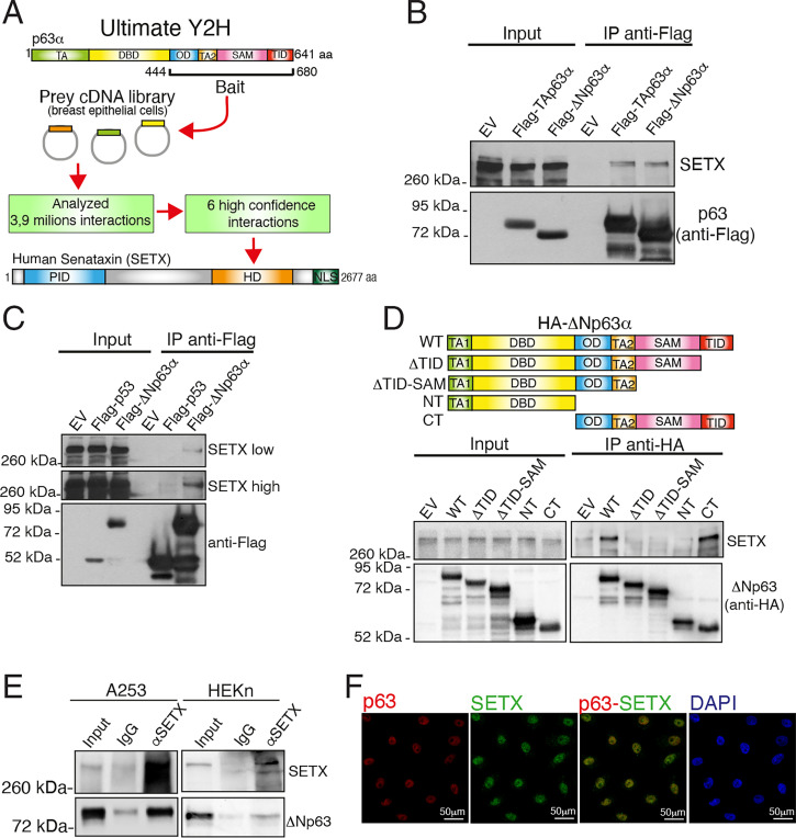
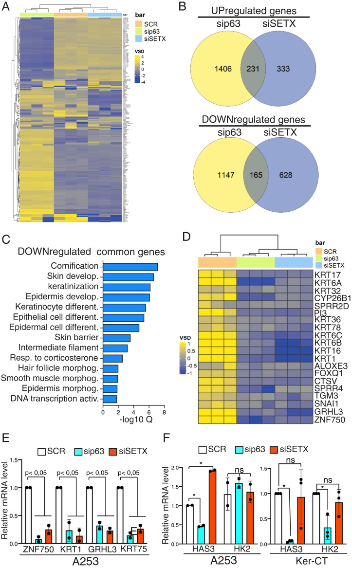
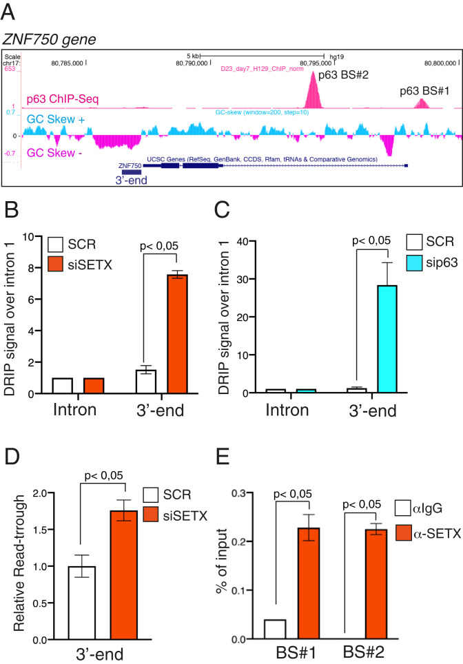
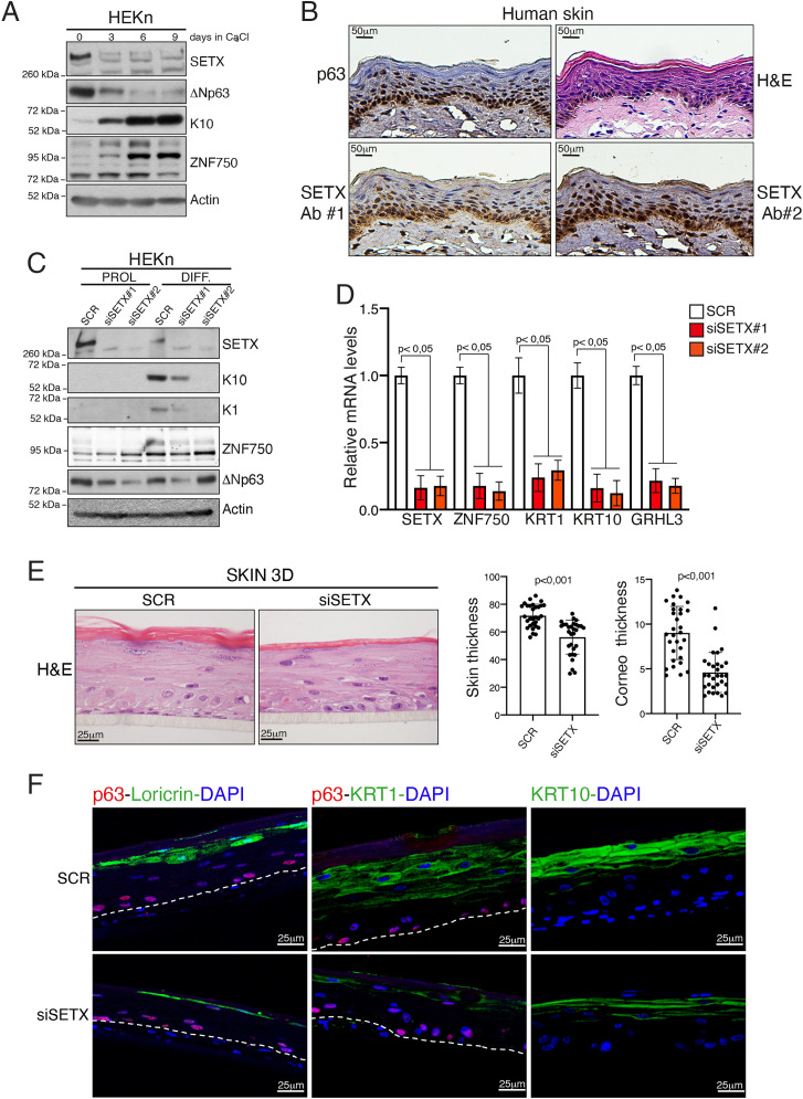
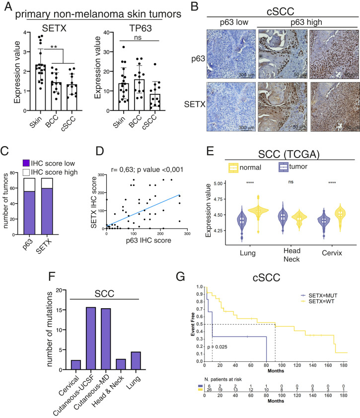
SignificanceΔNp63 is a master regulator of skin homeostasis since it finely controls keratinocyte differentiation and proliferation. Here, we provide cellular and molecular evidence demonstrating the functional role of a ΔNp63 interactor, the R-loop-resolving enzyme Senataxin (SETX), in fine-tuning keratinocyte differentiation. We found that SETX physically binds the p63 DNA-binding motif present in two early epidermal differentiation genes, Keratin 1 (KRT1) and ZNF750, facilitating R-loop removal over their 3' ends and thus allowing efficient transcriptional termination and gene expression. These molecular events translate into the inability of SETX-depleted keratinocytes to undergo the correct epidermal differentiation program. Remarkably, SETX is dysregulated in cutaneous squamous cell carcinoma, suggesting its potential involvement in the pathogenesis of skin disorders.

Keywords: Senataxin; p63; skin differentiation.


Conflict of interest statement

The authors declare no competing interest.

Figures

Fig. 1.
Fig. 1.
SETX is a p63 interactor. (A) Schematic workflow recapitulating the Ultimate Yeast two-hybrid screening approach utilized to identify p63 interactors. The p63α isoform contains the following protein domains: the transactivation domain (TA), DNA-binding domain (DBD), oligomerization domain (OD), transactivation domain 2 (TA2), sterile alpha motif (SAM), and transactivation inhibitory domain (TID). The following protein domains of SETX are also indicated: the protein interaction domain (PID), helicase domain (HD), and nuclear localization signals (NLS). (B) H1299 cells were transfected with the indicated FLAG-tagged p63 constructs or the empty vector (EV). Exogenous proteins were immunoprecipitated (IP) from cell extracts with anti-FLAG antibody, and immunocomplexes were analyzed with the indicated antibodies. (C) H1299 cells were transfected with the indicated FLAG-tagged constructs or the EV. Exogenous proteins were subjected to IP with an anti-FLAG antibody, and immunocomplexes were analyzed with anti-Flag and anti-SETX antibodies. (D) H1299 cells were transfected with EV, HA-tagged ΔNp63 (wild type, WT), or the HA-tagged deletion mutants (ΔTID, ΔTID-ΔSAM, NT, CT) as shown in the schematic model (Upper). Cell extracts were subjected to IP with an anti-HA antibody followed by immunoblotting with antibodies to the indicated protein. (E) Whole-cell extracts from A253 tumor cell line and human primary keratinocytes HEKn cells were subjected to IP with an anti-SETX antibody followed by immunoblotting with the indicated antibodies. (F) Immunofluorescence staining of SETX (green) and p63 (red) in immortalized keratinocytes (Ker-CT). DAPI (blue) was used to visualize nuclei.
Fig. 2.
Fig. 2.
SETX and ΔNp63 control a common transcriptional signature involved in epidermal differentiation. (A) Heatmap showing the unsupervised hierarchical clustering of the most variables (Deseq2 VSD) genes (n = 200) in the SCR (orange), sip63 (green), and siSETX (blue) transfected cells. Color scheme: violet (highest), yellow (lowest) VSD score. (B) Venn diagram showing the shared down-regulated genes (Top) and up-regulated genes (Bottom) in sip63 (yellow) and siSTEX (violet) transfected cells. (C) Bar plot showing the top GO terms for Biological Process, Molecular Function, and Cell Compartment ordered by false discovery rate (FDR). (D) Heatmap showing the unsupervised hierarchical clustering of genes associated to the top 5 GO terms in SCR (orange), sip63 (green), and siSETX (blue) transfected cells. Color scheme: violet (highest), yellow (lowest) VSD score. (E) A253 cells were transfected with siRNA targeting p63 (sip63), SETX (siSETX), or a nonrelevant mRNA (scramble, SCR), and mRNA levels of the indicated genes were quantified by RT-qPCR. Data are shown as the mean ± SD of two biological replicates (n = 2). P value was calculated by Student’s t test. (F) A253 and immortalized keratinocytes (Ker-CT) were transfected as in E, and mRNA levels of the indicated genes were quantified by RT-qPCR. Data are shown as the mean ± SD of two biological replicates (n = 2) for A253 and as the mean ± SD of three biological replicates (n = 3) for Ker-CT. P value was calculated by Student’s t test. *P < 0.05, ns = not significant.
Fig. 3.
Fig. 3.
SETX regulates R-loop formation on the 3′ end of the p63 target gene ZNF750. (A) Schematic representation of ZNF750 genetic locus (modified from Genome Browser, https://genome.ucsc.edu) showing from top to bottom p63 ChIP-seq peaks in human keratinocytes (12), GC skew signal, and gene structure with exons (boxes) and introns (lines). (B) Quantification by qPCR of DRIP signal in differentiated keratinocytes treated with SCR or SETX siRNAs. Values at 3′ ends were normalized to ZNF750 intron 1 signal. Bars represent the mean of three replicates (n = 3, PCR runs) ± SD and are representative of three independent experiments (n = 3 biological replicates). P value was calculated by Student’s t test. (C) Quantification by qPCR of DRIP signal in differentiated keratinocytes treated with SCR or p63 siRNAs. Values at 3′ ends were normalized to ZNF750 intron 1 signal. Bars represent the mean of three replicates (n = 3, PCR runs) ± SD and are representative of two independent experiments (n = 2 biological replicates). P value was calculated by Student’s t test. (D) RT-qPCR analysis of readthrough transcripts at the ZNF750 3′ region in SCR or siSETX differentiated keratinocytes. Values are normalized to ZNF750 intron 1 and showed as fold enrichment of siSETX over SCR. Bars represent the mean of three replicates (n = 3, PCR runs) ± SD and are representative of three independent experiments (n = 3 biological replicates). P value was calculated by Student’s t test. (E) Quantification by qPCR of ChIP analysis of endogenous SETX occupancy at p63-binding sites (BS#1 and BS#2) in differentiated human keratinocytes. Bars represent the mean of three replicates (n = 3, PCR runs) ± SD and are representative of three independent experiments (n = 3 biological replicates). P value was calculated by Student’s t test.
Fig. 4.
Fig. 4.
SETX depletion impairs keratinocyte differentiation. (A) Immunoblotting analysis of cell lysates extracted from HEKn at different time points (0, 3, 6, and 9 d) upon CaCl2 treatment. (B) Representative images of hematoxylin/eosin (H&E) staining or IHC analysis of p63 and SETX expression in normal skin samples. SETX antibody #1 (SETX Ab#1) is from Novus Biologicals (NBP1-94712), and SETX antibody #2 (SETX Ab#2) is a kind gift from Domenico Delia (University of Milan, Italy). (C) HEKn cells transfected with nonrelevant siRNA (SCR) or two different siRNAs targeting SETX mRNA (siSETX#1 or siSETX#2) were treated with CaCl2 to induce differentiation. Protein lysates from proliferating (PROL) or differentiating keratinocytes (DIFF, 4 d of differentiation) were analyzed by Western blotting using antibodies to the indicated proteins. (D) mRNA extracted from differentiated keratinocytes was used for RT-qPCR quantification of different epithelial differentiation markers. Bars represent the mean of three replicates (n = 3, PCR runs) ± SD and are representative of two independent experiments (n = 2 biological replicates). (E) H&E staining of SETX-depleted (siSETX) organotypic human epidermis compared to scramble control organotypic epidermis (SCR). Histograms on Right show quantification of whole skin or stratum corneum thickness in SCR (n = 30 measurement) and siSETX (n = 30 measurement) organotypic epidermis. (F) Immunofluorescence staining of the differentiation markers loricrin, KRT1, and KRT10 (green) in SCR and siSETX organotypic skin cultures. p63 staining (red) and DAPI (blue) were used to visualize the basal layer and nuclei, respectively. Dotted lines underline the keratinocyte–fibroblast border.
Fig. 5.
Fig. 5.
SETX is mutated or downmodulated in SCC, and SETX gene mutation is a negative prognostic factor for cutaneous SCC patient survival. (A) Total RNA extracted from normal skin (n = 18), basal cell carcinoma (BCC, n = 13), and cutaneous SCC (n = 12) samples was utilized for RT-qPCR quantification of SETX and p63 mRNA. **P < 0.01, ns = not significant. (B) Representative images of IHC analysis of SETX and p63 expression in skin SCC. (C) IHC score for p63 and SETX staining was calculated for each tumor sample. Distribution among high- and low-expressing tumors is shown for SETX and p63. (D) Correlation plot of SETX and p63 IHC scores in 73 human cSCC. Pearson’s correlation coefficient (r) and the P value of the correlation study are indicated. (E) Violin plot illustrating SETX expression values in SCCs and in the respective normal tissues. Asterisks indicate statistically significant differences (****P < 0.05), ns = not significant. (F) Bar plot showing the number of SETX mutations in the indicated types of SCCs. (G) Kaplan–Meier survival curves of Event Free related cSCC patients harboring wild-type (WT) or mutated SETX allele.

References

    1. Candi E., Schmidt R., Melino G., The cornified envelope: A model of cell death in the skin. Nat. Rev. Mol. Cell Biol. 6, 328–340 (2005). - PubMed
    1. Botchkarev V. A., The molecular revolution in cutaneous biology: Chromosomal territories, higher-order chromatin remodeling, and the control of gene expression in keratinocytes. J. Invest. Dermatol. 137, e93–e99 (2017). - PMC - PubMed
    1. Soares E., Zhou H., Master regulatory role of p63 in epidermal development and disease. Cell. Mol. Life Sci. 75, 1179–1190 (2018). - PMC - PubMed
    1. Candi E., et al. , p63 in epithelial development. Cell. Mol. Life Sci. 65, 3126–3133 (2008). - PMC - PubMed
    1. Medawar A., et al. , DeltaNp63 is essential for epidermal commitment of embryonic stem cells. PLoS One 3, e3441 (2008). - PMC - PubMed

MeSH terms