Spinal cord lesion mimicking a dysimmune myelitis revealing CANVAS syndrome
- PMID: 35235501
- PMCID: PMC9987767
- DOI: 10.1080/10790268.2022.2033936
Spinal cord lesion mimicking a dysimmune myelitis revealing CANVAS syndrome
Abstract
Context: Posterior spinal cord lesions are found in patients with ganglionopathy. These are normally found in later stages of the neuronopathy as a consequence of dorsal root ganglia degeneration. Cerebellar Ataxia, Neuropathy, Vestibular Areflexia Syndrome (CANVAS) is an emerging neurological disorder. Myelitis lesions have been described in confirmed CANVAS cases.
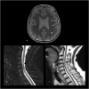
Findings: We describe a case of a 68-year-old woman with slowly progressive ataxia with paresthesia. Laboratory tests were normal. Total spine MRI showed a C4 posterior spinal cord lesion. Lumbar puncture was positive for oligoclonal bands with normal IgG index and protein level. Paraneoplastic antibodies were not detected. Electromyography showed nonlength dependent sensory neuropathy. The patient was treated with intravenous immunoglobulin for suspected dysimmune myelitis. Over 6 years, she progressively developed other neurological manifestations evoking CANVAS. Nerve conduction study showed isolated sensory impairment over the years and peripheral nerve ultrasound revealed abnormally small nerves. Further genetic testing confirmed the diagnosis.
Conclusion: This is the first case of CANVAS syndrome presenting initially with an isolated spinal cord lesion mimicking dysimmune myelitis. The purpose of this case report is to add to the current literature about this evolving neurological syndrome and to aid clinicians in their diagnostic approach in clinical practice.
Keywords: CANVAS; Myelitis; Neuronopathy; Peripheral nerve ultrasound.
Figures


References
-
- Gwathmey KG. Sensory neuronopathies: sensory neuronopathies. Muscle Nerve 2016 Jan;53(1):8–19. - PubMed
-
- Casseb RF, Martinez ARM, de Paiva JLR, França MC.. Neuroimaging in Sensory Neuronopathy: Neuroimaging in sensory neuronopathy. J Neuroimaging 2015 Sep;25(5):704–709. - PubMed
-
- Paisán-Ruiz C, Jen JC.. CANVAS with cerebellar/sensory/vestibular dysfunction from RFC1 intronic pentanucleotide expansion. Brain 2020 Feb 1;143(2):386–390. - PubMed
-
- Pelosi L, Mulroy E, Leadbetter R, Kilfoyle D, Chancellor AM, Mossman S, et al. Peripheral nerves are pathologically small in cerebellar ataxia neuropathy vestibular areflexia syndrome: a controlled ultrasound study. Eur J Neurol 2018 Apr;25(4):659–665. - PubMed
Publication types
MeSH terms
LinkOut - more resources
Full Text Sources
Medical
Miscellaneous
