Review
. 2022 Mar 2;11(1):11.
doi: 10.1186/s40164-022-00263-4.

Targeting PD-1/PD-L1 pathway in myelodysplastic syndromes and acute myeloid leukemia

Affiliations
Review

Targeting PD-1/PD-L1 pathway in myelodysplastic syndromes and acute myeloid leukemia

Xingcheng Yang et al. Exp Hematol Oncol. .

Abstract

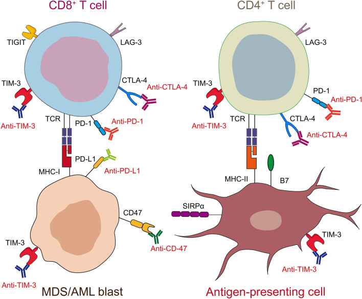
Myelodysplastic syndromes (MDS) and acute myeloid leukemia (AML) are clonal hematopoietic stem cell diseases arising from the bone marrow (BM), and approximately 30% of MDS eventually progress to AML, associated with increasingly aggressive neoplastic hematopoietic clones and poor survival. Dysregulated immune microenvironment has been recognized as a key pathogenic driver of MDS and AML, causing high rate of intramedullary apoptosis in lower-risk MDS to immunosuppression in higher-risk MDS and AML. Immune checkpoint molecules, including programmed cell death-1 (PD-1) and programmed cell death ligand-1 (PD-L1), play important roles in oncogenesis by maintaining an immunosuppressive tumor microenvironment. Recently, both molecules have been examined in MDS and AML. Abnormal inflammatory signaling, genetic and/or epigenetic alterations, interactions between cells, and treatment of patients all have been involved in dysregulating PD-1/PD-L1 signaling in these two diseases. Furthermore, with the PD-1/PD-L1 pathway activated in immune microenvironment, the milieu of BM shift to immunosuppressive, contributing to a clonal evolution of blasts. Nevertheless, numerous preclinical studies have suggested a potential response of patients to PD-1/PD-L1 blocker. Current clinical trials employing these drugs in MDS and AML have reported mixed clinical responses. In this paper, we focus on the recent preclinical advances of the PD-1/PD-L1 signaling in MDS and AML, and available and ongoing outcomes of PD-1/PD-L1 inhibitor in patients. We also discuss the novel PD-1/PD-L1 blocker-based immunotherapeutic strategies and challenges, including identifying reliable biomarkers, determining settings, and exploring optimal combination therapies.

Keywords: AML transformation; Acute myeloid leukemia; Hypomethylating agent; Immune checkpoint; Myelodysplastic syndrome; Programmed cell death ligand-1; Programmed cell death-1.


Conflict of interest statement

The authors declare no competing financial interests.

Figures

Fig. 1
Fig. 1
Function of dysregulated PD-1/PD-L1 pathway in MDS. Upon exposure to IFN-γ and TNF-α, PD-L1 levels are increased in MDS blasts via NF-κB and pSTAT1/pSTAT3 activation. Further, TP53 mutation also implicated in PD-L1 upregulation via MYC upregulation and miR-34a downregulation, thus regulating PD-L1 levels at a post-transcriptional level. In CD34+ HSPCs, TP53 mutation and S100A9 upregulate PD-1 via MYC. Furthermore, PD-L1+ MDS blasts mediate pathogenesis through PD-1/PD-L1 signaling, by the following mechanisms: ① blasts expressing PD-L1 confer proliferative advantages, expressing higher levels of CyclinD1/D2/D3 and growing more actively; ② the binding of PD-L1 on MDS blasts with PD-1 on CD34+ HSPCs result in PD-1+CD34+ HSPC apoptosis. ③ the binding of PD-L1 on MDS blasts with PD-1 on CD4+/CD8+ T cells inhibit the activation and proliferation of these effector T cells. MHC, major histocompatibility complex; TNFR, TNF receptor; MT, mutation; pSTAT, phosphorylated signal transducer and activator of transcription
Fig. 2
Fig. 2
Function of dysregulated PD-1/PD-L1 pathway in AML. Upon exposure to IFN-γ and TNF-α, PD-L1 levels are increased in MDS blasts via MEK and pSTAT1/pSTAT3 activation. Similar to MDS blasts, TP53 mutation also plays important roles in PD-L1 upregulation via MYC upregulation and miR-34a downregulation, thus regulating PD-L1 levels at a post-transcriptional level. In addition, miR-34a and miR200c are regulated by DICER, cJUN and MUC1. Furthermore, PD-L1+ AML blast-mediated pathogenesis occurs through PD-1/PD-L1 signaling, by the following mechanisms: ① blasts expressing PD-L1 confer proliferative advantages, including enhanced cell glycolysis and higher levels of Cyclin D2, via activation of pJNK, resulting in more active growth; ② the interaction of CD200 on AML blasts with CD200R on effectors leads to the upregulation of PD-1, which is also regulated by increased Bmilp-1, promoting the inaction of these effector T cells; ③ the binding of PD-L1 on AML blasts with PD-1 on effector T cells suppress activation of these effector T cells, and promote conversion of Tregs from conventional CD4+ T cells, which triggers the secretion of IL-35 and upregulates PD-L1 on AML blasts via pAkt activation
Fig. 3
Fig. 3
Immune microenvironment of the BM in HMA-failed MDS/AML patients. Following HMA therapy, a portion of MDS/AML blasts died, while other MDS/AML blasts survived and acquired HMA resistance, which can be further eradicated by PD-1 or PD-L1 inhibitors. The underlying mechanisms are as follows: ① following HMA therapy, PD-1 promoter methylation in CD8+ T cells is decreased, resulting in PD-1 upregulation; ② the activation of CD8+ T cells is suppressed by the binding of PD-1 expressed on CD8+ T cells and PD-L1 expressed on MDS/AML blasts; ③ further administration with PD-1/PD-L1 antibodies prevents the interaction of these two molecules, alleviating the activation of CD8+ T cells, which induces apoptosis in the remaining MDS/AML blasts
Fig. 4
Fig. 4
The prospect of novel immune checkpoint targets in MDS/AML treatment. An overview of the interactions between ICIs and immune checkpoints expressed on CD4+/CD8+ T cells, antigen-presenting cells and MDS/AML blasts in bone marrow of patients

References

    1. Arber DA, Orazi A, Hasserjian R, Thiele J, Borowitz MJ, Le Beau MM, et al. The 2016 revision to the World Health Organization classification of myeloid neoplasms and acute leukemia. Blood. 2016;127(20):2391–2405. - PubMed
    1. Yi M, Li A, Zhou L, Chu Q, Song Y, Wu K. The global burden and attributable risk factor analysis of acute myeloid leukemia in 195 countries and territories from 1990 to 2017: estimates based on the global burden of disease study 2017. J Hematol Oncol. 2020;13(1):72. - PMC - PubMed
    1. Menssen AJ, Walter MJ. Genetics of progression from MDS to secondary leukemia. Blood. 2020;136(1):50–60. - PMC - PubMed
    1. da Silva-Coelho P, Kroeze LI, Yoshida K, Koorenhof-Scheele TN, Knops R, van de Locht LT, et al. Clonal evolution in myelodysplastic syndromes. Nat Commun. 2017;8:15099. - PMC - PubMed
    1. Ghobrial IM, Detappe A, Anderson KC, Steensma DP. The bone-marrow niche in MDS and MGUS: implications for AML and MM. Nat Rev Clin Oncol. 2018;15(4):219–233. - PubMed