. 2022 Mar;10(3):e003725.
doi: 10.1136/jitc-2021-003725.

Optimizing therapeutic outcomes of immune checkpoint blockade by a microbial tryptophan metabolite

Affiliations

Optimizing therapeutic outcomes of immune checkpoint blockade by a microbial tryptophan metabolite

Giorgia Renga et al. J Immunother Cancer. 2022 Mar.

Abstract

Background: Despite the great success, the therapeutic benefits of immune checkpoint inhibitors (ICIs) in cancer immunotherapy are limited by either various resistance mechanisms or ICI-associated toxic effects including gastrointestinal toxicity. Thus, novel therapeutic strategies that provide manageable side effects to existing ICIs would enhance and expand their therapeutic efficacy and application. Due to its proven role in cancer development and immune regulation, gut microbiome has gained increasing expectation as a potential armamentarium to optimize immunotherapy with ICI. However, much has to be learned to fully harness gut microbiome for clinical applicability. Here we have assessed whether microbial metabolites working at the interface between microbes and the host immune system may optimize ICI therapy.

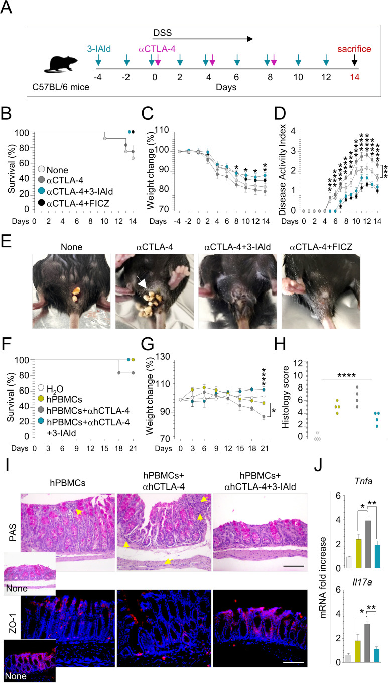
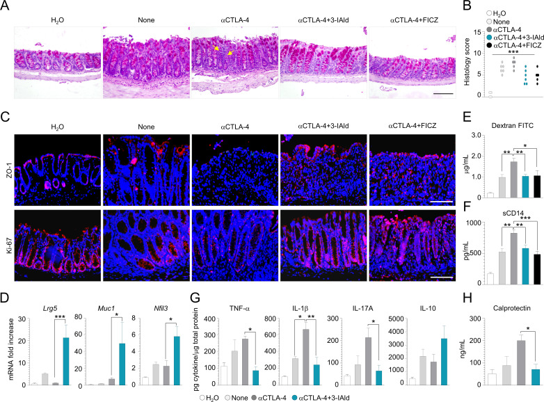
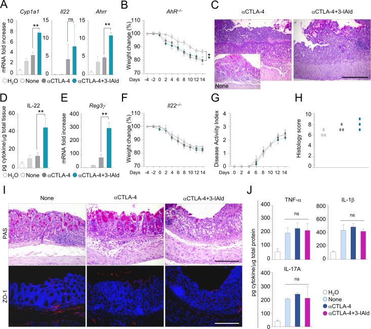
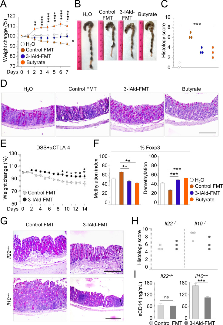
Methods: To this purpose, we have tested indole-3-carboxaldehyde (3-IAld), a microbial tryptophan catabolite known to contribute to epithelial barrier function and immune homeostasis in the gut via the aryl hydrocarbon receptor (AhR), in different murine models of ICI-induced colitis. Epithelial barrier integrity, inflammation and changes in gut microbiome composition and function were analyzed. AhR, indoleamine 2,3-dioxygenase 1, interleukin (IL)-10 and IL-22 knockout mice were used to investigate the mechanism of 3-IAld activity. The function of the microbiome changes induced by 3-IAld was evaluated on fecal microbiome transplantation (FMT). Finally, murine tumor models were used to assess the effect of 3-IAld treatment on the antitumor activity of ICI.

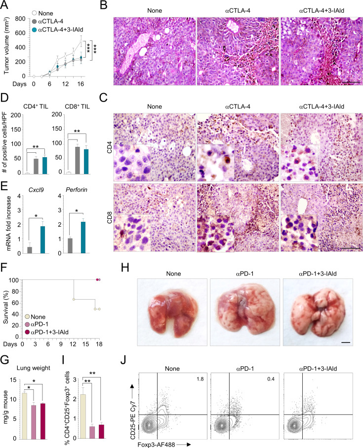
Results: On administration to mice with ICI-induced colitis, 3-IAld protected mice from intestinal damage via a dual action on both the host and the microbes. Indeed, paralleling the activation of the host AhR/IL-22-dependent pathway, 3-IAld also affected the composition and function of the microbiota such that FMT from 3-IAld-treated mice protected against ICI-induced colitis with the contribution of butyrate-producing bacteria. Importantly, while preventing intestinal damage, 3-IAld did not impair the antitumor activity of ICI.

Conclusions: This study provides a proof-of-concept demonstration that moving past bacterial phylogeny and focusing on bacterial metabolome may lead to a new class of discrete molecules, and that working at the interface between microbes and the host immune system may optimize ICI therapy.

Keywords: cytotoxicity; immune tolerance; immunologic; immunotherapy; inflammation; melanoma.


Conflict of interest statement

Competing interests: None declared.

Figures

Figure 1
Figure 1
3-IAld protects mice from ICI-induced colitis. C57BL/6 mice were treated with DSS in drinking water for 1 week followed by a recovery period of another week, and administered 100 µg of anti-CTLA-4 mAb or isotype control two times (at days 0, 4, and 8 following DSS administration). 3-IAld (18 mg/kg) was administered intragastrically every other day starting 4 days before DSS treatment. FICZ was used as control as depicted in the experimental schedule (A). Mice were evaluated for (B) % survival, (C) % weight change, (D) disease activity index, and (E) rectal bleeding. NSG mice infused intraperitoneally with hPBMCs were treated with αhCTLA-4 and 3-IAld. Mice were sacrificed at 21 days and evaluated for (F) % survival, (G) % weight change, (H) histology score, (I) colon histology (PAS staining) and ZO-1 protein expression and (J) inflammatory cytokine expression. Photographs were taken with a high-resolution microscope (Olympus BX51), ×20 magnification (scale bars, 200 µm). White arrow indicates rectal bleeding. Yellow arrows indicate inflammatory cells recruitment. For histology, data are representative of two or three independent experiments. Each in vivo experiment includes four to six mice per group (16–24 mice in each experiment). Data are represented as mean±SEM. (B–D) Anti-CTLA-4-treated mice versus none (isotype control), anti-CTLA-4+3-IAld- versus anti-CTLA-4-treated mice, (F–J) hPBMCs+αhCTLA-4 versus hPBMCs (+human IgG) or hPBMCs+αhCTLA-4+3 IAld. Two-way analysis of variance, Bonferroni post hoc test. H2O, untreated mice. *P<0.05, **P<0.01, ***P<0.001, ****P<0.0001. 3-IAld, indole-3-carboxaldehyde; FICZ, 6-formylindolo(3,2-b)carbazole; hPBMC, human peripheral blood mononuclear cell; ICI, immune checkpoint inhibitor; PAS, periodic acid–Schiff.
Figure 2
Figure 2
3-IAld prevents epithelial damage in DSS plus anti-CTLA-4-induced colitis. C57BL/6 mice were subjected to DSS plus anti-CTLA-4-induced colitis and administered 3-IAld as described in the legend of figure 1. Mice were evaluated for (A) colon histology (periodic acid–Schiff staining), (B) histology score, (C) ZO-1 and Ki-67 protein expression, (D) intestinal stem cells genes expression, (E) Dextran-FITC and (F) sCD14 levels in the serum, (G) cytokine and (H) calprotectin levels in colon homogenates. For immunofluorescence, nuclei were counterstained with Hoechst 33342. Photographs were taken with a high-resolution microscope (Olympus BX51), ×40 magnification (scale bars, 100 µm). Yellow arrows indicate inflammatory cell recruitment. For histology and immunofluorescence, data are representative of three independent experiments. Each in vivo experiment includes three to six mice per group (15–30 mice in each experiment). Data are represented as mean±SEM, (B) Kruskal-Wallis test. (D–H) Anti-CTLA-4- versus none (isotype control) or anti-CTLA-4+3-IAld-treated mice. One-way analysis of variance, Bonferroni post hoc test. H2O, untreated mice. *P<0.05, **P<0.01, ***P<0.001. 3-IAld, indole-3-carboxaldehyde; FICZ, 6-formylindolo(3,2-b)carbazole; IL, interleukin; TNF-α, tumour necrosis factor alpha.
Figure 3
Figure 3
Protective activity of 3-IAld is not dependent on IDO1 and IL-10. C57BL/6 (A–D, G), Indo1 –/– (E–F) and Il10 –/– (G–J) mice were subjected to anti-CTLA-4 with (A–F, H–J) or without (G) DSS and administered 3-IAld as described in the legend of figure 1. Mice were evaluated for (A) IDO1 gene and (B) protein expression, (C) Trp, Kyn levels and Kyn:Trp ratio, (D) Haao and Kynu expression, (E, H) % weight change, (F–J) colon histology (periodic acid–Schiff staining), (I) histology score. For immunofluorescence, nuclei were counterstained with Hoechst 33342. Photographs were taken with a high-resolution microscope (Olympus BX51), ×20 magnification (scale bar, 200 µm). For histology and immunofluorescence, data are representative of three independent experiments. Each in vivo experiment includes 3 mice per group (9–18 mice total). Data are represented as mean±SEM. Anti-CTLA-4+3-IAld- versus anti-CTLA-4-treated mice. One-way analysis of variance, Bonferroni post hoc test, (I) Kruskal-Wallis test. H2O, untreated mice. *P<0.05, **P<0.01. 3-IAld, indole-3-carboxaldehyde; IDO1, indoleamine 2,3-dioxygenase 1; IL, interleukin; Kyn, kynurenine; ns, not significant; Trp, tryptophan.
Figure 4
Figure 4
Beneficial effect of 3-IAld involves the AhR/IL-22 axis. C57BL/6 (A, D, E), AhR –/– (B, C), Il22 –/– (F–J) mice were subjected to DSS plus anti-CTLA-4-induced colitis and administered 3-IAld as described in the legend of figure 1. Mice were evaluated for (A) AhR-related genes, (B, F) % weight change, (C, I) colon histology (periodic acid–Schiff staining), (D) IL-22 levels, (E) Reg3γ gene expression, (G) disease activity index, (H) histology score, (I) ZO-1 expression, and (J) cytokine production in colon homogenates. For immunofluorescence, nuclei were counterstained with Hoechst 33342. Photographs were taken with a high-resolution microscope (Olympus BX51), ×20 magnification (scale bars, 200 µm). For histology and immunofluorescence, data are representative of three independent experiments. Each in vivo experiment includes 3 mice per group (9–12 mice in each experiment). Data are represented as mean±SEM. Anti-CTLA-4+3-IAld- versus anti-CTLA-4-treated mice. One-way analysis of variance, Bonferroni post hoc test, (F) Kruskal-Wallis test. H2O, untreated mice. None, DSS+isotype control. **P<0.01. 3-IAld, indole-3-carboxaldehyde; IL, interleukin; ns, not significant; TNF-α, tumour necrosis factor alpha.
Figure 5
Figure 5
3-IAld modifies intestinal microbiota composition and function. (A) Barplot showing bacterial composition (abundance percentage) of each sample at phylum level. Taxa are differentiated by colors. Samples are ranked based on the abundance of the most abundant Phylum (Bacteroidota) and grouped by None and 3-IAld-treated. (B) Boxplots of observed features, Chao1 and Shannon alpha diversity indexes. Significance was evaluated by applying a Kruskal-Wallis test (ns). (C) Boxplots of Jaccard and Bray-Curtis beta diversity indexes evaluating distances within or between none and 3-IAld-treated samples. Significance was evaluated by applying a Kruskal-Wallis test (the p value is indicated). (D, E) LEfSe at genus level. LEfSe emphasizes a set of features that significantly discriminate between none and 3-IAld treatments. A p value of <0.05 and an LDA score of ≥3.5 were regarded as significant in Kruskall-Wallis and pairwise Wilcoxon tests, respectively. The cladogram (D) simultaneously highlights both phyla and specific genera. Taxa (circles) are colored green when significantly associated to none, red when significantly associated with 3-IAld-treated samples, and yellow when not significantly associated to either groups. The size of each circle is proportional to the abundance of the corresponding taxon in all samples. The barplot of the LDA scores (E) shows genera significantly associated with either none and 3-IAld-treated samples accordingly to legend colors. LEfSe has been applied with default alpha values. (F) LEfSe on KEGG modules. Barplot of the LDA score computed on metabolic functions inferred by PICRUSt2 analysis (KEGG modules) and significantly associated with either none or 3-IAld-treated samples accordingly to legend colors. The threshold value of the logarithmic LDA score was set to 2.0. (G) Levels of SCFA measured in the feces and serum by mass spectrometry. (A–F) Six mice per group (12 mice total); (G) 3–6 mice per group (9 mice total). Data are represented as mean±SD. None versus 3-IAld, t test. *P<0.05, **P<0.01. 3-IAld, indole-3-carboxaldehyde; LDA, linear discriminant analysis; LEfSe, linear discriminant analysis together with effect size evaluation; ns; not significant.
Figure 6
Figure 6
3-IAld-modified microbiota provides protection to ICI-induced colitis. C57BL/6 (A–F), Il22 –/– and Il10 –/– (G–I) mice were subjected to DSS-colitis with (E) or without (A–D, F–I) anti-CTLA-4 and transplanted with fresh fecal pellets from control or 3-IAld-treated mice 1 day before and 2 days after colitis induction. One group of mice was treated with 1% butyrate in drinking water 1 week before starting DSS administration. Mice were sacrificed 7 (A–D, F–I) or 14 (E) days after colitis induction and evaluated for (A, E) % weight change, (B) gross pathology, (C, H) histology score, (D, G) colon histopathology (periodic acid–Schiff staining), (F) methylation/demethylation status of Foxp3 promoter in mesenteric lymph nodes, and (I) sCD14 serum levels. Photographs were taken with a high-resolution microscope (Olympus BX51), ×10 and ×20 magnification (scale bars, 500 and 200 µm). For histology, data are representative of three independent experiments. Each in vivo experiment includes 3 mice per group (6–12 mice in each experiment). Data are represented as mean±SD. Treated versus control FMT mice. One-way analysis of variance, Bonferroni post hoc test, (C) Kruskal-Wallis test. H2O, untreated mice. *P<0.05, **P<0.01, ***P<0.001, ****P<0.0001. 3-IAld, indole-3-carboxaldehyde; FMT, fecal microbiota transplantation. ns, not significant.
Figure 7
Figure 7
3-IAld does not interfere with the antitumor activity of anti-CTLA-4 antibody. (A–E) C57BL/6 mice were subcutaneously injected with B16 tumor cells and administered 100 µg anti-CTLA-4 mAb or isotype control intraperitoneally four times at 3-day intervals up to 16 days. 3-IAld was administered intragastrically every other day. Mice were evaluated for (A) tumor growth, (B) histology (periodic acid–Schiff staining), (C) immunohistochemistry for CD8+ and CD4+ tumor-infiltrating cells and (D) number of positive cells per HPF, and (E) Cxcl9 and Perforin expression in melanoma. (F–J) C57BL/6 mice were orthotopically injected with LLC cells and administered 200 µg anti-PD-1 mAb or isotype control intraperitoneally five times at 3-day intervals up to 18 days. 3-IAld was administered intragastrically every other day. Mice were evaluated for (F) % survival, (G) lung weight, (H) lung gross pathology, (I, J) CD4+CD25+Foxp3+ cells. Photographs were taken with a high-resolution microscope (Olympus BX51), ×40 magnification (scale bars, 100 µm). For histology, data are representative of two independent experiments. Each in vivo experiment includes three mice per group (nine mice in each experiment). Data are represented as mean±SEM. Treated versus none (isotype control) or αCTLA-4/αPD-1+3-IAld-treated versus αCTLA-4/αPD-1-treated mice. One-way analysis of variance, Bonferroni post hoc test. *P<0.05, **P<0.01, ***P<0.001. 3-IAld, indole-3-carboxaldehyde; HPF, high-power field; TIL, tumor-infiltrating lymphocyte.

References

    1. Schoenfeld AJ, Hellmann MD. Acquired resistance to immune checkpoint inhibitors. Cancer Cell 2020;37:443–55. 10.1016/j.ccell.2020.03.017 - DOI - PMC - PubMed
    1. Choi J, Lee SY. Clinical characteristics and treatment of immune-related adverse events of immune checkpoint inhibitors. Immune Netw 2020;20:e9. 10.4110/in.2020.20.e9 - DOI - PMC - PubMed
    1. Bauché D, Mauze S, Kochel C, et al. . Antitumor efficacy of combined CTLA4/PD-1 blockade without intestinal inflammation is achieved by elimination of FcγR interactions. J Immunother Cancer 2020;8. 10.1136/jitc-2020-001584 - DOI - PMC - PubMed
    1. Pitt JM, Vétizou M, Daillère R, et al. . Resistance mechanisms to immune-checkpoint blockade in cancer: tumor-intrinsic and -Extrinsic factors. Immunity 2016;44:1255–69. 10.1016/j.immuni.2016.06.001 - DOI - PubMed
    1. Roy S, Trinchieri G. Microbiota: a key orchestrator of cancer therapy. Nat Rev Cancer 2017;17:271–85. 10.1038/nrc.2017.13 - DOI - PubMed

Publication types