. 2022 Mar 2;12(1):3446.
doi: 10.1038/s41598-022-07341-x.

Gramicidin S and melittin: potential anti-viral therapeutic peptides to treat SARS-CoV-2 infection

Affiliations

Gramicidin S and melittin: potential anti-viral therapeutic peptides to treat SARS-CoV-2 infection

Mohammed Ghalib Enayathullah et al. Sci Rep. .

Abstract

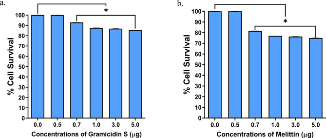
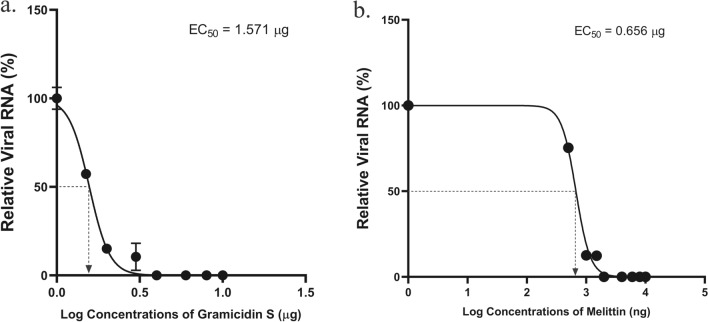
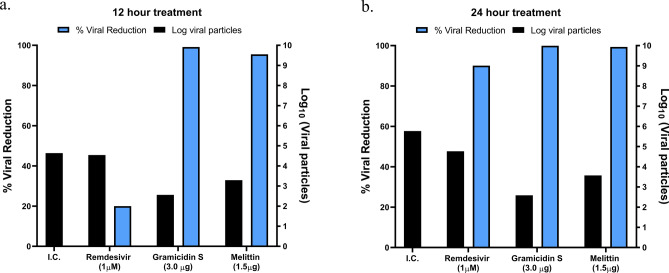
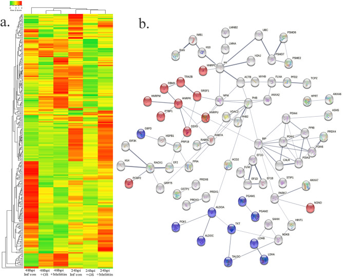
The COVID19 pandemic has led to multipronged approaches for treatment of the disease. Since de novo discovery of drugs is time consuming, repurposing of molecules is now considered as one of the alternative strategies to treat COVID19. Antibacterial peptides are being recognized as attractive candidates for repurposing to treat viral infections. In this study, we describe the anti-SARS-CoV-2 activity of the well-studied antibacterial peptides gramicidin S and melittin obtained from Bacillus brevis and bee venom respectively. The EC50 values for gramicidin S and melittin were 1.571 µg and 0.656 µg respectively based on in vitro antiviral assay. Significant decrease in the viral load as compared to the untreated group with no/very less cytotoxicity was observed. Both the peptides treated to the SARS-CoV-2 infected Vero cells showed viral clearance from 12 h onwards with a maximal viral clearance after 24 h post infection. Proteomics analysis indicated that more than 250 proteins were differentially regulated in the gramicidin S and melittin treated SARS-CoV-2 infected Vero cells against control SARS-CoV-2 infected Vero cells after 24 and 48 h post infection. The identified proteins were found to be associated in the metabolic and mRNA processing of the Vero cells post-treatment and infection. Both these peptides could be attractive candidates for repurposing to treat SARS-CoV-2 infection.


Conflict of interest statement

The authors declare no competing interests.

Figures

Figure 1
Figure 1
Measurement of Cytolytic activity of gramicidin S and melittin using MTT Assay: The graphs represent the percentage of cell viability vs. concentrations of (a) Gramicidin S (µg). (b) Melittin (µg). The values represent Mean ± SD of atleast three independent experiments. The data analysis and graphs were generated using GraphPad Prism (Ver 8.4.2). Significance of variance, p < 0.05 is considered statistically significant.
Figure 2
Figure 2
Anti SARS-CoV-2 activity of gramicidin S and melittin in vitro: (a). Relative viral RNA (%) vs. Log concentrations of gramicidin S (µg). (b) Relative viral RNA (%) vs. Log concentrations of melittin (ng). The graphs represent the Ct values of N-gene calculated using RT-qPCR in the supernatants. The Log EC50 value of gramicidin S (0.1963) corresponds to 1.571 µg and Log EC50 value (2.826) of melittin corresponds to 0.656 µg as shown in the graph.
Figure 3
Figure 3
The anti SARS-CoV2 activity of remdesivir gramicidin S and melittin at 12 (a) and 24 (b) h post infection. The X axis represents different experimental groups: I.C (infection control), remdesivir (1 µM), gramicidin S (3.0 µg), and melittin (1.5 µg). The Y axis (Blue bars) represents % of viral reduction and Y’-Axis (black bars) represents the Log10 viral particles.
Figure 4
Figure 4
Immunofluorescence staining images against RBD protein expression specific to SARS-CoV-2 in Vero cells. (A) (I) Vero cells without SARS-CoV-2 infection (mock), (II) DAPI staining representing nucleus (blue); (III) Merged image of f-Actin (Phalloidin staining, red), nucleus (DAPI, blue). (B) Vero cells infected with SARS-CoV-2, (I) represents RBD protein expression (green) of SARS-CoV-2 virus at 12 h; (II) DAPI staining representing nucleus (blue) along with RBD protein expression of SARS-CoV-2 (green) at 12 h; (III) Merged image of f-Actin (red) (Phalloidin staining), nucleus (DAPI) and RBD protein of SARS-CoV-2 (green) at 12 h; (IV) represents RBD protein expression (green) of SARS-CoV-2 virus at 24 h; (V) DAPI staining representing nucleus (blue) along with RBD protein expression of SARS-CoV-2 (green) at 24 h; (VI) Merged image of f-Actin (Phalloidin staining, red), nucleus (DAPI, blue) and RBD protein of SARS-CoV-2 (green) at 24 h. (C) Vero cells infected with SARS-CoV-2 treated with gramicidin S, (I) RBD protein expression (green) of SARS-CoV-2 virus at 12 h; (II) DAPI staining representing nucleus (blue) along with RBD protein expression of SARS-CoV-2 (green) at 12 h; (III) Merged image of f-Actin (red) (Phalloidin staining), nucleus (DAPI) and RBD protein of SARS-CoV-2 (green) at 12 h; (IV) represents RBD protein expression (green) of SARS-CoV-2 virus at 24 h; (V) DAPI staining representing nucleus (blue) along with RBD protein expression of SARS-CoV-2 (green) at 24 h; (VI) Merged image f-Actin (Phalloidin staining, red), nucleus (DAPI, blue) and RBD protein of SARS-CoV-2 (green) at 24 h. (D) Vero cells infected with SARS-CoV-2 treated with melittin. (I) represents RBD protein expression (green) of SARS-CoV-2 virus at 12 h; (II) DAPI staining representing nucleus (blue) along with RBD protein expression of SARS-CoV-2 (green) at 12 h; (III) Merged image of f-Actin (red) (Phalloidin staining), nucleus (DAPI) and RBD protein of SARS-CoV-2 (green) at 12 h; (IV) represents RBD protein expression (green) of SARS-CoV-2 virus at 24 h; (V) DAPI staining representing nucleus (blue) along with RBD protein expression of SARS-CoV-2 (green) at 24 h; (VI) Merged image of f-Actin (Phalloidin staining, red), nucleus (DAPI, blue) and RBD protein of SARS-CoV-2 (green) at 24 h. (E) Vero cells infected with SARS-CoV-2 treated with remdesivir, (I) represents RBD protein expression (green) of SARS-CoV-2 virus at 12 h; (II) DAPI staining representing nucleus (blue) along with RBD protein expression of SARS-CoV-2 (green) at 12 h; (III) Merged image of f-Actin (red) (Phalloidin staining), nucleus (DAPI) and RBD protein of SARS-CoV-2 (green) at 12 h; (IV) represents RBD protein expression (green) of SARS-CoV-2 virus at 24 h; (V) DAPI staining representing nucleus (blue) along with RBD protein expression of SARS-CoV-2 (green)at 24 h; (VI) Merged image of f-Actin (Phalloidin staining, red), nucleus (DAPI, blue) and RBD protein of SARS-CoV-2 (green) at 24 h. Scale bars, 20 µm (40 × image).
Figure 5
Figure 5
(a) Heat map expression of proteins in Vero cells post infection and peptide treatment. The image was created using web-enabled heatmapper software. (b) Network pathway analysis of the differentially expressed protein based on STRING analysis. The image was created using web-enabled STRING v11 software.
Figure 6
Figure 6
Structures of RBD of spike proteins and peptides and RBD binding region of ACE 2. (a) RBD and ACE, (b) RBD and gramicidin S, (c) RBD and melittin. The ACE2 binding region in RBD is colored violet. In ACE2, gramicidin S and melittin, the peptide regions binding to RBD are represented as sticks. LigPlots of interaction between ACE2 binding domain of the spike protein and (d) gramicidin S and (e) melittin. The residues of the RBD involved in binding to ACE2 are in yellow. The structures in panel (ac) were generated using Discovery Studio v19.1.0.18287. The figures in panel (d, e) were generated using LigPlot.

References

    1. Chilamakuri R, Agarwal S. COVID-19: Characteristics and therapeutics. Cells. 2021;10:206. - PMC - PubMed
    1. Creech CB, Walker SC, Samuels RJ. SARS-CoV-2 vaccines. JAMA. 2021;325:1318–1320. - PubMed
    1. Bok K, Sitar S, Graham BS, Mascola JR. Accelerated COVID-19 vaccine development: Milestones, lessons, and prospects. Immunity. 2021;54:1636–1651. - PMC - PubMed
    1. Hacisuleyman E, Hale C, Saito Y, Blachere NE, Bergh M, Conlon EG, Schaefer-Babajew DJ, DaSilva J, Muecksch F, Gaebler C, Lifton R, Nussenzweig MC, Hatziioannou T, Bieniasz PD, Darnell RB. Vaccine breakthrough infections with SARS-CoV-2 variants. N. Engl. J. Med. 2021;384:2212–2218. - PMC - PubMed
    1. Taylor PC, Adams AC, Hufford MM, de la Torre I, Winthrop K, Gottlieb RL. Neutralizing monoclonal antibodies for treatment of COVID-19. Nat. Rev. Immunol. 2021;1:382–393. - PMC - PubMed

Publication types