. 2022 Mar;603(7900):343-347.
doi: 10.1038/s41586-022-04470-1. Epub 2022 Mar 2.

Structural basis for mismatch surveillance by CRISPR-Cas9

Affiliations

Structural basis for mismatch surveillance by CRISPR-Cas9

Jack P K Bravo et al. Nature. 2022 Mar.

Erratum in

Abstract

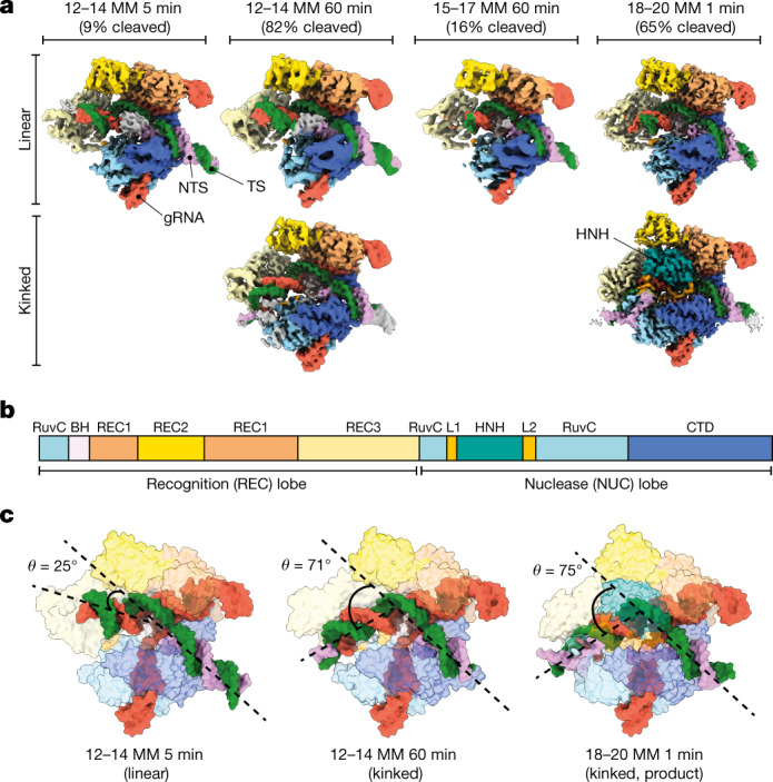
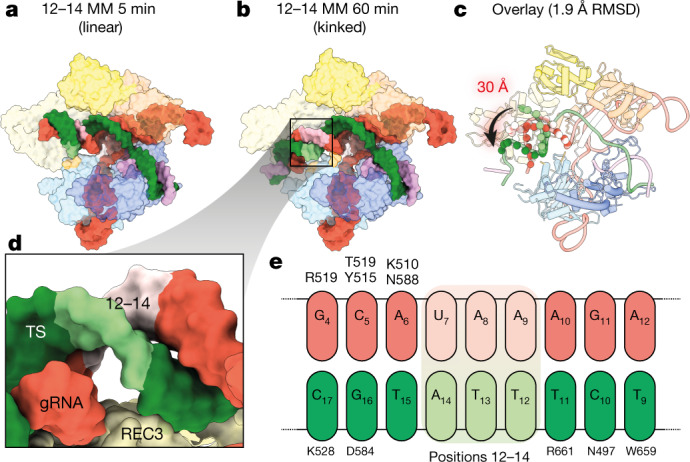
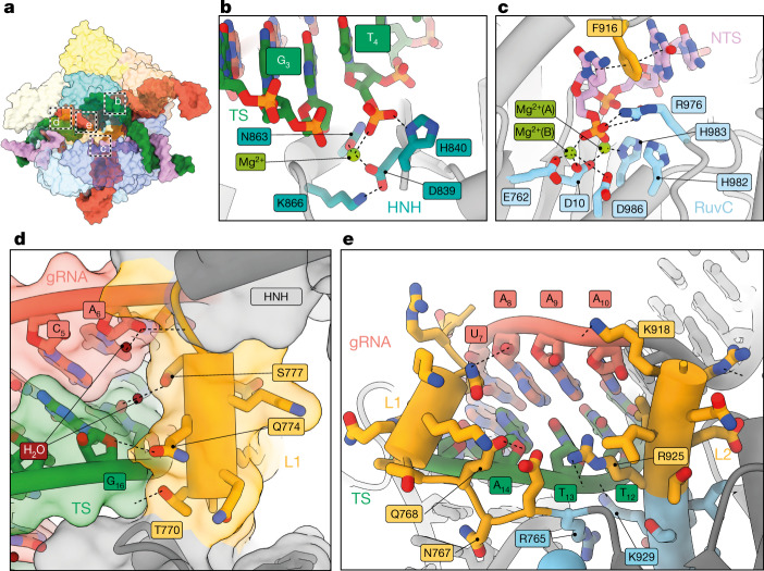
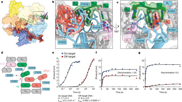
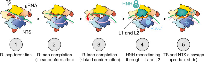
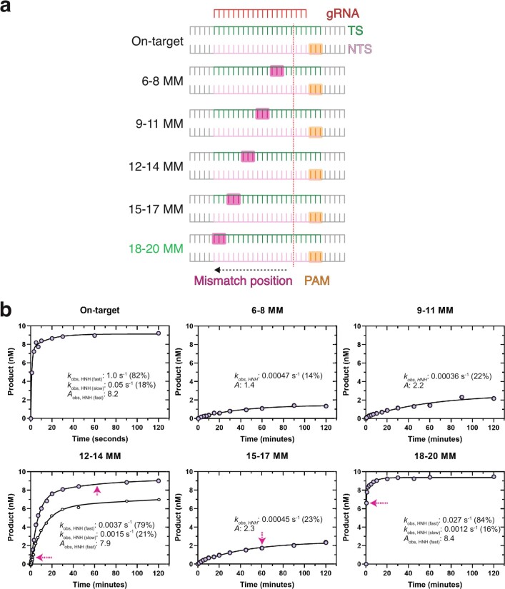
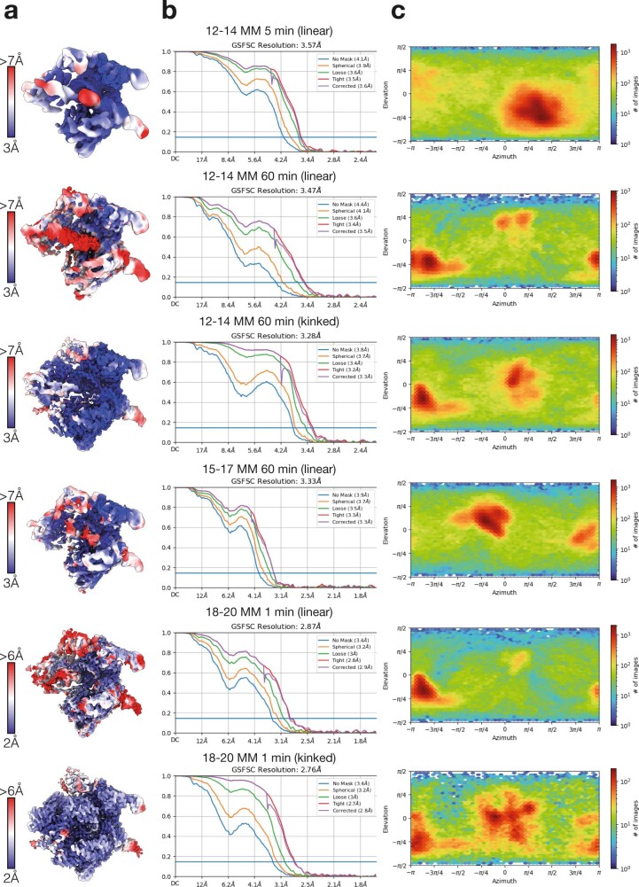
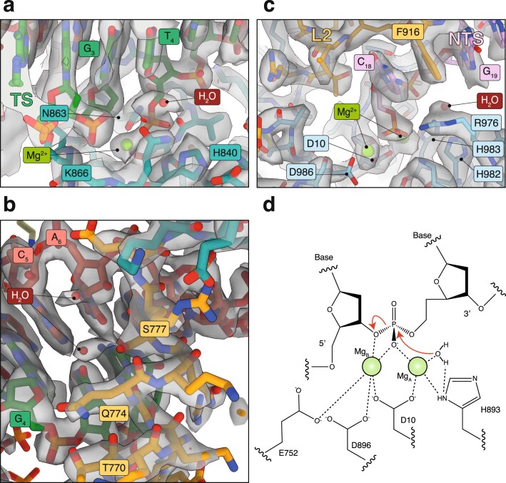
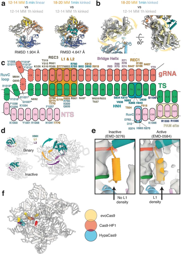
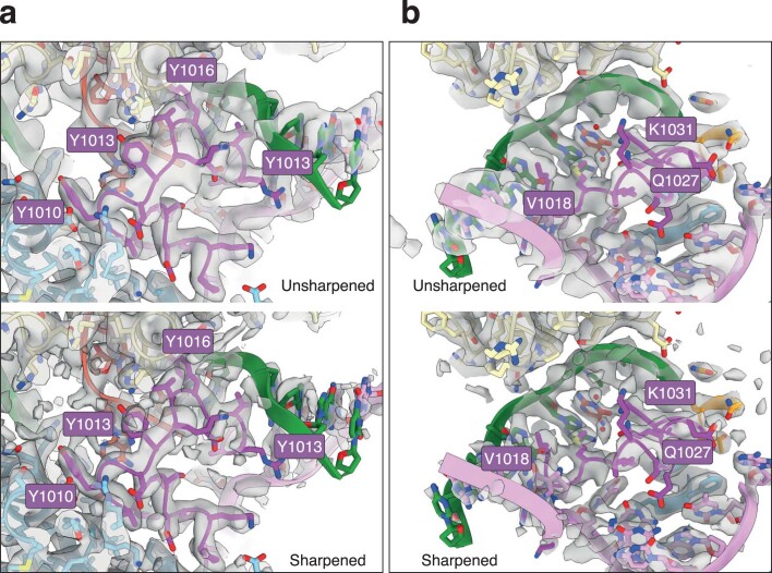
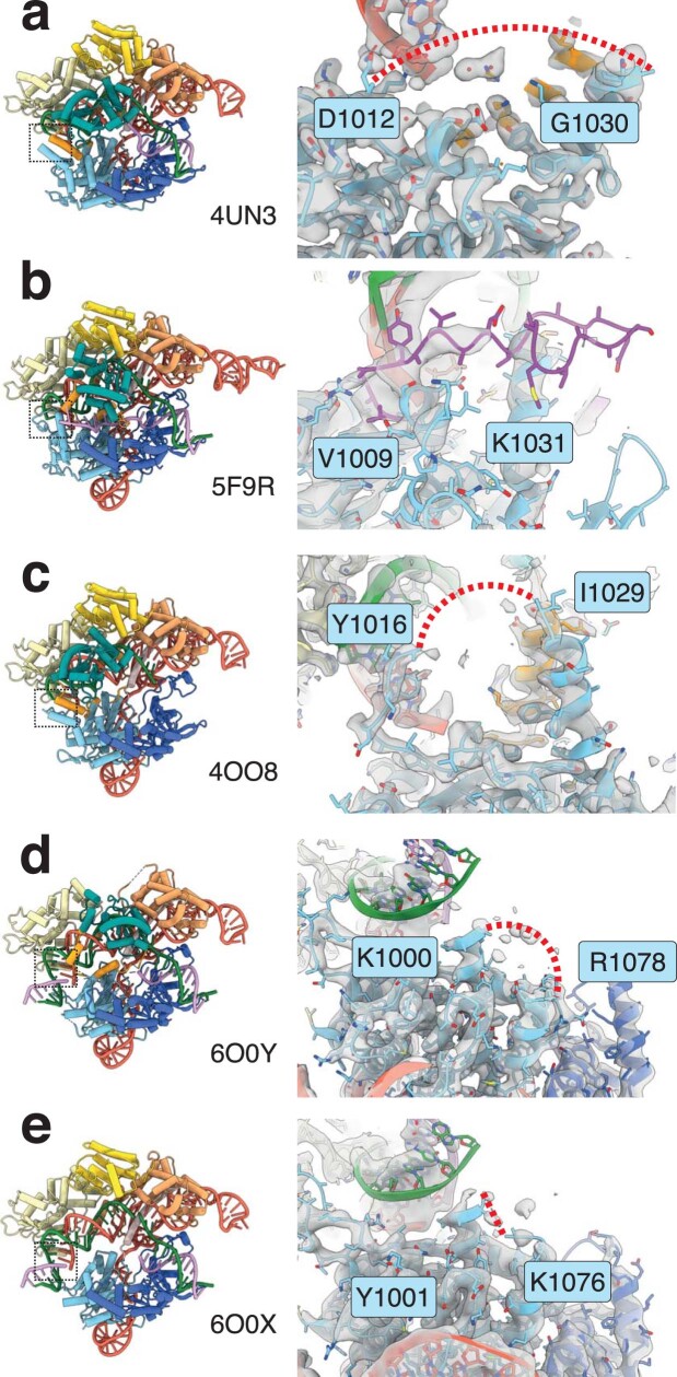
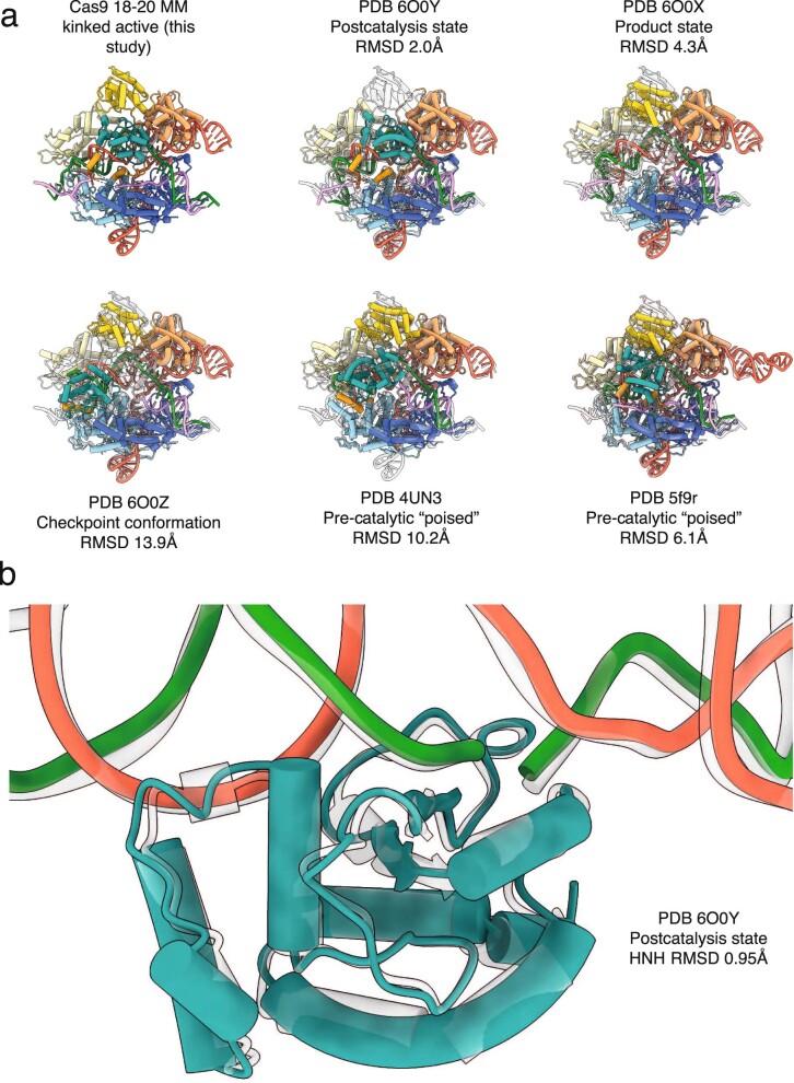
CRISPR-Cas9 as a programmable genome editing tool is hindered by off-target DNA cleavage1-4, and the underlying mechanisms by which Cas9 recognizes mismatches are poorly understood5-7. Although Cas9 variants with greater discrimination against mismatches have been designed8-10, these suffer from substantially reduced rates of on-target DNA cleavage5,11. Here we used kinetics-guided cryo-electron microscopy to determine the structure of Cas9 at different stages of mismatch cleavage. We observed a distinct, linear conformation of the guide RNA-DNA duplex formed in the presence of mismatches, which prevents Cas9 activation. Although the canonical kinked guide RNA-DNA duplex conformation facilitates DNA cleavage, we observe that substrates that contain mismatches distal to the protospacer adjacent motif are stabilized by reorganization of a loop in the RuvC domain. Mutagenesis of mismatch-stabilizing residues reduces off-target DNA cleavage but maintains rapid on-target DNA cleavage. By targeting regions that are exclusively involved in mismatch tolerance, we provide a proof of concept for the design of next-generation high-fidelity Cas9 variants.


Conflict of interest statement

J.P.K.B., M.-S.L., G.N.H., T.L.D., K.A.J. and D.W.T. are inventors on a patent application based on this research titled ‘Methods and compositions for improved Cas9 specificity’ filed by the Board of Regents, The University of Texas System. The US Patent and Trademark Office (USPTO) has assigned US application no. 63/243,481 to this application, and the filing date of 13 September 2021. K.A.J. is the president of KinTek, which provided the chemical-quench flow instruments and the KinTek Explorer software used in this study.

Figures

Fig. 1
Fig. 1. Mismatch-induced Cas9 conformational intermediates.
a, Cryo-EM reconstructions of Cas9 in complex with various partially mismatched DNA substrates, determined at nominal resolutions ranging from 2.8 to 3.6 Å. Cryo-EM structures are coloured according to the domain map for Cas9. Nucleotides are coloured: target strand (TS), green; NTS, pink; and gRNA, red. The fraction of target strand DNA cleaved by Cas9 containing contiguous triple mismatches at the position and time point used for structural determination is shown above each structure. b, Domain organization of SpCas9. CTD, C-terminal domain. c, Models of Cas9 in complex with mismatched DNA substrates shown as isosurface representations. The angle between PAM-proximal and PAM-distal duplexes (θ) is shown. θ is equivalent to around 25º for all linear conformations observed.
Fig. 2
Fig. 2. Positions 12–14 of the gRNA–TS duplex occupy a blind spot for REC3 mismatch detection.
a, b, Structures of 12–14 MM at 5 min (a) and 1 h (b) in linear and kinked conformations, respectively. The position of the 12–14 MM is shown as light green and light pink for the gRNA and the target strand, respectively. Models are shown as isosurface representations. c, Conformational change of the PAM-distal gRNA–TS duplex. The Cas9 protein structure is largely unchanged (root-mean-square deviation (RMSD) of less than 2 Å for equivalent C-alpha atoms), but the PAM-distal gRNA–TS duplex end undergoes a 30 Å conformational change, docking with REC3. d, Close-up view of positions 12–14, showing that because of the phase of the gRNA–TS duplex, REC3 makes no contacts with these base pairs. e, Schematic of interactions between REC3 and positions 9–17 of the gRNA–TS duplex. No interactions occur between Cas9 REC3 and positions 12–14 MM. Position 1 of the duplex is the first base of the target strand that hybridizes with the gRNA spacer.
Fig. 3
Fig. 3. Linkers L1 and L2 mediate the structural transition to the active state.
a, Overview of the 18–20 MM active conformation. b, c, Detailed view of HNH (b) and RuvC (c) active sites. d, Docking of the L1 linker helix against the PAM-distal gRNA–TS duplex, shown as an isosurface representation. e, Interactions of L1 and L2 regions with the minor groove of the gRNA–TS duplex. HNH extending from L1 and L2 linkers has been removed for clarity and does not interact with this region of the gRNA–TS duplex.
Fig. 4
Fig. 4. Stabilization of distorted 18–20 MM by the RuvC domain and improved fidelity of SuperFi-Cas9.
a, Overall structure of the 18–20 MM active conformation viewed from the back. b, c, Magnified views of Cas9 interacting with the distal end of the duplex. Flipped gRNA base position 2 is accommodated by stacking interactions and hydrogen bonding with RuvC tyrosine side-chains, whereas a network of interactions (including a water-mediated hydrogen bond) stabilizes the stretched target strand configuration, which allows TS(20) to resume base-pairing with the NTS. d, Schematic of distorted PAM-distal gRNA–TS duplex. Red circles correspond to water molecules. e, Kinetics of on-target and off-target (18–20 MM) Mg2+-initiated cleavage by the 7-D Cas9 mutant (SuperFi-Cas9). f, g, Cleavage competition assay for wild-type Cas9 (f) and SuperFi-Cas9 (g). 25 nM of either Cas9 variant was mixed with 50 nM of each substrate and the cleaved DNA product was monitored. Discrimination in favour of the on-target DNA is defined by the ratio of amplitudes for on-target and off-target product formed.
Fig. 5
Fig. 5. Model for Cas9 activation.
During R-loop propagation (step 1), the gRNA–TS duplex adopts a linear conformation. After R-loop completion, the PAM-distal end of the linear duplex is captured by REC3 (steps 2 and 3). Mismatches in the PAM-distal region appear to prevent REC3 docking and thereby block subsequent steps of Cas9 activation. Once the kinked R-loop conformation has been formed, L1 and L2 linkers use the gRNA–TS duplex as a scaffold to position the HNH domain at the scissile phosphate of the target strand and to position the NTS in the RuvC site (step 4), which enables Cas9 to make a double-strand break (step 5). According to this model, mutations in the RuvC loop (corresponding to SuperFi-Cas9) inhibit formation of the kinked conformation and subsequent cleavage of the gRNA–TS duplex with mismatches at the PAM-distal end.
Extended Data Fig. 1
Extended Data Fig. 1. Kinetic basis for mismatch discrimination by Cas9.
a, Schematic representation of mismatch constructs used for kinetic analysis. b, Time course of cleavage of on-target and mismatched DNA (10 nM) by Cas9. Magenta arrows correspond to time-points used to prepare cryo-EM samples. Aobs corresponds to amplitude of product formed (i.e. total cleavage). For 12–14 MM, target strand cleavage is shown with larger filled circles, while NTS cleavage is given with smaller open circles. For other mismatches we only show target strand cleavage. We previously reported NTS cleavage data for on-target and 18–20 MM substrates.
Extended Data Fig. 2
Extended Data Fig. 2. Resolution estimates and orientation distributions of cryo-EM maps.
a, Unsharpened maps coloured according to local resolution. b, Gold-standard FSC curves for cryo-EM reconstructions. Resolutions were estimated at FSC=0.143. c, Euler diagrams showing orientation distributions of cryo-EM reconstructions.
Extended Data Fig. 3
Extended Data Fig. 3. Representative cryo-EM densities for 18–20 MM 1-min kinked (product) structure.
a, HNH active site, showing cleaved target strand. b, L1 linker docked on PAM-distal kinked gRNA–TS duplex. Two water molecules are involved within the network of interactions that stabilize the L1 helix conformation. c, RuvC active site, showing cleaved NTS, and positioning of two Mg2+ ions. d, RuvC DNA cleavage mechanism. This is a typical two-metal-ion mechanism as described in and agrees with QM/MM simulations for histidine-mediated activation, and the proposed mechanisms of Cas12j and Cas12i,.
Extended Data Fig. 4
Extended Data Fig. 4. Structural analysis of Cas9.
a, Left, comparison of Cas9 protein only between 12–14 MM 60 min linear (colour) and 12–14 MM 1-h kinked (grey) models. Right, comparison of Cas9 protein only active conformation (18–20 MM 1 min linear, colour) and kinked pre-active (12–14 MM 60 min kinked, grey) models. While there is no significant conformational change associated between transition from linear to kinked pre-active (root-mean standard deviation (RMSD) between equivalent Cα atoms of 1.904 Å), the change from kinked pre-active to active conformations is associated with a larger conformational change (4.647 Å, most of which occurs within the REC3 domain). b, Close-up view of REC3 conformational changes that occur upon activation, as viewed from one angle. REC3 moves forwards towards the kinked duplex by ~15 Å upon activation and HNH repositioning. c, Schematic representation of Cas9–nucleic acid contacts in the context of 18–20 MM. Residues mutated in SuperFi-Cas9 are denoted by an asterisk. d, Conformations of HNH domain (green) and L1 (gold) and L2 (purple) linkers in the context of Cas9 binary complex (i.e. with gRNA, PDB 4ZT0), Cas9–gRNA complex bound to dsDNA in an inactive conformation (PDB 5F9R), and in the active Cas9 18–20 MM structure presented in this work. Upon activation, HNH is repositioned at the target strand cleavage site, driven by large conformational changes in the L1 and L2 linkers. e, Comparison with the active Cas9 18–20 MM structure presented in this work and previously determined cryo-EM maps (transparent grey) of inactive (left, EMD-3276) and active (right, EMD-0584) Cas9 bound to on-target dsDNA. The inactive Cas9 has no density for L1 helix at the kinked distal-docked gRNA–TS site, whereas there is clear density for L1 at this site in the active Cas9 cryo-EM map. f, Mapping of residues mutated to alanine in selected high-fidelity Cas9 variants. EvoCas9 (yellow) – M495, Y515, R661, K526. Cas9-HF1 (red) – N497, R661, Q695, Q926. HypaCas9 (blue) – N692, M694, Q695, H698. Residues shared between Cas9-HF-1 and either EvoCas9 or HypaCas9 are shown as orange and purple, respectively.
Extended Data Fig. 5
Extended Data Fig. 5. Representative cryo-EM density for the RuvC loop.
Two different views are shown (a, b). Unsharpened and B-factor sharpened maps are shown for each view with the RuvC loop shown as dark magenta. Key residues involved in stabilizing this distorted conformation are labelled.
Extended Data Fig. 6
Extended Data Fig. 6. RuvC loop in on-target SpCas9 structures.
a, On-target inactive Cas9 bound to dsDNA (PDB 4UN3). RuvC loop is missing between 1013–1029. b, On-target inactive (primed – HNH rearranged and adjacent to target strand scissile phosphate) Cas9 bound to dsDNA (PDB 5F9R). RuvC loop has been built primarily as alanine ‘stub’ residues, but electron density is very poor and diffuse for this region. c, On-target inactive Cas9 bound to dsDNA (PDB 4OO8). RuvC loop is missing between 1017–1028. d, On-target active Cas9 bound to dsDNA in postcatalysis state. RuvC loop is missing between 1001–1077. e, On-target active Cas9 bound to dsDNA in product state. RuvC loop is missing between 1000–1075. In ac, electron density is displayed as a grey surface, and in d, e cryo-EM density is shown as a grey surface. In all structures, missing residues are depicted as a red dashed line with the RuvC loop in b shown as magenta. Position of RuvC loop is denoted by a black dashed box in the left panel for each model.
Extended Data Fig. 7
Extended Data Fig. 7. Comparison of Cas9 with previous structures.
a, Comparison of 18–20 MM kinked product state Cas9 with a selection of previously determined structures. RMSD between equivalent C-alpha atoms is shown. b, Alignment of HNH from the 18–20 MM kinked product state presented here (transparent grey) and the previously determined ‘post-catalysis’ state (PDB 6O0Y). The catalytically competent HNH conformation between these two structures is highly similar.

Comment in

References

    1. Jinek M, et al. RNA-programmed genome editing in human cells. eLife. 2013;2:e00471. doi: 10.7554/eLife.00471. - DOI - PMC - PubMed
    1. Cong L, et al. Multiplex genome engineering using CRISPR/Cas systems. Science. 2013;339:819–823. doi: 10.1126/science.1231143. - DOI - PMC - PubMed
    1. Fu Y, et al. High-frequency off-target mutagenesis induced by CRISPR–Cas nucleases in human cells. Nat. Biotechnol. 2013;31:822–826. doi: 10.1038/nbt.2623. - DOI - PMC - PubMed
    1. Doudna JA. The promise and challenge of therapeutic genome editing. Nature. 2020;578:229–236. doi: 10.1038/s41586-020-1978-5. - DOI - PMC - PubMed
    1. Liu M, et al. Engineered CRISPR/Cas9 enzymes improve discrimination by slowing DNA cleavage to allow release of off-target DNA. Nat. Commun. 2020;11:3576. doi: 10.1038/s41467-020-17411-1. - DOI - PMC - PubMed

Publication types