. 2022 Mar;603(7901):470-476.
doi: 10.1038/s41586-022-04463-0. Epub 2022 Mar 2.

FSH blockade improves cognition in mice with Alzheimer's disease

Affiliations

FSH blockade improves cognition in mice with Alzheimer's disease

Jing Xiong et al. Nature. 2022 Mar.

Abstract

Alzheimer's disease has a higher incidence in older women, with a spike in cognitive decline that tracks with visceral adiposity, dysregulated energy homeostasis and bone loss during the menopausal transition1,2. Inhibiting the action of follicle-stimulating hormone (FSH) reduces body fat, enhances thermogenesis, increases bone mass and lowers serum cholesterol in mice3-7. Here we show that FSH acts directly on hippocampal and cortical neurons to accelerate amyloid-β and Tau deposition and impair cognition in mice displaying features of Alzheimer's disease. Blocking FSH action in these mice abrogates the Alzheimer's disease-like phenotype by inhibiting the neuronal C/EBPβ-δ-secretase pathway. These data not only suggest a causal role for rising serum FSH levels in the exaggerated Alzheimer's disease pathophysiology during menopause, but also reveal an opportunity for treating Alzheimer's disease, obesity, osteoporosis and dyslipidaemia with a single FSH-blocking agent.


Conflict of interest statement

Competing interests M.Z. is listed as an inventor on issued patents on inhibiting FSH for the prevention and treatment of osteoporosis and obesity: US patent numbers 5,436,285 (1995, awarded to Icahn School of Medicine at Mount Sinai (ISMMS)), 5,674,887 (1997, awarded to ISMMS and University of Pittsburgh), 8,435,948 (2013, awarded to ISMMS) and 11,034,761 (2021, awarded to ISMMS). M.Z. is also listed as an inventor on a pending patent application on composition and use of humanized monoclonal anti-FSH antibodies. These patents are owned by ISMMS, and M.Z. would be recipient of royalties according to institutional policy. M.Z. and K.Y. are listed as inventors of a pending patent application on the use of FSH as a target for preventing Alzheimer’s disease. The latter patent is jointly owned by ISMMS and Emory University, and M.Z. and K.Y. would be recipient of royalties according to institutional policy. The other authors declare no competing interests.

Figures

Extended Data Fig. 1 |
Extended Data Fig. 1 |. Effects of Anti-FSHβ Antibody in Reversing Ovariectomy-Induced Neuropathology in 3xTg Mice.
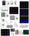
a, Ovariectomized 3xTg mice displayed hypoplastic thread-like uteri and elevated serum FSH and LH levels. FSH-Ab (200 μg/mouse, every 2 days, i.p., 8 weeks) did not alter total serum FSH, LH or 17β-estradiol levels. Statistics: mean ± s.e.m., N = 8 mice per group, one-way ANOVA. b, Immunofluorescent micrographs showing enhanced labelling in the following pairs: amyloid β (Aβ, red) and thioflavin-S (Thio-S, green); Aβ (red) and cleaved APPC586 (green); and pTau (red) and Tau1–368 (green) in the hippocampus after OVX, and its amelioration with FSH-Ab (scale bar, 20 μm). c, Upregulation of Cebpb, Lgmn, App and Mapt in OVX mouse brains, with reversal to near-baseline with FSH-Ab. Statistics: mean ± s.e.m., N = 3 mice per group, one-way AVOVA. d, Immunofluorescence micrographs showing that OVX induces apoptosis (TUNEL, green) in hippocampal NeuN-positive neurons (red); this apoptosis is abolished by FSH-Ab (scale bar, 20 μm). e, Golgi staining on brain sections from CA1 region post-OVX shows a substantial reduction in spine numbers, which is corrected with the FSH-Ab (scale bar, 5 μm). Statistics: mean ± s.e.m., N = 3 mice per group (10 sections), one-way AVOVA. f, Transmission electron micrographic images and quantitative analysis of synapses in hippocampal sections post-OVX treated with IgG or FSH-Ab (scale bar, 1 μm). Statistics: mean ± s.e.m., N = 3 mice per group (8 sections), one-way AVOVA. g, Morris Water Maze testing shows no differences in swim speed. Statistics: mean ± s.e.m., N = 9 mice per group, one-way ANOVA. h, Cognitive testing using the Novel Object Recognition test revealed the absence of significant difference between APP/PS1 and non-transgenic mice in Discrimination Index [(Novel Object Head Entry – Familiar Object Head Entry)/Total Head Entry]; the result is expected at 9 months of age in APP/PS1 mice. Thus, no effect of FSH-Ab was noted at this age, despite the reduction in Aβ40 and Aβ42 accumulation shown in Fig. 1f. Statistics: mean ± s.e.m., mice per group 9, 10, 4 and 4 from left to right; Whisker plot, upper and lower ends of the whiskers show maxima and minima, line in box shows median, and upper and lower box boundaries show 75th and 25th percentile, respectively; unpaired two-tailed Student’s t-test; i, ELISA showing no cross-reactivity of FSH-Ab with LH. j, Western immunoblot showing no change in expression of the LHCGR in whole brain lysates upon OVX or FSH-Ab treatment (N = 2 mice per group). k, IVIS imaging of isolated tissues from mice injected with AlexaFluor750–FSH, i.v., showing localization of FSH in the brain (N = 3 mice per group). l, Western immunoblots of whole brain lysates showing that i.p. injection of human FSH (5 IU) causes an elevation of brain FSH (N = 3 mice per group). m, Immunofluorescence micrographs showing the detection of peripherally injected (i.p.) biotinylated FSH-Ab (red) and biotinylated goat IgG (red) in brain sections (scale bar, 20 μm). Note the absence of cellular or nuclear co-localization with MAP2 or DAPI, respectively. n, Representative PET image shows that 89Zr-labelled humanized monoclonal FSH-Ab (89Zr-Hu6), injected i.v., is localized to live brain (arrows). γ-counting in perfused tissue shows presence of 89Zr-Hu6 in dissected brain tissue at 24 and 48 h post-injection (N = 4 mice). o, IVIS imaging and quantitation with AlexaFluor750-labelled Hu6, given i.v. shows localization in perfused whole brain tissue; N = 3 mice per group. Control (Ctrl): phosphate-buffered saline (PBS). p, Confirmatory immunofluorescence on the same mice (o) using anti-human IgG showing Hu6 localization (red) in proximity to CD31+ endothelial cells (green) (scale bar, 100 μm). For gel source data, see Supplementary Fig. 1.
Extended Data Fig. 2 |
Extended Data Fig. 2 |. FSHR Activation Triggers Amyloidogenic Protein Accumulation.
a, Western immunoblots showing the effect of activating neuronal FSHRs by FSH (30 ng/mL) in human SH-SY5Y and primary rat neuronal cells on the expression of C/EBPβ, AEP, as well as the cleavage of amyloid precursor protein (APP) and Tau using antibodies noted in ‘Methods’. FSH (30 ng/mL) likewise stimulated the expression of CEBPB, LGMN, APP and MAPT (qPCR) (b); AEP activity (c); and certain inflammatory cytokines (ELISA), namely IL-6 and IL-1β (d). Statistics: mean ± s.e.m.; Mice per group, (b) 3, (c) 6, and (d) 6; one-way ANOVA. e, Western immunoblotting showing C/EBPβ, AEP, APP, cleaved APP1–585, total Tau, and cleaved Tau1–368 in response to FSH or PBS following transfection with human FSHR siRNA (si-FSHR) for SH-SY5Y cells or rat Fshr siRNA (si-Fshr) for primary rat neurons, or appropriate scrambled siRNAs. f, mRNA levels of CEBPB, LGMN, APP and MAPT in SH-SY5Y cells incubated with FSH after control or si-FSHR transfection. g, AEP activity after incubation with FSH in control or si-FSHR-transfected SH-SY5Y cells. h, IL-6 and IL-1β levels (ELISAs) in SH-SY5Y cells incubated with FSH following control or si-FSHR infection. Statistics: mean ± s.e.m.; (f) 3 biological replicates; (g, h) 6 mice per group; one-way ANOVA.
Extended Data Fig. 3 |
Extended Data Fig. 3 |. FSH Induces APP and Tau Cleavage Through C/EBPβ and AEP/δ-Secretase Activation in Human SH-SY5Y Cells and Rat Cortical Neurons.
a, Western immunoblots showing the effect of FSH (30 ng/mL) on Tau, APP, AEP and FSHR of knocking down C/EBPβ expression by lentiviral infection with shRNA-Cebpb (sh-Cebpb) or reducing δ-secretase activity by adeno-associated virus infection of AEPC189S in both human SH-SY5Y cells and rat cortical neurons. The stimulatory action of FSH was reversed at 48 h. b, c, Effect of FSH (30 ng/mL) on APP, APPC586, pTau and Tau1–368 accumulation (immunofluorescence, scale bar, 40 μm, b) and AEP activity (c) in rat cortical neurons infected with sh-Cebpb or AAV-AEPC189S. Statistics: (c) mean ± s.e.m.; N = 6 mice per group; one-way ANOVA. d, Western immunoblots showing the time course of FSH effects on C/EBPβ, phosphorylated C/EBPβ (pC/EBPβ), AEP, pAEPS226, total AKT, pAKTS473, total ERK1/2, pERK1/2, total SRPK2, pSRPK2T492 and pNFκB-p65. e, Western immunoblots showing the effect of a 30-minute incubation with FSH (30 ng/mL) on levels of C/EBPβ, AEP, pAEPS226, total AKT and pAKTS473, total ERK1/2 and pERK1/2, total SRPK2 and pSRPK2T492 in the presence or absence of the cAMP inhibitor SQ22536 (100 μM), Gαi inhibitor pertussis toxin (PTX, 50 ng/ml), AKTi-1/2 inhibitor (10 μM) and ERK1/2 inhibitor PD98059 (10 μM).
Extended Data Fig. 4 |
Extended Data Fig. 4 |. Targeted Knockdown of Fshr in the Hippocampus Diminishes AD Pathologies.
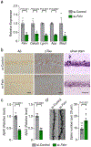
a, Quantitative PCR shows significantly reduced expression of Fshr, Cebpb, Lgmn, App and Mapt. b, Immunohistochemistry of the hippocampus shows reduced accumulation of Aβ and pTau, as well as of proteinaceous deposits (silver staining) in si-Fshr-injected OVX mice (scale bar, 50 μm). c, The two isoforms of Aβ, namely Aβ40 and Aβ42, were also reduced. d, Notable is the marked increase in dendritic spines (Golgi staining) (scale bar, 5 μm). Statistics: mean ± s.e.m., (a) 3 biological replicates; (c) 5 mice per group; (d) 10 sections from 3 mice per group; unpaired two-tailed Student’s t-test.
Extended Data Fig. 5 |
Extended Data Fig. 5 |. Recombinant FSH Triggers AD Pathology in 3xTg Mice.
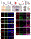
a, Serum FSH levels—both mouse (endogenous) and human (exogenous)—24 h after i.p. injection of 2, 5 or 10 IU human recombinant FSH. b, Serum LH levels also shown. Female 3xTg mice were injected with recombinant FSH (5 IU per mouse, daily, i.p., 3 months). c, Immunohistochemistry for Aβ or pTau in hippocampus post-FSH injection (scale bar, 50 μm). d, Silver staining of the prefrontal cortex, and hippocampal CA1 and dentate gyrus (DG) regions showing enhanced proteinaceous deposits in FSH-injected mice (scale bar, 50 μm). e, Brain mRNA levels of Cebpb, Lgmn, App and Mapt. f, Golgi staining of brain sections from the CA1 region shows reduced spine numbers in FSH-injected mice (scale bar, 5 μm). g, Transmission electron micrographs of hippocampal sections showing reduced synapse numbers post-FSH (scale bar, 1 μm). Immunofluorescence micrographs showing the following image pairs in the hippocampus and/or cortex post-FSH: (h) Aβ (red) and cleaved APPC586 (green); (i) pTau (red) and cleaved Tau1–368 (green); (j) Aβ (red) and thioflavin-S (green); and (k) NeuN (red) and TUNEL (green) (scale bar, 20 μm). l, Immunofluorescence showing co-localization of C/EBPβ, AEP, Aβ and pTau to NeuN-positive neurons upon FSH stimulation [10x (scale bar, 300 μm) and 40x (scale bar, 50 μm) magnifications]. Statistics: mean ± s.e.m., (a, b) 3 mice per group; (e) 3 biological replicates, (f, g) 10 sections from 3 mice per group; unpaired two-tailed Student’s t-test.
Extended Data Fig. 6 |
Extended Data Fig. 6 |. Effect of Recombinant FSH in Triggering AD Pathology in Ovariectomized 3xTg Mice With Oestrogen Replacement.
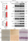
3xTg mice were ovariectomized at 3 months and supplemented with 17β-estradiol using 90-day-release pellets (E2, 0.36 mg) to render them biochemically eugonadal. The mice were randomly divided to be injected with PBS or recombinant human FSH (5 IU per mouse, daily, i.p., 3 months). a, Serum level of FSH and 17β-estradiol. b, Western immunoblotting showing increased C/EBPβ, AEP, cleaved APP1–373 and APP1–585, total Tau, cleaved Tau1–368 and pTau in the brain after FSH injection. c, Brain AEP enzymatic activity also shown. d, Immunohistochemistry of the hippocampus shows increased expression of Aβ and pTau in the FSH group. Silver staining showed increased proteinaceous deposits in FSH-treated mice (scale bar, 50 μm). Statistics: mean ± s.e.m., mice per group; (a) 4 and (c) 5; unpaired two-tailed Student’s t-test.
Extended Data Fig. 7 |
Extended Data Fig. 7 |. Effect of Recombinant FSH in Triggering AD Pathology and Cognitive Decline in Male Mice.
Male 3xTg mice were injected with recombinant FSH at 5 IU per mouse daily, i.p. for 3 months. a, Western immunoblots showing increased C/EBPβ, AEP, cleaved APP1–373 and APP1–585, total Tau, cleaved Tau1–368 and pTau in the brain (3 mice per group). b, c, Brain AEP activity (b) and Aβ isoforms, Aβ40 and Aβ42 (c) were also increased with FSH. d, Morris Water Maze test shows enhanced escape latency to mount the platform (seconds). Also shown are integrated escape latency (area under the curve, AUC) and percentage of time spent in the target quadrant (Probe Trial Test). e, Silver staining of the prefrontal cortex, and hippocampus CA1 and dentate gyrus (DG) regions showing enhanced proteinaceous deposits in FSH-injected mice (scale bar, 50 μm). f, Immunohistochemistry for Aβ or pTau in the hippocampus post-FSH injection (scale bar, 50 μm). g, Brain mRNA levels of Cebpb, Lgmn, App and Mapt. h, Golgi staining of brain sections from the CA1 region of the hippocampus showing reduced spine numbers in FSH-injected mice (scale bar, 5 μm). i, Transmission electron micrographs of hippocampal sections showing reduced synapse numbers post-FSH (scale bar, 1 μm). Immunofluorescence micrographs showing the following image pairs: (j) Aβ (red) and cleaved APPC586 (green); (k) pTau (red) and cleaved Tau1–368 (green); (l) Aβ (red) and thioflavin-S (green); and (m) NeuN (red) and TUNEL (green) in the hippocampus and/or cortex of male 3xTg mice after FSH (scale bar, 20 μm). Statistics: mean ± s.e.m., mice per group, (b, c) 5, (d) 7, (g) 3, (h, i), 3 (10 sections); unpaired two-tailed Student’s t-test.
Extended Data Fig. 8 |
Extended Data Fig. 8 |. Effect of FSH in Triggering AD Pathology and Cognitive Decline in Female Wild Type and APP-KI Mice.
In APP-KI mice, three amino acid substitutions (G601R;F606Y;R609H) are knocked into Aβ-coding exon 14 of the APP gene—this results in the non-transgenic expression at basal levels of oligomerizable human Aβ. Female wild type and APP-KI mice were injected with recombinant FSH (5 IU, daily, i.p. 3 months). a–d, In wild type mice, Western immunoblotting showing increased C/EBPβ, AEP, cleaved APP1–373 and APP1–585, total Tau, and cleaved Tau1–368 in whole brain (3 mice per group) (a), as well as increased silver staining (b), elevated AEP activity (c) and increases Aβ isoforms, Aβ40 and Aβ42 (d) upon FSH treatment. e, Morris Water Maze test, however, showed no difference in escape latency to mount the platform (seconds). Also shown are no differences in integrated escape latency (area under the curve, AUC) and percentage of time spent in the target quadrant (Probe Trial Test). f–I, Western immunoblotting showing elevations in C/EBPβ, AEP, cleaved APP1–373 and APP1–585, and cleaved Tau1–368 in whole brain (f), along with enhancements in silver staining (g), AEP activity (h), and Aβ isoforms (i) in APP-KI mice in response to FSH injection. j, There was also a significant spatial memory deficit on the Morris Water Maze test. k, Immunofluorescence micrographs showed increases in the hippocampus and/or cortex of female APP-KI mice post-FSH injected in the following pairs: Aβ (red) and cleaved APPC586 (green); Aβ (red) and thioflavin-S (green); and NeuN (red) and TUNEL (green). Scale bar: (b, g) 50 μm, (k) 100 μm (magnified view, 10 μm). Statistics: mean ± s.e.m.; mice per group, (c, d, h, i) 5, (e, j) 8; unpaired two-tailed Student’s t-test.
Extended Data Fig. 9 |
Extended Data Fig. 9 |. C/EBPβ Mediates FSH-Induced AD Neuropathology and Cognitive Decline in 3xTg Mice.
a, Cebpb, Lgmn, App and Mapt mRNA expression following FSH injection to 3xTg or Cebpb+/− 3xTg mice. Statistics: mean ± s.e.m., N = 3 biological replicates, one-way ANOVA. b, Immunohistochemistry for Aβ and pTau and silver staining for proteinaceous deposits (scale bar, 50 μm). c–e, Immunofluorescence staining for Aβ (red) and C/EBPβ (green) (c) and for pTau (red) and C/EBPβ (green) (d) (scale bar, 20 μm), and Golgi staining for dendritic spines (e) in the hippocampus in female 3xTg or Cebpb+/− 3xTg mice, post-FSH (scale bar, 5 μm). Statistics: mean ± s.e.m., 10 sections from 3 mice per group, one-way ANOVA. f, Morris Water Maze testing showed no difference in swim speed. Statistics: 9 mice per group, one-way ANOVA.
Extended Data Fig. 10 |
Extended Data Fig. 10 |. C/EBPβ Mediates Ovariectomy-Induced AD Neuropathology and Cognitive Decline in 3xTg Mice.
a, Cebpb, Lgmn, App and Mapt mRNA expression following ovariectomy of 3xTg or Cebpb+/− 3xTg mice. Statistics: mean ± s.e.m., 3 biological replicates, one-way ANOVA. b, Immunohistochemistry for Aβ and pTau and silver staining for proteinaceous deposits (scale bar, 50 μm). c–e, Immunofluorescence staining for Aβ (red) and C/EBPβ (green) (c) and for pTau (red) and C/EBPβ (green) (d) (scale bar, 20 μm), and Golgi staining for dendritic spines (e) in the hippocampus in female 3xTg or Cebpb+/− 3xTg mice (scale bar, 5 μm), post-OVX. Statistics: mean ± s.e.m., 10 sections from 3 mice per group, one-way ANOVA. f, Morris Water Maze test showed no difference in the swim speed between 3xTg and Cebpb+/− 3xTg mice. Statistics: left to right: 7, 8, 8 mice per group, one-way ANOVA.
Fig. 1 |
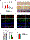
Fig. 1 |. FSH-blocking antibody reverses AD neuropathology and cognitive decline in Alzheimer’s mice.
ae, The effects of FSH-blocking antibody (FSH-Ab) or goat IgG after ovariectomy (OVX) or sham operation (Sham) of 3xTg mice (aged 3 months) on hippocampal Aβ, pTau and proteinaceous deposits (a); Aβ40 and Aβ42 (b); C/EBPβ, AEP, cleaved APP, Tau and pTau (c); AEP activity (d); and parameters of spatial memory determined using Morris water maze testing (e). For a, scale bars, 50 μm. f, Separate experiments show the effect of FSH-Ab or goat IgG given to male APP/PS1 mice (aged 5 months) over 4 months on hippocampal and cortical Aβ40 and Aβ42. Data are mean ± s.e.m. n = 6 (b and d); n = 9 (e); and, from left to right, n = 9, n = 10, n = 4 and n = 4 (f) mice per group. Statistical analysis was performed using one-way analysis of variance (ANOVA) (b, d and e) or unpaired two-tailed Student’s t-tests (f). Gel source data are provided in Supplementary Fig. 1.
Fig. 2 |
Fig. 2 |. Neuronal FSH receptors in mouse and human brain.
ac, Fshr expression in human cortex, neuroblastoma cells (SH-SY5Y), mouse cortex and hippocampus, rat cortical neurons and/or mouse ovaries, determined by end-point PCR (a), qPCR (b) and/or western blotting (c). d, RNAscope signals (red arrows) in haematoxylin-stained cells from testes, and Nissl-stained neurons in the hippocampal dentate gyrus and entorhinal cortex of Fshr+/+ and Fshr−/− mice. Scale bars, 50 μm. e, Transcript counts and density in multiple brain regions from 34 sections. f, Co-staining of FSHR, NeuN, GFAP and IBA1 in the hippocampus after stereotactic siControl or siFshr injection in wild-type mice or in uninjected Fshr−/− mice. Scale bars, 100 μm (main images), 10 μm (magnified images). g, ViewRNA signals (FSHR, dark blue; MALAT1, red) in cells from the hippocampal dentate gyrus (granular layer) (left) and parahippocampal cortex (layers V–VI) (right) from post-mortem human brain. Scale bars, 500 μm (left, main image), 50 μm (left, inset), 200 μm (right, main image), 20 μm (right, inset). For b, data are mean ± s.e.m. The number of mice per group is shown.
Fig. 3 |
Fig. 3 |. Targeted Fshr knockdown in the hippocampus ameliorates AD neuropathology and impaired spatial memory.
ae, The effect of stereotactic injection of siFshr versus siControl into ovariectomized 3xTg mice on total C/EBPβ, AEP, cleaved APP and Tau, and FSHR levels in the whole brain (a); AEP activity (b); synapse number (transmission electron micrographs, red arrows) (c); cell viability (NeuN-positivity and TUNEL) (d); and parameters of spatial memory determined using Morris water maze testing (e). Scale bars, 1 μm (c), 20 μm (d). Data are mean ± s.e.m. n = 3 (a), n = 5 (b), n = 3 (10 sections) (c), and n = 7 or n = 8 (e) mice per group. Statistical analysis was performed using unpaired two-tailed Student’s t-tests.
Fig. 4 |
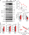
Fig. 4 |. Recombinant FSH induces AD pathologies and cognitive decline in 3xTg Mice.
ad, The effect of injecting female 3xTg mice with recombinant human FSH on whole-brain C/EBPβ, AEP, cleaved APP and Tau proteins (a); AEP activity (b); Aβ40 and Aβ42 (c); and parameters of spatial memory determined using Morris water maze testing (d). e, The effect of FSH on long-term potentiation (LTP) shown as fEPSPs before (grey) and 60 min after (red) theta-burst stimulation. Data are mean ± s.e.m. n = 5 (b and c), n = 7 (d) and n = 4 (e) mice per group. Statistical analysis was performed using unpaired two-tailed Student’s t-tests.
Fig. 5 |
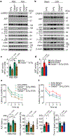
Fig. 5 |. FSH-induced AD pathology is dampened in Cebpb+/− 3xTg mice.
af, The effect of injecting recombinant human FSH (a, c and e) or ovariectomy (b, d, f) in 3xTg mice or compound mutant Cebpb+/− 3xTg mice on C/EBPβ, AEP, cleaved APP, cleaved Tau, pTau and FSHR (a, b); AEP activity (c, d); and parameters of spatial memory determined using Morris water maze testing (e, f). Data are mean ± s.e.m. n = 6 (c), n = 5 (d), n = 9 (e), and n = 7 or n = 8 (f) mice per group. Statistical analysis was performed using one-way ANOVA.

Comment in

References

    1. Sowers M et al. Changes in body composition in women over six years at midlife: ovarian and chronological aging. J. Clin. Endocrinol. Metab 92, 895–901 (2007). - PMC - PubMed
    1. Sowers MR et al. Hormone predictors of bone mineral density changes during the menopausal transition. J. Clin. Endocrinol. Metab 91, 1261–1267 (2006). - PubMed
    1. Guo Y et al. Blocking FSH inhibits hepatic cholesterol biosynthesis and reduces serum cholesterol. Cell Res. 29, 151–166 (2019). - PMC - PubMed
    1. Han X et al. A novel follicle-stimulating hormone vaccine for controlling fat accumulation. Theriogenology 148, 103–111 (2020). - PubMed
    1. Ji Y et al. Epitope-specific monoclonal antibodies to FSHβ increase bone mass. Proc. Natl Acad. Sci. USA 115, 2192–2197 (2018). - PMC - PubMed

Publication types

Substances