Review
. 2022 Feb 14:13:782199.
doi: 10.3389/fphar.2022.782199. eCollection 2022.

Aryl Hydrocarbon Receptor Mechanisms Affecting Chronic Kidney Disease

Affiliations
Review

Aryl Hydrocarbon Receptor Mechanisms Affecting Chronic Kidney Disease

Colleen S Curran et al. Front Pharmacol. .

Abstract

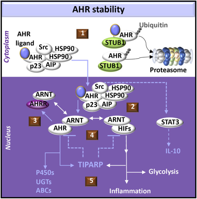
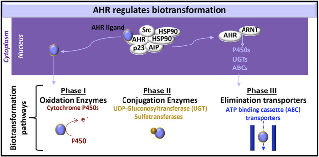
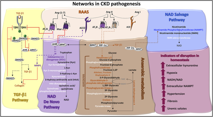
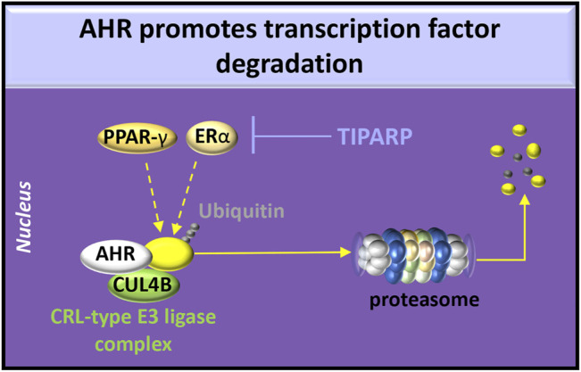
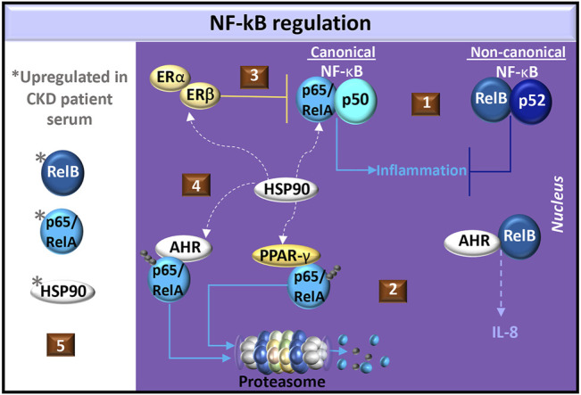
The aryl hydrocarbon receptor (AHR) is a basic helix-loop-helix transcription factor that binds diverse endogenous and xenobiotic ligands, which regulate AHR stability, transcriptional activity, and cell signaling. AHR activity is strongly implicated throughout the course of chronic kidney disease (CKD). Many diverse organic molecules bind and activate AHR and these ligands are reported to either promote glomerular and tubular damage or protect against kidney injury. AHR crosstalk with estrogen, peroxisome proliferator-activated receptor-γ, and NF-κB pathways may contribute to the diversity of AHR responses during the various forms and stages of CKD. The roles of AHR in kidney fibrosis, metabolism and the renin angiotensin system are described to offer insight into CKD pathogenesis and therapies.

Keywords: PPAR γ; RAAS; TGF—β1; aryl hydrocarbon (Ah) receptor; hypoxia; kynurenine.


Conflict of interest statement

The authors declare that the research was conducted in the absence of any commercial or financial relationships that could be construed as a potential conflict of interest.

Figures

FIGURE 1
FIGURE 1
AHR-initiated cell signaling pathways. (1) The aryl hydrocarbon receptor (AHR) forms a complex with chaperone molecules in the cytoplasm. Ligand binding may promote transport of the complex into the nucleus. Alternatively, release of AHR from the complex may promote interactions with an E3 ligase (e.g., STUB1) which acts as a platform for AHR ubiquitination and targeted degradation via the proteasome. Both ligand-bound and unliganded AHR can be targeted to the proteasome. (2) AHR dissociates from its cytoplasmic complex to bind the AHR nuclear translocator (ARNT), which alternatively may dimerize with hypoxia inducible transcription factors (HIFs). AHR induces activation of signal-transducer and activator of transcription (STAT3) through Src signaling, acting as a tyrosine protein kinase involved in the production of IL-10. (3) AHR promotes the expression of the AHR repressor (AHRR), which also dimerizes with ARNT and negatively regulates AHR functions by competing with AHR binding sites in DNA regulatory sequences. (4) Activation of AHR or HIFs induces transcription and translation of PARP7 (poly ADP-ribosyl transferase 7), also known as TIPARP (TCDD-inducible poly-ADP-ribose polymerase). TIPARP further promotes polyADP-ribosylation and subsequent degradation of AHR and HIFs (5) Downstream responses of AHR can include activation of the biotransformation enzymes, including cytochrome P450 enzymes (P450s), UDP-gluconosyltransferases (UGTs), and ATP-binding cassette transporters (ABCs). Downstream responses of HIFs can include increased glycolysis and the expression of immunomodulatory genes that provoke inflammation.
FIGURE 2
FIGURE 2
AHR-initiated biotransformation. AHR induces the transcription of certain genes whose products are involved in each of the three phases of drug metabolism.
FIGURE 3
FIGURE 3
Networks in CKD pathogenesis. TGF-β1 pathway: TGF-β1 signaling is initiated through serine/threonine kinase receptors, TGF-β1 receptor (TβR)-I and TβRII. TGF-β binding to TβRII recruits TβRI to form a receptor heterodimer, which is phosphorylated. SMAD2 and SMAD3 are recruited to the receptor heterodimer and are also phosphorylated. SMAD2 and SMAD3 co-localize with SMAD4 and translocate to the nucleus to activate genes, such as collagen and TGF-β1. ACE2 activity promotes the production of SMAD7, potentially via Ang-(1–7)-induced Mas or AT2R receptor signals. SMAD7 is a negative regulator of TGF-β1 by recruiting E3 ligases to TβRI and blocking TβRI-induced SMAD2/3 phosphorylation. Ligand activated AHR antagonizes TGF-β1 and collagen gene expression and protein production, which are associated with fibrosis. Ligand activated AHR also induces the degradation of collagen through the production of matrix metalloproteinase-1 (MMP1). Renin angiotensin aldosterone system (RAAS): Angiotensin I (Ang I) is cleaved by angiotensin converting enzyme (ACE) into Ang II. The binding of Ang II to AT1R or AT2R promotes the stable expression of HIF-1α and hypoxic responses. The ACE homolog, ACE2, inactivates Ang II by cleaving and processing Ang I and Ang II into Ang-(1–7), which is a ligand for the Mas receptor and AT2R. AHR regulates the expression of ACE2 and Mas. AT2R activation promotes TβRII degradation, inhibiting TGF-β1 signals. NAD de novo biosynthesis pathway (also known as the kynurenine pathway): Tryptophan catabolism is the defining feature of this pathway. The rate limiting enzymes are indoleamine dioxygenase (IDO) and tryptophan dioxygenase (TDO). IDO1 is induced by AHR:ARNT transcriptional activation of the IDO promoter and promotes the production of kynurenine, which can be released as a cytokine. A series of additional enzymes (highlighted in white) catalyze the production of immunomodulatory and neuroregulatory molecules that are further processed into NAD. Anaerobic metabolism: The production of adenosine triphosphate (ATP) in the absence of oxygen occurs through enzymatic reactions in glycolysis and results in the production of lactate and the increased formation of NADH relative to NAD. Enzymes in this pathway are regulated by HIF-1α activated genes (highlighted in orange), which can be stabilized by the RAAS. NAD salvage pathway: The primary source of mammalian NAD is from the recycling nicotinamide, which is the amide version of vitamin B3 and a by-product from the enzymatic activity of Poly-ADP-ribose polymerases (PARPs) and sirtuins. The rate limiting enzyme is nicotinamide phosphoribosyltransferase (NAMPT), which is transcriptionally activated by HIF-2α and functions as an extracellular cytokine. Indicators of disruption in homeostasis: Factors and conditions along these pathways are induced during CKD pathogenesis.
FIGURE 4
FIGURE 4
AHR degradative functions. AHR participates in the cullin/RING ubiquitin ligase (CRL-type E3 ligase) complex involving chaperones [e.g., cullin 4b (CUL4B)] to promote ubiquitination of estrogen receptor (ER)-α or peroxisome proliferator-activated receptor (PPAR)-γ. Activation of AHR or HIFs induces transcription and translation of PARP7 (poly ADP-ribosyl transferase 7), also known as TIPARP (TCDD-inducible poly-ADP-ribose polymerase). TIPARP promotes polyADP-ribosylation and subsequent degradation of ER-α.
FIGURE 5
FIGURE 5
Regulation of NF-κB in CKD. (1) Common NF-κB dimer pairs include p65/RelA and p50 in the canonical pathway and RelB and p52 in the non-canonical pathway. Non-canonical cell signals may antagonize canonical cell signals in fibroblasts. (2) AHR dimerizes with RelB in the production of IL-8. AHR or PPAR-γ activation promotes ubiquitination and degradation of p65/RelA. (3) Estrogen activates estrogen receptors (ER-α/β) to produce the NF-κB inhibitor, IκBα, and promotes the recruitment of ER-β to p65 binding sites, which blocks p65 transcriptional activity. (4) Heat shock protein (HSP)-90 is a chaperone shared by ER-α/β, PPAR-γ, AHR, and canonical NF-κB. (5) Elevated levels of p65/RelA, RelB, and HSP90 are found in CKD patient serum.

Similar articles

Cited by

References

    1. Addi T., Poitevin S., McKay N., El Mecherfi K. E., Kheroua O., Jourde-Chiche N., et al. (2019). Mechanisms of Tissue Factor Induction by the Uremic Toxin Indole-3 Acetic Acid through Aryl Hydrocarbon Receptor/nuclear Factor-Kappa B Signaling Pathway in Human Endothelial Cells. Arch. Toxicol. 93 (1), 121–136. 10.1007/s00204-018-2328-3 - DOI - PubMed
    1. Almaimani M., Sridhar V. S., Cherney D. Z. I. (2021). Sodium-glucose Cotransporter 2 Inhibition in Non-diabetic Kidney Disease. Curr. Opin. Nephrol. Hypertens. 30, 474–481. 10.1097/MNH.0000000000000724 - DOI - PubMed
    1. Angelos M. G., Kaufman D. S. (2018). Advances in the Role of the Aryl Hydrocarbon Receptor to Regulate Early Hematopoietic Development. Curr. Opin. Hematol. 25 (4), 273–278. 10.1097/MOH.0000000000000432 - DOI - PubMed
    1. Asai H., Hirata J., Watanabe-Akanuma M. (2018). Indoxyl Glucuronide, a Protein-Bound Uremic Toxin, Inhibits Hypoxia-Inducible Factor‒dependent Erythropoietin Expression through Activation of Aryl Hydrocarbon Receptor. Biochem. Biophys. Res. Commun. 504 (2), 538–544. 10.1016/j.bbrc.2018.09.018 - DOI - PubMed
    1. Assefa E. G., Yan Q., Gezahegn S. B., Salissou M. T. M., He S., Wu N., et al. (20192019). Role of Resveratrol on Indoxyl Sulfate-Induced Endothelial Hyperpermeability via Aryl Hydrocarbon Receptor (AHR)/Src-Dependent Pathway. Oxid Med. Cel Longev 2019, 5847040. 10.1155/2019/5847040 - DOI - PMC - PubMed

LinkOut - more resources