. 2022 Feb 14:13:827048.
doi: 10.3389/fimmu.2022.827048. eCollection 2022.

Antigen-Specific CD4+ T-Cell Activation in Primary Antibody Deficiency After BNT162b2 mRNA COVID-19 Vaccination

Affiliations

Antigen-Specific CD4+ T-Cell Activation in Primary Antibody Deficiency After BNT162b2 mRNA COVID-19 Vaccination

Kai M T Sauerwein et al. Front Immunol. .

Abstract

Previous studies on immune responses following COVID-19 vaccination in patients with common variable immunodeficiency (CVID) were inconclusive with respect to the ability of the patients to produce vaccine-specific IgG antibodies, while patients with milder forms of primary antibody deficiency such as immunoglobulin isotype deficiency or selective antibody deficiency have not been studied at all. In this study we examined antigen-specific activation of CXCR5-positive and CXCR5-negative CD4+ memory cells and also isotype-specific and functional antibody responses in patients with CVID as compared to other milder forms of primary antibody deficiency and healthy controls six weeks after the second dose of BNT162b2 vaccine against SARS-CoV-2. Expression of the activation markers CD25 and CD134 was examined by multi-color flow cytometry on CD4+ T cell subsets stimulated with SARS-CoV-2 spike peptides, while in parallel IgG and IgA antibodies and surrogate virus neutralization antibodies against SARS-CoV-2 spike protein were measured by ELISA. The results show that in CVID and patients with other milder forms of antibody deficiency normal IgG responses (titers of spike protein-specific IgG three times the detection limit or more) were associated with intact vaccine-specific activation of CXCR5-negative CD4+ memory T cells, despite defective activation of circulating T follicular helper cells. In contrast, CVID IgG nonresponders showed defective vaccine-specific and superantigen-induced activation of both CD4+T cell subsets. In conclusion, impaired TCR-mediated activation of CXCR5-negative CD4+ memory T cells following stimulation with vaccine antigen or superantigen identifies patients with primary antibody deficiency and impaired IgG responses after BNT162b2 vaccination.

Keywords: CXCR5-negative CD4+ memory T cells; activation induced marker assay; circulating follicular T helper cells; common variable immunodeficiency; primary immunoglobulin isotype deficiency; surrogate virus neutralization assay.


Conflict of interest statement

Authors KS and ME were employed by the company Biomedizinische Forschung & Bio-Produkte AG that had no role in the design of this study or during its execution, analyses, interpretation of the data and decision to submit the present manuscript. The remaining authors declare that the research was conducted in the absence of any commercial or financial relationships that could be construed as a potential conflict of interest.

Figures
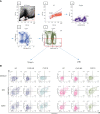
Figure 1
Figure 1
Gating strategy and representative FACS plots of CD4+ T memory cells responsive to SARS-CoV-2 spike peptides. Panel (A) A lymphocyte gate was applied to a forward - sideward scatter plot of all events (1), followed by doublet exclusion using forward scatter area vs height for cells within the lymphocyte gate (LG) (2). CD3+cells were then selected out of singlets (3) and CD3+ T-cells were examined for CD4 staining (4). Finally CD3+CD4+ CD45RA- T-memory cells were divided into CXCR5- (Tmem) and CXCR5+ (cTfh) cells (5). OX40(CD134)+CD25+ cells were finally defined as activated T-cells. Panel (B) Activation of Tmem and cTfh cells after stimulation of the cells with SEB or overlapping peptides of SARS-CoV-2 spike protein is shown in a representative healthy control (HC), a CVID patient IgG-responsive to BNT162b2 vaccination (CVID R, SARS-CoV-2 IgG antibody level following the second vaccination more than 33 RE/ml) and a CVID patient IgG-non-responsive following mRNA vaccination (CVID NR, SARS-CoV-2 IgG below 33 RE/ml following the second vaccination). Unstimulated control cells were incubated in medium alone (Medium). The percentages of OX40(CD134)+ and CD25+ double-positive activated CD4 T cell subsets are shown in the upper right panel of the respective FACS plots.
Figure 2
Figure 2
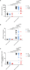
Antibody response following a second BNT162b2 mRNA vaccination in patients with CVID and oPAD. Serum IgG (A) and IgA (B) antibodies against spike protein of SARS-CoV-2 were examined by ELISA in patients with CVID, patients with milder forms of primary antibody deficiency (other predominantly antibody deficiency, oPAD) and healthy controls (HC) before COVID-19 vaccination and after the second vaccination with the Pfizer-BioNTech COVID-19 vaccine BNT162b2 (Comirnaty). Results for IgG antibodies are expressed as relative units (RE)/ml, the dotted line indicates 11 RE/ml that were used as the cutoff for positivity. Semiquantitative measurements of IgA antibodies are expressed as the ratio between the extinction of patient samples and the extinction of a calibrator provided with the kit; the dotted line indicates a ratio of 1.1 that was considered as the cutoff for positivity. Surrogate virus neutralizing antibodies (C) were assessed by testing the ability of serum antibodies (irrespective of isotype) to inhibit the interaction between recombinant SARS-CoV-2 receptor-binding domain and angiotensin-converting enzyme 2 using a blocking ELISA. Results are expressed as percent inhibition; the dotted horizontal line depicts 30% inhibition used as the cutoff for positive SARS-CoV-2 neutralizing antibody. Statistical differences between the two groups depicted in the figure and were determined with a non-parametric two-tailed Mann–Whitney U-test (Kruskal–Wallis H test for all groups: p <0.0001), the median is represented by a horizontal bar.
Figure 3
Figure 3
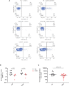
Detection of SARS-CoV-2 spike protein-specific CXCR5+ circulating follicular T-helper cells (cTfh), CXCR3-negative cTfh and CXCR5 CD4+ T-memory cells (Tmem) by flow cytometry. Human peripheral blood mononuclear cells (PBMC) from healthy controls (HC), CVID patients and patients with other, milder forms of primary antibody deficiency (oPAD), before COVID-19 vaccination and after the second vaccination with BNT162b2, were stimulated for two days using overlapping peptides of immunogenic regions of SARS-CoV2 spike protein (1 µg of peptides/ml). Activation of circulating follicular T-helper cells [CD3+CD4+CD45RA-CXCR5+, cTfh, panel (A)] and CXCR5-negative CD4 memory T cells [Tmem, panel (B)] was determined by measuring upregulation of CD25 and CD134 (OX40) by flow cytometry. Results are expressed as percent CD134 (OX40) and CD25 double positive cells relative to the respective CD4+ T cell subpopulation. Unstimulated control cells were incubated in parallel in culture medium only (percent CD134 and CD25 double positive unstimulated cells, median [IQR], before vaccination: HC (n = 12), Tfh 0.04 [0.04], Tmem 0.11 [0.42]; CVID (n = 4), Tfh 0.06 [0.15], Tmem 0.1 [0.2]; oPAD (n = 8), Tfh 0.06 [0.13], Tmem 0.05 [0.01]; after the second vaccination: HC (n = 14), Tfh 0.13 [0.11], Tmem 0.11 [0.42]; CVID (n = 19), Tfh: 0.03 [0.11], Tmem 0.08 [0.14]; oPAD (n = 31), Tfh 0.08 [0.11], Tmem 0.07 [0.53]; Kruskal–Wallis H test: n.s.). Panel (C) shows percent of activated, CD134 (OX40) and CD25 double positive cTfh following stimulation of PBMC for two days using overlapping peptides of immunogenic regions of SARS-CoV2 spike protein (1 µg of peptides/ml). PBMC were obtained from healthy controls (HC), CVID responders (anti-spike protein IgG antibody following second vaccination > three times cutoff = 33 RE/ml) and CVID non-responders after the second vaccination against COVID-19. Panel (D) shows the percentage of activated CD134 (OX40) and CD25 double positive CXCR3-negative cTfh activated in response to stimulation with SARS-CoV-2 spike peptides. Panel (E) shows activation of cTfh and Tmem as assessed by measuring OX40- and CD25-expression following prolonged stimulation with SARS-CoV-2 spike peptides for 4 days [panel (E.1); black circles: healthy controls, red squares:CVID]. In two healthy controls and two CVID patients, cTfh and Tmem activation was examined after two, three and four days of stimulation with SARS-CoV-2 spike peptides [panel (E.2)]. Statistical differences between two groups are depicted in the figure and were determined with a non-parametric two-tailed Mann–Whitney U-test (Kruskal–Wallis H test for all groups: p <0.0001), the median is represented by a horizontal bar.
Figure 4
Figure 4
Induction of early activation events such as CD69 expression and TNF-alpha production as well as cell proliferation as a late activation event is reduced in CD4-positive T cells from CVID patients stimulated with SARS-CoV2 spike peptides. TNF-alpha induction was measured as an early activation marker by flow cytometry in CD3+CD4+CD45RA cells from CVID patients responding with IgG antibody production following BNT162b2 mRNA COVID-19 vaccination (CVID R), CVID nonresponders (CVID NR) and healthy controls (HC). The figures depicted in panel (A) are representative for a total number of four CVID patients and three healthy controls investigated. Unstimulated cells incubated in medium alone were examined in parallel. In panel (B) CD69 expression was examined by flow cytometry on cTfh and Tmem of CVID and healthy controls stimulated with SARS-CoV-2 spike peptides; median CD69 expression in unstimulated cells incubated in medium alone was 1.25% cTfh and 0.41% Tmem in healthy controls, and 0.66% cTfh and 0.28% Tmem in CVID patients. In panel (C), PBMCs from healthy controls (HC) and CVID patients were stimulated for seven days using SARS-CoV2 Spike peptides before cell proliferation was examined by measuring 3H-thymidine incorporation. Results are expressed as netto dpm of 3H-thymindine incorporation, calculated by subtracting dpm of unstimulated cells from dpm of stimulated cells. In unstimulated PBMCs, 3H-thymidine incorporation [dpm, median (IQR)] was 932.85 (1,838.28) in healthy controls (n = 14) and 185.15 (463.85) in CVID (n = 16). Statistical differences between two groups were determined with a non-parametric two-tailed Mann–Whitney U-test, the median is represented by a horizontal bar.
Figure 5
Figure 5
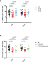
Defective SEB-induced activation of circulating follicular T helper cells and CXCR5-negative CD4 memory T cells from CVID patients. Panel (A) human peripheral blood mononuclear cells (PBMC) from healthy controls (HC), CVID patients and patients with other, milder forms of primary antibody deficiency (oPAD) were stimulated for two days with the bacterial superantigen Staphylococcus enterotoxin B (SEB, final concentration 1 µg/ml) before activation of circulating follicular T-helper cells (CD3+CD4+CD45RACXCR5+, cTfh) and CXCR5-negative CD4 memory T cells (Tmem) was determined by measuring upregulation of CD25 and CD134 (OX40) with flow cytometry. Results are expressed as percent CD134 (OX40) and CD25 double positive cells relative to the respective CD4+ T cell subpopulation. Panel (B) human peripheral blood mononuclear cells (PBMC) from healthy controls (HC), CVID IgG antibody responders (anti-spike protein IgG antibody following second vaccination > three times cutoff = 33 RE(/ml) and CVID IgG antibody non-responders were stimulated for two days with the bacterial superantigen Staphylococcus enterotoxin B (SEB, final concentration 1 µg/ml) before activation of circulating follicular T-helper cells (CD3+CD4+CD45RACXCR5+, cTfh) and CXCR5 CD4 memory T cells (Tmem) was determined by measuring upregulation of CD25 and CD134 (OX40) with flow cytometry. Results are expressed as percent CD134 (OX40) and CD25 double positive cells relative to the respective CD4+ T cell subpopulation. Statistical differences between two groups given in the figure were determined with a non-parametric two-tailed Mann–Whitney U-test (KruskalWallis H test for all groups: p <0.0001), the median is represented by a horizontal bar.

References

    1. Zhu N, Zhang D, Wang W, Li X, Yang B, Song J, et al. . A Novel Coronavirus From Patients With Pneumonia in China, 2019. N Engl J Med (2020) 382(8):727–33. doi: 10.1056/NEJMoa2001017 - DOI - PMC - PubMed
    1. Hu B, Guo H, Zhou P, Shi ZL. Characteristics of SARS-CoV-2 and COVID-19. Nat Rev Microbiol (2021) 19(3):141–54. doi: 10.1038/s41579-020-00459-7 - DOI - PMC - PubMed
    1. Wiersinga WJ, Rhodes A, Cheng AC, Peacock SJ, Prescott HC. Pathophysiology, Transmission, Diagnosis, and Treatment of Coronavirus Disease 2019 (COVID-19): A Review. JAMA J Am Med Assoc (2020) 324(8):782–93. doi: 10.1001/jama.2020.12839 - DOI - PubMed
    1. Zhang Q, Bastard P, Liu Z, Le Pen J, Moncada-Velez M, Chen J, et al. . Inborn Errors of Type I IFN Immunity in Patients With Life-Threatening COVID-19. Science (80 ) (2020) 370(6515):eabd4570. doi: 10.1126/science.abd4570 - DOI - PMC - PubMed
    1. Van Der Made CI, Simons A, Schuurs-Hoeijmakers J, van den Heuvel G, Mantere T, Kersten S, et al. . Presence of Genetic Variants Among Young Men With Severe COVID-19. JAMA J Am Med Assoc (2020) 324(7):663–73. doi: 10.1001/jama.2020.13719 - DOI - PMC - PubMed

Publication types

MeSH terms