. 2022 Oct;71(10):2421-2431.
doi: 10.1007/s00262-022-03170-z. Epub 2022 Mar 3.

Fc-independent functions of anti-CTLA-4 antibodies contribute to anti-tumor efficacy

Affiliations

Fc-independent functions of anti-CTLA-4 antibodies contribute to anti-tumor efficacy

Yosuke Sato et al. Cancer Immunol Immunother. 2022 Oct.

Abstract

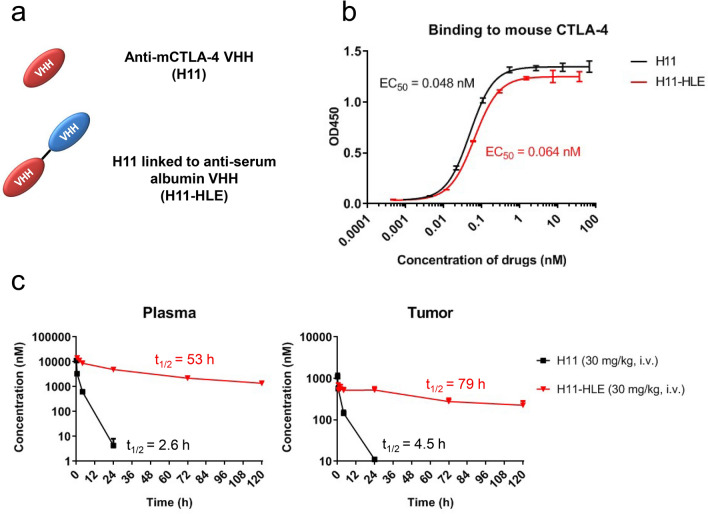
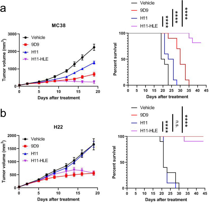
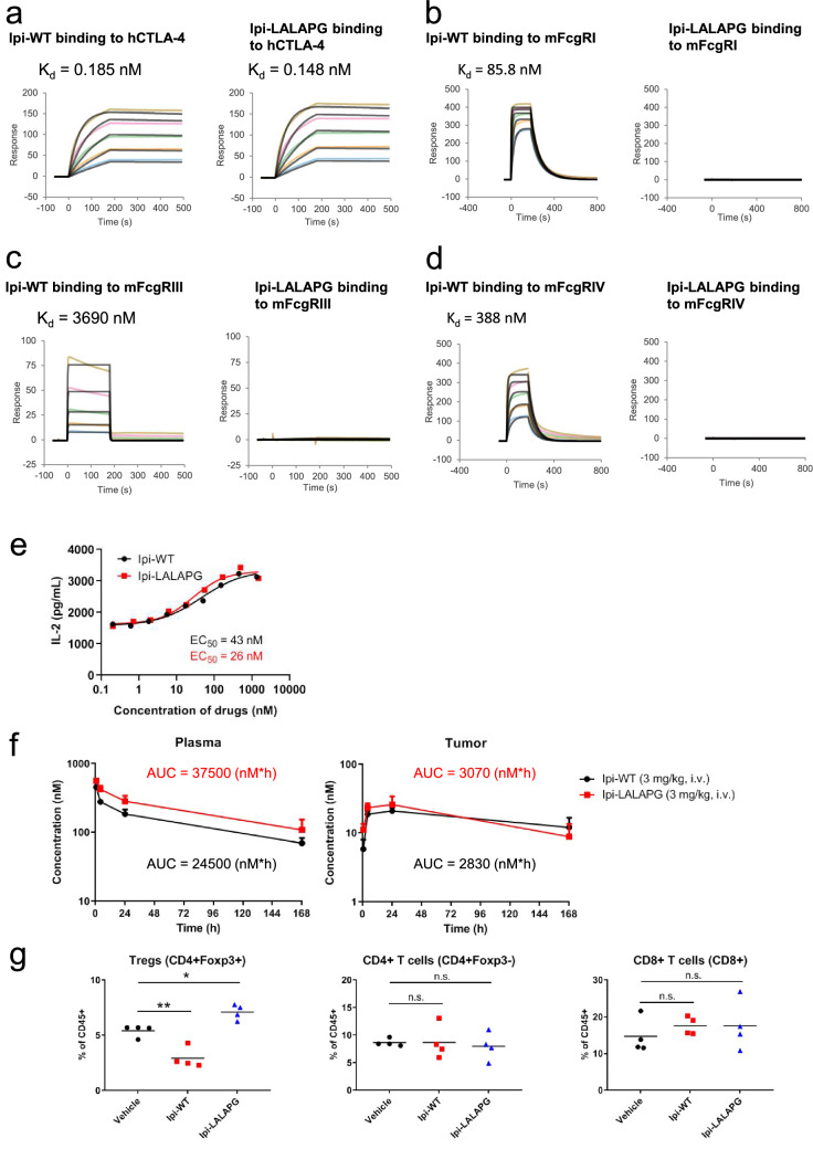
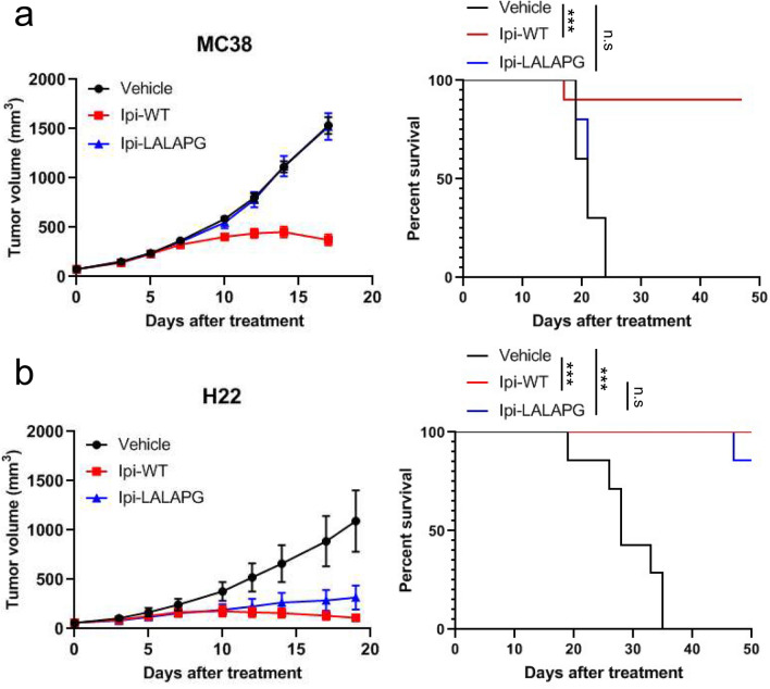
Ipilimumab, a monoclonal antibody that recognizes cytotoxic T-lymphocyte associated protein 4 (CTLA-4), was the first immune checkpoint inhibitor approved by the FDA to treat metastatic melanoma patients. Multiple preclinical studies have proposed that Fc effector functions of anti-CTLA-4 therapy are required for anti-tumor efficacy, in part, through the depletion of intratumoral regulatory T cells (Tregs). However, the contribution of the Fc-independent functions of anti-CTLA-4 antibodies to the observed efficacy is not fully understood. H11, a non-Fc-containing single-domain antibody (VHH) against CTLA-4, has previously been demonstrated to block CTLA-4-ligand interaction. However, in vivo studies demonstrated lack of anti-tumor efficacy with H11 treatment. Here, we show that a half-life extended H11 (H11-HLE), despite the lack of Fc effector functions, induced potent anti-tumor efficacy in mouse syngeneic tumor models. In addition, a non-Fc receptor binding version of ipilimumab (Ipi-LALAPG) also demonstrated anti-tumor activity in the absence of Treg depletion. Thus, we demonstrate that Fc-independent functions of anti-CTLA-4 antibodies contributed to anti-tumor efficacy, which may indicate that non-Treg depleting activity of anti-CTLA-4 therapy could benefit cancer patients in the clinic.

Keywords: Anti-CTLA-4 antibody; Anti-tumor efficacy; Fc effector functions; Syngeneic tumor model.


Conflict of interest statement

The authors declare that they have no conflict of interest.

Figures

Fig. 1
Fig. 1
Half-life extended H11 shows similar binding to mouse CTLA-4 as H11 but longer half-life than H11. a Schematic representation of anti-mouse CTLA-4 VHH (H11) and H11 linked to anti-serum albumin VHH (H11-HLE). b Binding to mCTLA-4 of H11 and H11-HLE and their EC50 values. His-tagged H11 or H11-HLE was incubated with plate-bound mCTLA-4-Fc. Binding to mCTLA-4 was detected by using HRP-conjugated anti-His tag antibodies and tetramethylbenzidine (TMB). Data are represented as OD. Error bars show SD. c Plasma (left) and tumor (right) concentration–time profiles and half-life of H11 (30 mg/kg) or H11-HLE (30 mg/kg) in MC38 tumor-bearing C57BL/6 mice (n = 3) via intravenous administration. C57BL/6 mice were inoculated with 1 × 106 MC38 cells. When the mean tumor volume reached approximately 300–500 mm3, animals were randomized into treatment groups (n = 3/group) and dosing was initiated. At indicated time points after single dosing, mice were euthanized and plasma and tumor tissues were harvested. H11 or H11-HLE was quantified using ELISA using HRP-conjugated anti-His tag antibodies
Fig. 2
Fig. 2
Half-life extended H11 induces anti-tumor efficacy in syngeneic tumor models. a C57BL/6 mice were inoculated with 1 × 106 MC38 cells and were treated with PBS (vehicle), 10 mg/kg of anti-mCTLA-4 mIgG2b (9D9), 30 mg/kg of H11 and 30 mg/kg of H11-HLE twice weekly for 3 weeks. When the mean tumor volume reached approximately 60 mm3, animals were randomized into treatment groups (n = 10/group) and dosing was initiated on Day 0 of the study. b BALB/c mice were inoculated with 4 × 105 H22 cells and were treated with PBS (vehicle), 10 mg/kg of anti-mCTLA-4 mIgG2b (9D9), 30 mg/kg of H11 and 30 mg/kg of H11-HLE twice weekly for 3 weeks. When the mean tumor volume reached approximately 60 mm3, animals were randomized into treatment groups (n = 10/group) and dosing was initiated on Day 0 of the study. (Left) Tumor size as measured by vernier calipers, and the data shown in all panels are the mean (n = 10/group) ± SEM. (Right) Survival curve comparing treatment groups. Mice were euthanized when tumors reached 2000 mm3. n.s., nonsignificant. ****P < 0.0001 by log-rank test
Fig. 3
Fig. 3
Ipilimumab mIgG2a LALAPG exhibits similar binding to human CTLA-4 and similar plasma and tumor PK as ipilimumab mIgG2a wildtype but does not bind to mouse FcγRs or induce in vivo Fc-dependent intratumoral Treg depletion. Surface plasmon resonance measurements of Ipi-WT and Ipi-LALAPG to a human CTLA-4, b mouse FcγRI, c FcγRIII and d FcγRIV. Human CTLA-4 was injected at concentrations ranging from 3.125 to 50 nM, and mouse FcγRs were injected at concentrations ranging from 187.5 to 3000 nM. e Inhibitory activity against interaction between CTLA-4 and B7 ligands by Ipi-WT and Ipi-LALAPG and their EC50 values. PHA-stimulated human primary CD3 + T cells expressing huCTLA-4 were co-cultured with engineered Raji cells expressing CTLA-4 ligands CD80 and CD86 in the presence of Ipi-WT or Ipi-LALAPG. Data are representative of 4 donors. Human-CTLA-4 knock-in C57BL/6 mice were inoculated with 1 × 106 MC38 cells. When the mean tumor volume reached approximately 580 mm3, animals were randomized into treatment groups (n = 3/group) and dosing was initiated. f Plasma (left) and tumor (right) concentration–time profiles and the AUC of Ipi-WT (3 mg/kg) or Ipi-LALAPG (3 mg/kg) in MC38 tumor-bearing huCTLA-4 knock-in C57BL/6 mice (n = 3) via intravenous administration. Mice were treated with Ipi-WT or Ipi-LALAPG at day 0. At indicated time points after single dosing, mice were euthanized and plasma and tumor tissues were harvested. Ipi-WT or Ipi-LALAPG was quantified using ELISA. g Quantification of tumor-infiltrating Tregs, CD4 + T cells and CD8 + T cells. Mice (n = 4) were treated with PBS (vehicle), 3 mg/kg of Ipi-WT or 3 mg/kg of Ipi-LALAPG at day 0. The mice were euthanized 72 h after single dosing, and single-cells were isolated from resected tumors and analyzed by flow cytometry. n.s., nonsignificant. *P < 0.05 and **P < 0.01 by one-way ANOVA with Dunnett’s multiple comparisons test
Fig. 4
Fig. 4
Ipilimumab lacking Fc effector functions induces anti-tumor responses. a HuCTLA-4 KI C57BL/6 mice were inoculated with 1 × 106 MC38 cells and were treated with PBS (vehicle), 3 mg/kg of ipilimumab mIgG2a (Ipi-WT), or 3 mg/kg of ipilimumab mIgG2a LALAPG (Ipi-LALAPG) twice weekly for 3 weeks. When the mean tumor volume reached approximately 60 mm3, animals were randomized into treatment groups (n = 10/group) and dosing was initiated on Day 0 of the study. b HuCTLA-4 KI BALB/c mice were inoculated with 4 × 105 H22 cells and were treated with PBS (vehicle), 3 mg/kg of ipilimumab mIgG2a LALAPG (Ipi-LALAPG) twice weekly for 3 weeks. When the mean tumor volume reached approximately 60 mm3, animals were randomized into treatment groups (n = 7/group) and dosing was initiated on Day 0 of the study. (Left) Tumor size as measured by vernier calipers, and the data shown in all panels are the mean (n = 7–10/group) ± SEM. (Right) Survival curve comparing treatment groups. Mice were euthanized when tumors reached 2000 mm3. n.s., nonsignificant. ***P < 0.001 by log-rank test

References

    1. Hodi FS, O’Day SJ, McDermott DF, et al. Improved survival with ipilimumab in patients with metastatic melanoma. N Engl J Med. 2010;363:711–723. doi: 10.1056/nejmoa1003466. - DOI - PMC - PubMed
    1. Robert C, Thomas L, Bondarenko I, et al. Ipilimumab plus dacarbazine for previously untreated metastatic melanoma. N Engl J Med. 2011;364:2517–2526. doi: 10.1056/nejmoa1104621. - DOI - PubMed
    1. Sharma P, Allison JP. The future of immune checkpoint therapy. Science. 2015;348:56–61. doi: 10.1126/science.aaa8172. - DOI - PubMed
    1. Wei SC, Duffy CR, Allison JP. Fundamental mechanisms of immune checkpoint blockade therapy. Cancer Discov. 2018;8:1069–1086. doi: 10.1158/2159-8290.cd-18-0367. - DOI - PubMed
    1. Schildberg FA, Klein SR, Freeman GJ, et al. Coinhibitory pathways in the B7-CD28 ligand-receptor family. Immunity. 2016;44:955–972. doi: 10.1016/j.immuni.2016.05.002. - DOI - PMC - PubMed