. 2022 Mar 3;6(1):12.
doi: 10.1186/s41747-022-00264-2.

The effects of repeated brain MRI on chromosomal damage

Affiliations

The effects of repeated brain MRI on chromosomal damage

Cecile Herate et al. Eur Radiol Exp. .

Abstract

Background: Magnetic resonance imaging (MRI) is currently considered a safe imaging technique because, unlike computed tomography, MRI does not expose patients to ionising radiation. However, conflicting literature reports possible genotoxic effects of MRI. We herein examine the chromosomal effects of repeated MRI scans by performing a longitudinal follow-up of chromosomal integrity in volunteers.

Methods: This ethically approved study was performed on 13 healthy volunteers (mean age 33 years) exposed to up to 26 3-T MRI sessions. The characterisation of chromosome damage in peripheral blood lymphocytes was performed using the gold-standard biodosimetry technique augmented with telomere and centromere staining.

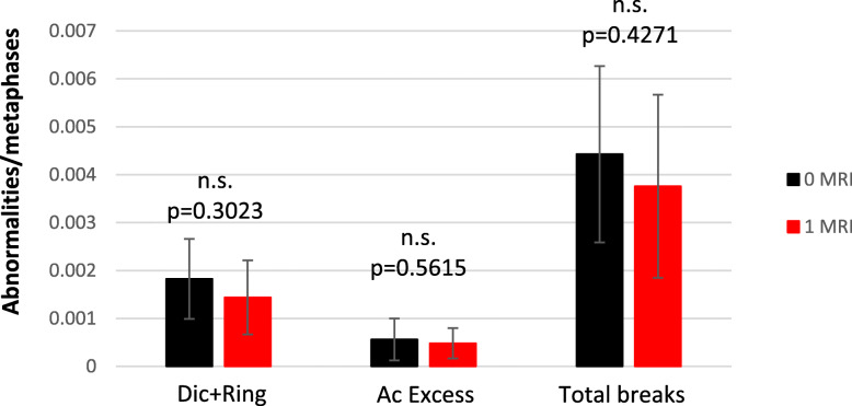
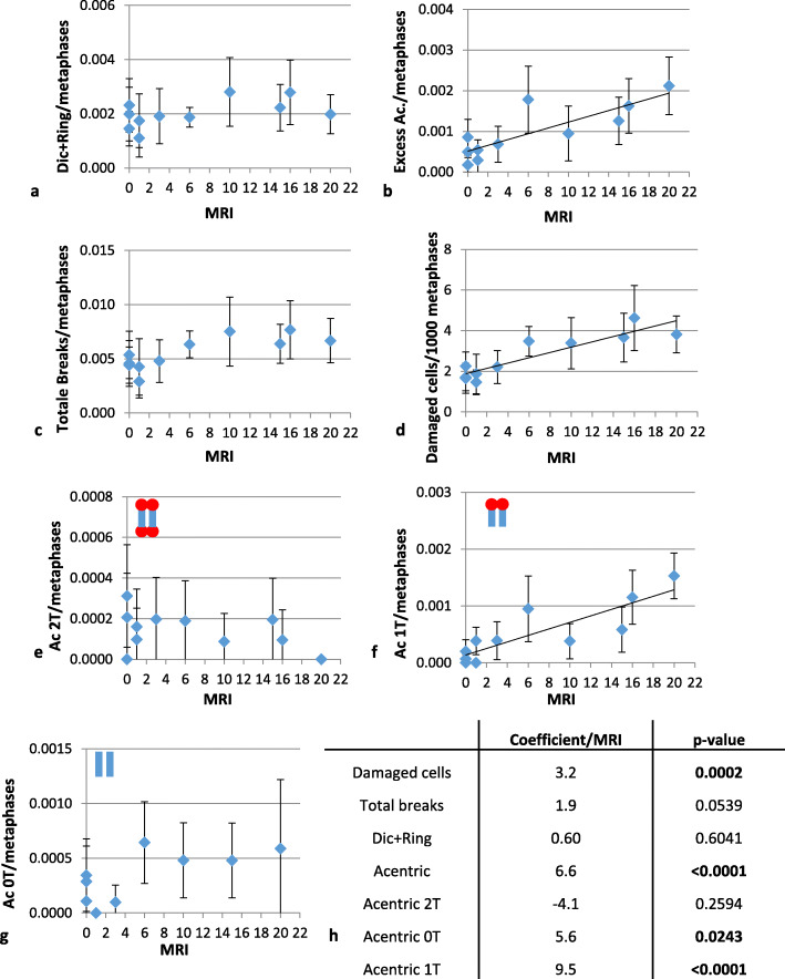
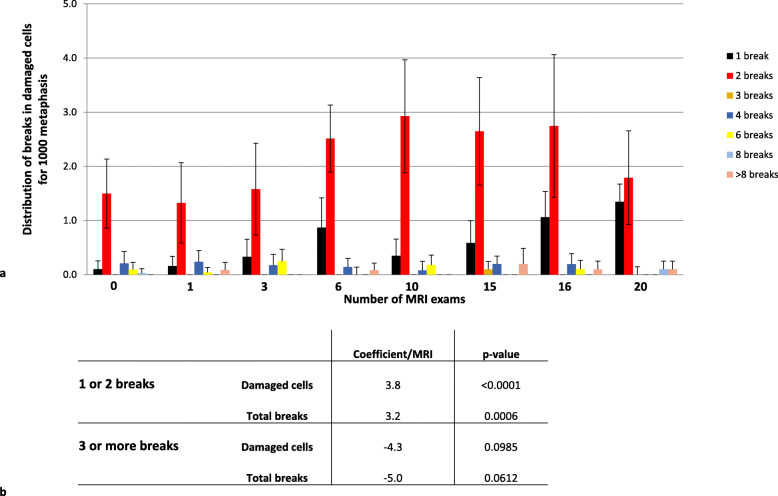
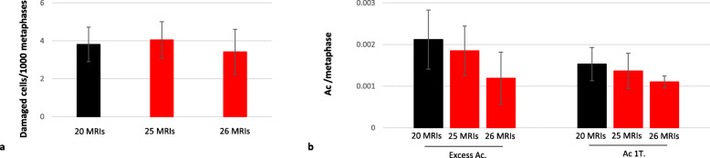
Results: Cytogenetic analysis showed no detectable effect after a single MRI scan. However, repeated MRI sessions (from 10 to 20 scans) were associated with a small but significant increase in chromosomal breaks with the accumulation of cells with chromosomal terminal deletions with a coefficient of 9.5% (95% confidence interval 6.5-12.5%) per MRI (p < 0.001). Additional exposure did not result in any further increase. This plateauing of damage suggests lymphocyte turnover. Additionally, there was no significant induction of dicentric chromosomes, in contrast to what is observed following exposure to ionising radiation.

Conclusions: Our study showed that MRI can affect chromosomal integrity. However, the amount of damage per cell might be so low that no chromosomal rearrangement by fusion of two deoxyribonucleic breaks is induced, unlike that seen after exposure to computed tomography. This study confirms that MRI is a safe imaging technique.

Keywords: Centromere; Chromosome aberrations; Cytogenetic analysis; Magnetic resonance imaging; Telomere.


Conflict of interest statement

The authors declare that they have no competing interests.

Figures

Fig. 1
Fig. 1
One MRI session does not trigger any genotoxic effect. The dicentric chromosome assay was performed on blood cells of volunteers sampled 30, 15, and 0 days before the exam to take into account the heterogeneity of the background (mean in black). The potential effect of one MRI session was assessed the day of the first MRI and prior to the second MRI (mean in red). Unstable aberrations (Dic + Tric + ring) were scored after telomere and centromere staining, as well as acentric fragments (type E acentrics, acentrics in excess), those resulting from Dics or ring being excluded). Total breaks were calculated from these data. Ac, Acentric fragment; Dic, Dicentric chromosome; MRI, Magnetic resonance imaging; n.s., No significant differences; Tric, Tricentric chromosome
Fig. 2
Fig. 2
Increasing chromosomal instability due to terminal deletions during the first 20 MRI sessions. Between 846 and 3,357 metaphases were observed for each time point and participant after telomere/centromere staining. Various cytogenetic parameters were scored: dicentric chromosomes and rings (a); total type E acentrics (excess acentric fragments) (b), total chromosomal breaks (c), damaged cells (d), 2-T acentric fragments resulting from two chromosomal breaks and fusion (e), 1-T acentric fragments equivalent to a terminal deletion (f), and 0-T acentric fragments resulting from two chromosomal breaks in the same chromosomal arm (g). h Table presenting the stratified statistical analysis (correlation coefficients and p values). Ac, Acentric fragment; Dic, Dicentric chromosomes; MRI, Magnetic resonance imaging; T, Telomere
Fig. 3
Fig. 3
Distribution of deoxyribonucleic acid breaks in damaged cells. a Only the data for damaged cells are shown and are clustered according to the number of breaks scored. In this graph, all damaged cells are considered, including rogue cells. b Statistical analysis of the damaged-cell phenotype. Cells with one or two breaks were separated from those with three or more breaks. The correlation between the number of MRI sessions and the breaks was calculated for both groups. The statistical analysis shows a strong correlation between the number of MRI sessions and the apparition of cells with few (one or two) breaks. However, there was no correlation between the number of MRI sessions and the number of cells with three or more chromosome breaks. MRI, Magnetic resonance imaging
Fig. 4
Fig. 4
Stabilisation of cytogenetic parameters after 20 MRI sessions. Further analysis was conducted after six supplementary MRI sessions. Evolution of damaged cells (a) and type E acentric fragments/1-T acentric fragments (b) between 20 and 25–26 MRI sessions. All parameters remained stable. Ac, Acentric fragment; MRI, Magnetic resonance imaging; T, Telomere
Fig. 5
Fig. 5
Increase in the frequency of dicentric chromosomes after CT. The frequency of dicentric and ring chromosomes is presented up until 26 MRI sessions for the ten MRI-only volunteers and for the subject number 8, a volunteer exposed to computed tomography examinations (8 between the 10 and 25 MRI sessions) for an unexpected diagnostic assessment of a non-brain pathology during the originally planned MRI sessions schedule. Dic, Dicentric chromosomes; MRI, Magnetic resonance imaging

References

    1. Lee JW, Kim MS, Kim YJ, Choi YJ, Lee Y, Chung HW. Genotoxic effects of 3 T magnetic resonance imaging in cultured human lymphocytes. Bioelectromagnetics. 2011;32:535–542. doi: 10.1002/bem.20664. - DOI - PubMed
    1. Szerencsi Á, Kubinyi G, Váliczkó É, et al. (2013) DNA integrity of human leukocytes after magnetic resonance imaging. Int J Radiat Biol 89:870–876. 10.3109/09553002.2013.804962 - PubMed
    1. Yildiz S, Cece H, Kaya I, et al. (2011) Impact of contrast enhanced MRI on lymphocyte DNA damage and serum visfatin level. Clin Biochem 44:975–979. 10.1016/j.clinbiochem.2011.05.005 - PubMed
    1. Fasshauer M, Krüwel T, Zapf A, et al. (2018) Absence of DNA double-strand breaks in human peripheral blood mononuclear cells after 3 Tesla magnetic resonance imaging assessed by γH2AX flow cytometry. Eur Radiol 28:1149–1156. 10.1007/s00330-017-5056-9 - PubMed
    1. Fatahi M, Reddig A, Vijayalaxmi null, et al (2016) DNA double-strand breaks and micronuclei in human blood lymphocytes after repeated whole body exposures to 7T magnetic resonance imaging. Neuroimage 133:288–293. 10.1016/j.neuroimage.2016.03.023 - PubMed

Publication types

LinkOut - more resources