. 2022 Mar 8;119(10):e2110415119.
doi: 10.1073/pnas.2110415119. Epub 2022 Mar 1.

Amino acid sensor conserved from bacteria to humans

Affiliations

Amino acid sensor conserved from bacteria to humans

Vadim M Gumerov et al. Proc Natl Acad Sci U S A. .

Abstract

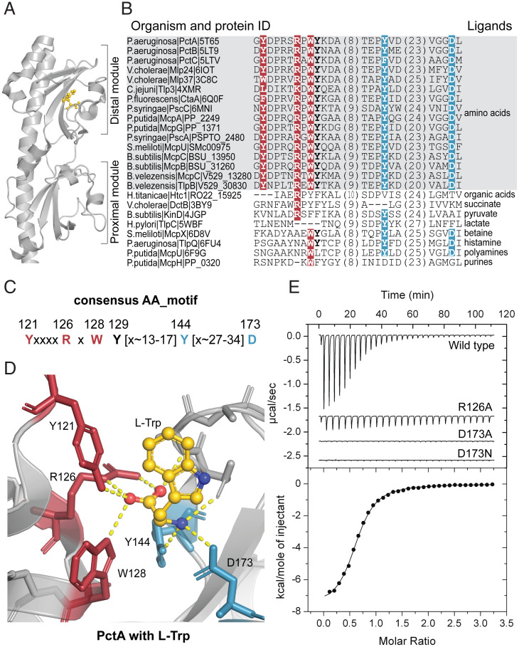
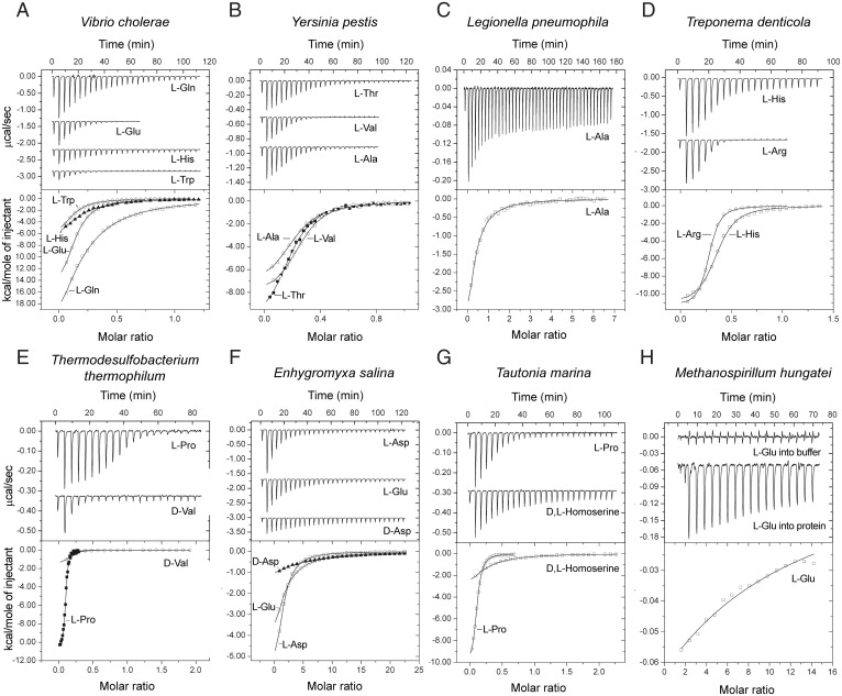
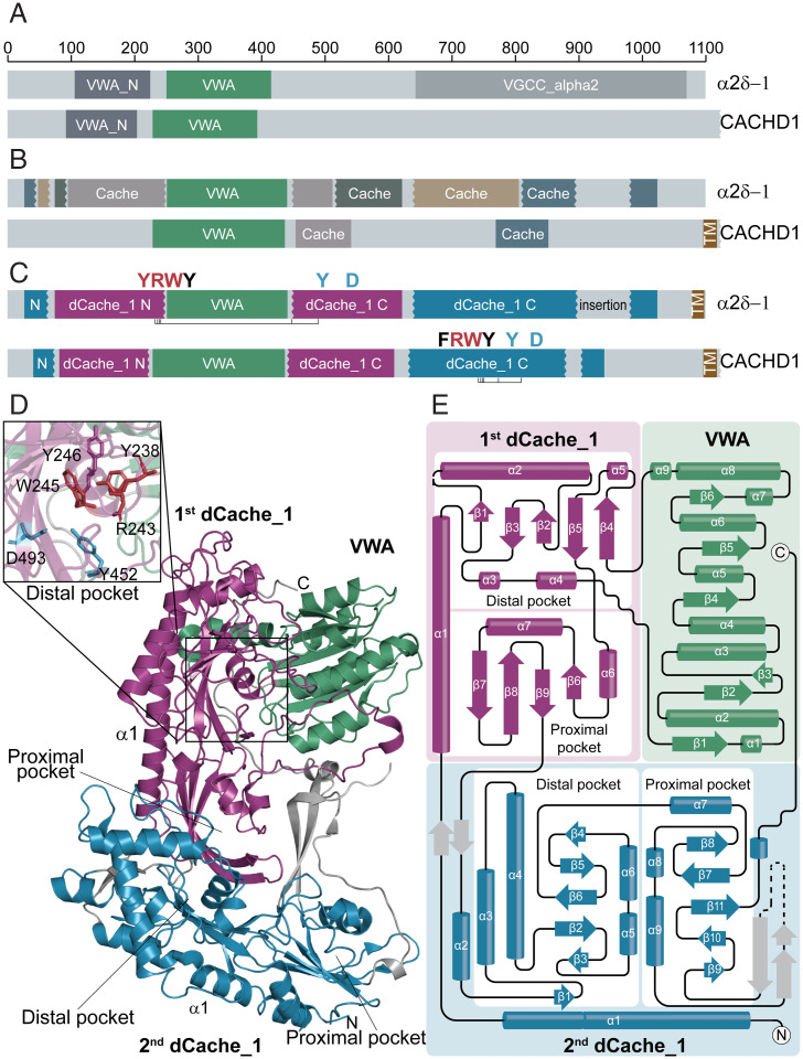
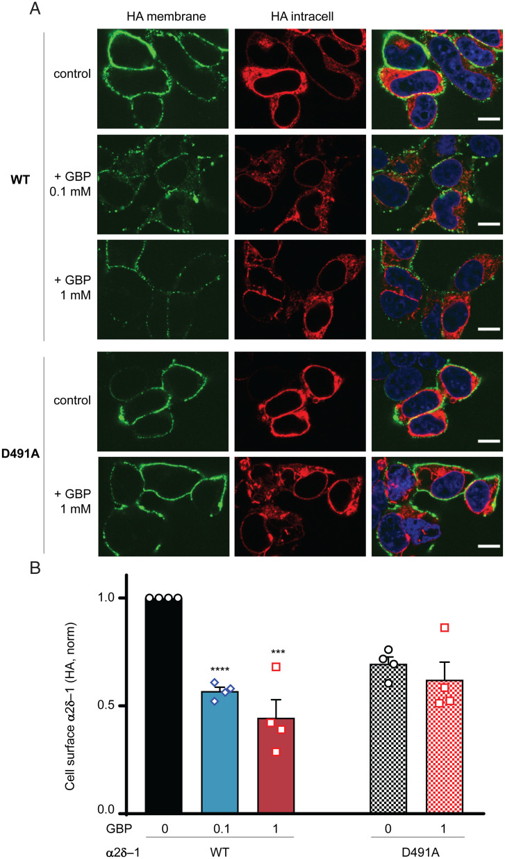
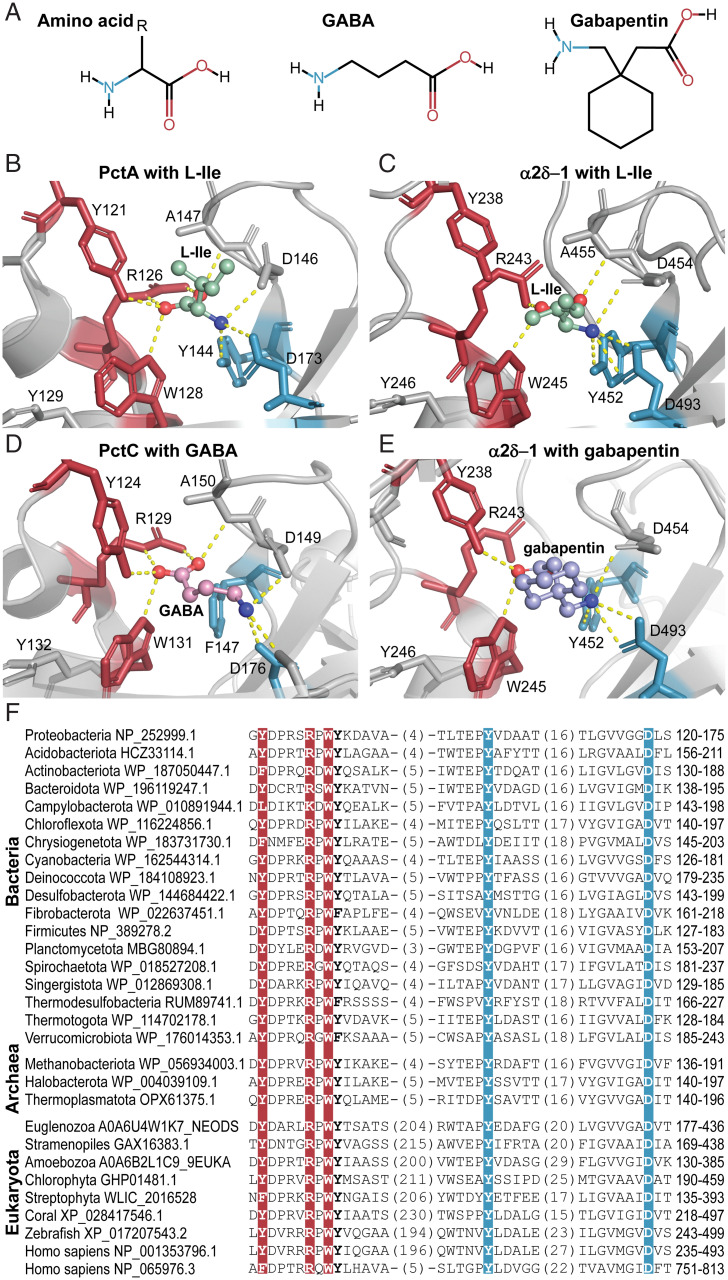
SignificanceAmino acids are the building blocks of life and important signaling molecules. Despite their common structure, no universal mechanism for amino acid recognition by cellular receptors is currently known. We discovered a simple motif, which binds amino acids in various receptor proteins from all major life-forms. In humans, this motif is found in subunits of calcium channels that are implicated in pain and neurodevelopmental disorders. Our findings suggest that γ-aminobutyric acid-derived drugs bind to the same motif in human proteins that binds natural ligands in bacterial receptors, thus enabling future improvement of important drugs.

Keywords: evolution; gabapentin; ion channels; serine/threonine kinases; signal transduction.


Conflict of interest statement

The authors declare no competing interest.

Figures

Fig. 1.
Fig. 1.
AA_motif in the dCache_1 domain. (A) dCache_1 domain of the PctA chemoreceptor from P. aeruginosa PAO1 with bound L-Trp (gold; PDB ID code 5T7M). (B) Protein sequence alignment of experimentally studied bacterial dCache_1 domains with respective ligands. The AA_motif (in bold) is present in all amino acid binding dCache_1 domains (gray background). (C) Consensus AA_motif. Numbers above the motif correspond to positions in PctA. (D) L-Trp interactions with AA_motif residues in the ligand binding pocket of PctA. (B–D) Here and in all figures, red indicates residues that coordinate the carboxyl group of the ligand, and blue indicates residues that make contacts with the amino group. (E) Isothermal titration calorimetry study of L-Ala binding to the wild type and the mutated dCache_1 domain of PctA.
Fig. 2.
Fig. 2.
Microcalorimetric titration of selected recombinant dCache_1AA domains with amino acids. In each panel, Upper shows raw titration data, and Lower shows integrated corrected peak areas of the titration data fit using the “one–binding site model.” Details of each experiment can be found in SI Appendix, Table S2. (A) V. cholerae (gammaproteobacteria) c-di-GMP phosphodiesterase (NP_233280.1). (B) Y. pestis (gammaproteobacteria) chemoreceptor (WP_016674185.1). (C) L. pneumophila (gammaproteobacteria) guanylate/adenylate cyclase (WP_154766400.1). (D) Treponema denticola (spirochaetota) chemoreceptor (WP_002687321.1). (E) Thermodesulfobacterium thermophilum (desulfobacterota) c-di-GMP cyclase (WP_162138226.1). (F) Enhygromyxa salina (myxococcota) serine/threonine kinase (WP_106093935.1). (G) Tautonia marina (planctomycetota) serine/threonine phosphatase (WP_152054232.1). (H) Methanospirillum hungatei (archaea, halobacteriota) sensor histidine kinase (WP_011449640.1).
Fig. 3.
Fig. 3.
dCache_1AA domains in α2δ-subunit and CACHD1 subunit of VGCC. (A and B) Domains that are currently recognized in α2δ-1 and CACHD1 proteins by the Pfam database (A) and experimental studies (27,29) (B). (C) Domain architectures of α2δ-1 and CACHD1 proteins revealed in the present study. The AA_motif is shown. (D) Structural composition of the α2δ-1–subunit uncovered in the present study shown on the solved structure [PDB ID code 6JPA (44)]. A close-up view of the dCache_1AA distal module (Upper Left) showing the spatial proximity of the AA_motif residues despite the VWA insertion. (E) The α2δ-1–subunit topology shows that the VWA domain is inserted into the first dCache_1 domain, which in turn, is inserted into the second dCache_1 domain.
Fig. 4.
Fig. 4.
Gabapentin impairs cell surface expression of wild-type (WT) α2δ-1 but not α2δ-1D491A. (A) Representative images of tsA-201 cells expressing hemagglutinin (HA) tagged α2δ-1-HA WT (rows 1 to 3) and α2δ-1D491A-HA (rows 4 and 5) in the absence (control; rows 1 and 4) or presence of gabapentin (GBP; 0.1 mM, row 2 [WT]; 1 mM, rows 3 [WT] and 5 [D491A]). Left shows cell surface HA staining in nonpermeabilized conditions (green). Center shows intracellular HA staining after permeabilization (red). Merged images with the nuclei stained with DAPI (blue) are shown in Right. (Scale bars: 10 μm.) (B) Bar chart (mean ± SEM for n = 4 independent experiments) for cell surface expression of α2δ-1-HA for WT (left three bars) and D491A (right two bars) in the absence (0) or presence of 0.1 or 1 mM GBP. For each experiment, HA staining was measured for over 50 cells and normalized to that of the WT control. Points for individual experiments are shown. Total numbers of cells measured for each condition are as follows; WT control: 379 (black); WT + 0.1 mM GBP: 360 (blue); WT + 1 mM GBP: 449 (red); D491A control: 351 (black hatched); and D491A + 1 mM GBP: 395 (red hatched). Statistical significance of the effect of GBP on cell surface expression was determined using one-way ANOVA and Dunnett’s post hoc test. ***P = 0.0003; ****P < 0.0001.
Fig. 5.
Fig. 5.
Bacterial and mammalian receptors bind amino acid ligands through the conserved AA_motif. (A) Structural comparison of the ligands found to bind dCache_1AA. (B–E) Ligand binding modes of bacterial and eukaryotic dCache_1AA: PctA with L-Ile (B; PDB ID code 5T65), α2δ-1 with docked L-Ile (C), PctC in complex with GABA (D; PDB ID code 5LTV), and α2δ-1 with docked gabapentin (E). (F) Protein sequence alignment of the dCache_1AA from representatives of major phyla of Bacteria, Archaea, and Eukaryota.
Fig. 6.
Fig. 6.
The AA_motif across the Tree of Life. (A) Distribution of dCache_1AA across major lineages of life. Thick lines with dots at the tips denote the presence of the AA_motif. Positions of relevant organisms are shown. The red circle indicates horizontal gene transfer of the dCache_1AA to Archaea. The orange circle indicates three events that happened around the same time (LECA): 1) horizontal transfer of dCache_1AA from Bacteria to Eukaryota, 2) VWA domain insertion, and 3) insertion of the first dCache_1 into the second dCache_1 domain. (B) Prevalent domain architectures of the dCache_1AA containing proteins found in each domain of life are shown. Domain definitions are according to the Pfam domain nomenclature: EAL (PF00563), a diguanylate phosphodiesterase; GGDEF (PF00990), a diguanylate cyclase; Guanylate_cyc (PF00211), an adenylate or guanylate cyclase; HATPase_c (PF02518), a histidine kinase; HD (PF01966), phosphohydrolase; MCPsignal (PF00015), methyl-accepting chemotaxis protein (chemoreceptor); Pkinase (PF00069), serine/threonine kinase; SpoIIE (PF07228), serine/threonine phosphatase.

References

    1. Chantranupong L., Wolfson R. L., Sabatini D. M., Nutrient-sensing mechanisms across evolution. Cell 161, 67–83 (2015). - PMC - PubMed
    1. Ellaithy A., Gonzalez-Maeso J., Logothetis D. A., Levitz J., Structural and biophysical mechanisms of class C G protein-coupled receptor function. Trends Biochem. Sci. 45, 1049–1064 (2020). - PMC - PubMed
    1. Laverty D., et al. , Cryo-EM structure of the human α1β3γ2 GABAA receptor in a lipid bilayer. Nature 565, 516–520 (2019). - PMC - PubMed
    1. Parkinson J. S., Hazelbauer G. L., Falke J. J., Signaling and sensory adaptation in Escherichia coli chemoreceptors: 2015 update. Trends Microbiol. 23, 257–266 (2015). - PMC - PubMed
    1. Ortega Á., Zhulin I. B., Krell T., Sensory repertoire of bacterial chemoreceptors. Microbiol. Mol. Biol. Rev. 81, e00033-17 (2017). - PMC - PubMed