. 2022 Mar 8;119(10):e2111537119.
doi: 10.1073/pnas.2111537119. Epub 2022 Mar 1.

Necroptosis triggers spatially restricted neutrophil-mediated vascular damage during lung ischemia reperfusion injury

Affiliations

Necroptosis triggers spatially restricted neutrophil-mediated vascular damage during lung ischemia reperfusion injury

Wenjun Li et al. Proc Natl Acad Sci U S A. .

Abstract

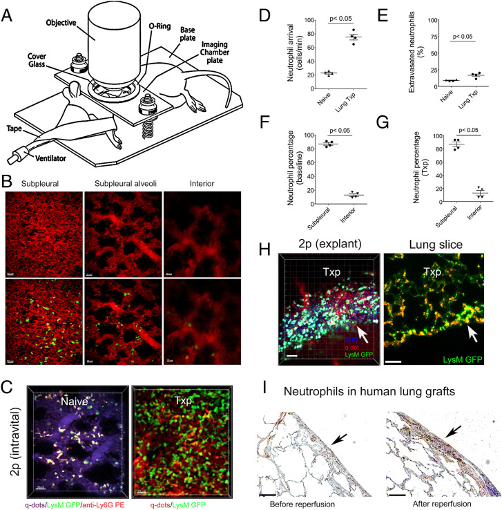
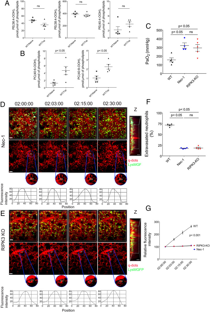
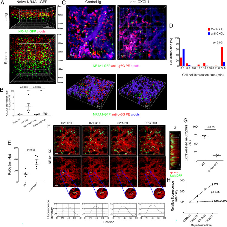
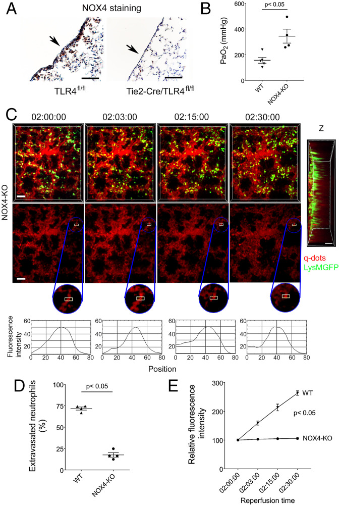
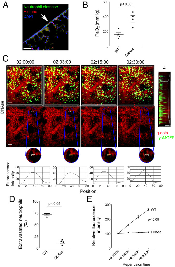
Ischemia reperfusion injury represents a common pathological condition that is triggered by the release of endogenous ligands. While neutrophils are known to play a critical role in its pathogenesis, the tissue-specific spatiotemporal regulation of ischemia-reperfusion injury is not understood. Here, using oxidative lipidomics and intravital imaging of transplanted mouse lungs that are subjected to severe ischemia reperfusion injury, we discovered that necroptosis, a nonapoptotic form of cell death, triggers the recruitment of neutrophils. During the initial stages of inflammation, neutrophils traffic predominantly to subpleural vessels, where their aggregation is directed by chemoattractants produced by nonclassical monocytes that are spatially restricted in this vascular compartment. Subsequent neutrophilic disruption of capillaries resulting in vascular leakage is associated with impaired graft function. We found that TLR4 signaling in vascular endothelial cells and downstream NADPH oxidase 4 expression mediate the arrest of neutrophils, a step upstream of their extravasation. Neutrophil extracellular traps formed in injured lungs and their disruption with DNase prevented vascular leakage and ameliorated primary graft dysfunction. Thus, we have uncovered mechanisms that regulate the initial recruitment of neutrophils to injured lungs, which result in selective damage to subpleural pulmonary vessels and primary graft dysfunction. Our findings could lead to the development of new therapeutics that protect lungs from ischemia reperfusion injury.

Keywords: intravital imaging; ischemia reperfusion injury; transplantation.


Conflict of interest statement

Competing interest statement: K.J.L. and D.K. have a pending patent entitled “Compositions and methods for detecting CCR2 receptors” (application No. 15/611,577).

Figures

Fig. 1.
Fig. 1.
Neutrophils accumulate in subpleural capillaries at baseline and after ischemia reperfusion injury. (A) Diagram depicting setup of mouse for intravital two-photon imaging. (B) Representative two-photon images of vessels above (Left) (10 μm) and below (Right) (80 μm) subpleural alveoli (Middle) (25 μm) in lungs of naïve B6 LysM-GFP mice. The Top panel depicts vessels after injection of quantum dots (red), and the Bottom panel shows vessels (red) and LysM-GFP+ cells (green) (Scale bars: 20 μm). (C) Two-photon images of neutrophils in subpleural capillaries of (Left) lungs of naïve B6 LysM-GFP mice and (Right) reperfused B6 lung grafts that were subjected to 60 min of cold storage 2 h after transplantation into syngeneic B6 LysM-GFP hosts. In naïve lungs, neutrophils are green with a red ring after labeling with intravenous anti-Ly6G antibody (PE). Pulmonary vessels are labeled after intravenous injection of quantum dots (Left: purple; Right: red) (Scale bars: 30 μm). (D) No. of neutrophils arriving in pulmonary arterioles per minute and (E) percentage of extravasated neutrophils in naïve B6 lungs versus B6 lung grafts 2 h after transplantation into syngeneic recipients. Percentage of neutrophils in subpleural versus interior perialveolar capillaries in (F) lungs of naïve B6 LysM-GFP mice and (G) reperfused B6 lung grafts that were subjected to 60 min of cold storage 2 h after transplantation into syngeneic B6 LysM-GFP hosts. (H) Two-photon imaging (Left) and immunofluorescent staining (Right) of neutrophils (green) of tissue slices of B6 lung grafts that were subjected to 60 min of cold storage 2 h after transplantation into syngeneic B6 LysM-GFP hosts. The arrow points to pleural surface (Scale bars: Left 30 μm, Right 100 μm). (I) Neutrophil staining (antineutrophil elastase) in human lung grafts before (Left) and 2 h after reperfusion (Right). The arrow points to pleural surface (Scale bars: 100 μm). The data in (DG) represent the mean ± SEM (n = 4).
Fig. 2.
Fig. 2.
Severe ischemia reperfusion injury results in neutrophil-mediated vascular leakage from subpleural capillaries. (A) Arterial blood oxygenation 2 h after transplantation of B6 lungs that were subjected to 60 min of cold ischemia (CIS) or 60 min of cold and 45 min of warm ischemia (WIS) into syngeneic recipients. (B) Percentage of neutrophils in subpleural versus interior perialveolar capillaries of WIS grafts. Time lapse intravital two-photon imaging of neutrophils (green) (Top), quantum dots (red) that were injected intravenously (Middle), quantification of disruption of vascular integrity as evidenced by extravascular quantum dot signal (boxed region and kymographs), and side projections of z stacks of (C) CIS and (D) WIS grafts. The x-axis in the kymographs refers to pixels in the x dimension along the boxed region (Scale bars: 20 μm). (E) Two-photon imaging of neutrophils (green) in lung explants, whole (Top [Scale bars: 30 μm]) or sliced (Middle, [Scale bars: 30 μm]) of B6 lung grafts that were subjected to WIS 2 h after transplantation into syngeneic B6 LysM-GFP hosts. The Bottom image depicts LysM-GFP+ cells in sliced lung explants 24 h after reperfusion. The arrow points to pleural surface (Scale bars: 20 μm). (F) Percentage of extravasated neutrophils in CIS versus WIS grafts 2 h after transplantation into syngeneic recipients. (G) Comparison of extravascular quantum dot intensity in subpleural space of CIS versus WIS grafts over time. (H) Neutrophil elastase and histone immunostaining (Top) and neutrophil elastase, histone, and DAPI colocalization by immunofluorescence (Bottom) in CIS and WIS grafts (Scale bars: 100 μm). The arrow points to pleural surface. (I) Arterial blood oxygenation 2 h after transplantation of B6 lungs that were subjected to WIS into syngeneic recipients that were treated with isotype control (control Ig) or neutrophil-depleting (anti-Ly6G) antibodies. (J) Time lapse intravital two-photon imaging of neutrophils (green) (Top [Scale bars: 30 μm]) and quantum dots (red) that were injected intravenously (Middle [Scale bars: 30 μm]), quantification of disruption of vascular integrity as evidenced by extravascular quantum dot signal (boxed region and kymographs), and side projections of z stacks (Scale bar: 20 μm) after transplantation of WIS grafts into neutrophil-depleted recipients. (K) Comparison of extravascular quantum dot intensity in subpleural space of WIS grafts over time after transplantation into recipients that were treated with isotype control (control Ig) versus neutrophil-depleting (anti-Ly6G) antibodies. The data in (AB), (F), (G), (I), and (K) represent the mean ± SEM (n = 4). Statistical analysis for (G) and (K) is for last time point. The Left side of z stacks in (C), (D), and (J) denotes pleural surface.
Fig. 3.
Fig. 3.
Graft necroptosis mediates neutrophil infiltration and loss of vascular integrity. Graft levels of (A) oxidized phosphatidylethanolamine and (B) oxidized phosphatidylcholine species in B6 naïve lungs and B6 lungs 2 h after transplantation into B6 mice (WIS). (C) Arterial blood oxygenation 2 h after transplantation of B6 lungs into control syngeneic recipients or recipients treated with Nec-1 as well as RIPK3-deficient B6 lungs into syngeneic hosts (WIS for all conditions). Time lapse intravital two-photon imaging of neutrophils (green [Scale bar: 30 μm]) (Top), quantum dots (red) that were injected intravenously (Middle [Scale bar: 30 μm]), quantification of disruption of vascular integrity as evidenced by extravascular quantum dot signal (boxed region and kymographs), and side projections of z stacks (Scale bar: 20 μm) of (D) WIS wild-type lungs transplanted into Nec-1–treated syngeneic hosts or (E) WIS RIPK3-deficient grafts after transplantation into syngeneic recipients. (F) Percentage of extravasated neutrophils and (G) comparison of extravascular quantum dot intensity in subpleural space of B6 lungs (WIS) over time after transplantation into control syngeneic recipients or recipients treated with Nec-1 (WIS) as well as in RIPK3-deficient B6 lungs (WIS) after transplantation into syngeneic hosts. The data in (A), (B), (C), (F), and (G) represent the mean ± SEM (n = 5 for A and B; n = 4 for C, F, and G). ns: not significant. Left side of z stacks in (D and E) denotes pleural surface. Statistical analysis for (G) is for last time point.
Fig. 4.
Fig. 4.
Nonclassical monocytes recruit neutrophils to subpleural vessels through production of CXCL1. (A) Two-photon imaging of nonclassical monocytes (green) in lungs (Top) and spleens (Bottom) of naïve B6 Nr4a1-GFP mice. Quantum dots (red) were injected intravenously prior to imaging. The surface of the organ is displayed at the Top of the image (Scale bars, 100 μm). (B) CXCL1 expression, determined by RT-PCR in nonclassical monocytes (NCM) (CD45.2+CD45.1Ly6GSiglec-FCD64CD11b+Ly6ClowMHC class II) of naïve B6 CD45.2 wild-type lungs (n = 4), naïve B6 RIPK3–deficient lungs (n = 4), and B6 CD45.2 (WT TXP) (n = 5) and B6 RIPK3–deficient (RIP3-KO TXP) (n = 6) lung grafts 2 h after transplantation into B6 CD45.1 recipients (WIS). (C) Intravital two-photon imaging of donor nonclassical monocytes (green), neutrophils (labeled red after intravenous injection of PE-conjugated anti-Ly6G antibodies), and subpleural vessels (labeled purple after intravenous injection of quantum dots) 2 h after transplantation of B6 Nr4a1-GFP lungs into B6 recipients (WIS) after injection of control Ig (Left) (n = 3) or anti–CXCL1-neutralizing antibodies (Right) (n = 3) (Scale bars, 20 μm). (D) Neutrophils have prolonged interaction times around donor nonclassical monocytes after injection of control Ig antibodies (red) compared to injection of anti–CXCL1-neutralizing antibodies (blue) (16.7 versus 6.4 min, P < 0.001). (E) Arterial blood oxygenation 2 h after transplantation of B6 wildtype and Nr4a1-deficient lungs (WIS) into syngeneic hosts. (F) Time lapse intravital two-photon imaging of neutrophils (green) (Top [Scale bars: 30 μm]), quantum dots (red) that were injected intravenously (Middle [Scale bars: 30 μm]), quantification of disruption of vascular integrity as evidenced by extravascular quantum dot signal (boxed region and kymographs), and side projections of z stacks (Scale bar: 20 μm) of WIS Nr4a1-deficient lungs after transplantation into syngeneic recipients. (G) Percentage of extravasated neutrophils and (H) comparison of extravascular quantum dot intensity in subpleural space of B6 wildtype (WIS) and Nr4a1-deficient lungs (WIS) over time after transplantation into syngeneic recipients. Data in (B), (E), (G), and (H) represent the mean ± SEM (n = 4). The Left side of z stacks in (F) denotes pleural surface. Statistical analysis for (H) is for last time point.
Fig. 5.
Fig. 5.
Graft endothelial expression of TLR4 mediates neutrophil infiltration and loss of vascular integrity. (A) HMGB-1 levels (ng/ml) in serum of naïve B6 mice and B6 recipients 2 h after receiving B6 lungs (WIS). (B) Arterial blood oxygenation 2 h after transplantation of B6 wildtype and TLR4-deficient lungs (WIS) into syngeneic hosts. (C) Time lapse intravital two-photon imaging of neutrophils (green) (Top [Scale bars: 30 μm]), quantum dots (red) that were injected intravenously (Middle [Scale bars: 30 μm)], quantification of disruption of vascular integrity as evidenced by extravascular quantum dot signal (boxed region and kymographs), and side projections of z stacks (Scale bar: 20 μm) of WIS TLR4-deficient lungs after transplantation into syngeneic recipients. (D) Percentage of extravasated neutrophils and (E) comparison of extravascular quantum dot intensity in subpleural space of B6 wildtype (WIS) and TLR4-deficient lungs (WIS) over time after transplantation into syngeneic recipients. (F) Arterial blood oxygenation 2 h after transplantation of B6 TLR4fl/fl (WIS) and Tie2-Cre/TLR4fl/fl (WIS) lungs into syngeneic recipients. (G) Time lapse intravital two-photon imaging of neutrophils (green) (Top [Scale bars: 30 μm]), quantum dots (red) that were injected intravenously (Middle [Scale bars: 30 μm]), quantification of disruption of vascular integrity as evidenced by extravascular quantum dot signal (boxed region and kymographs), and side projections of z stacks (Scale bar: 20 μm) of Tie2-Cre/TLR4fl/fl (WIS) lungs after transplantation into syngeneic recipients. (H) Percentage of extravasated neutrophils and (I) comparison of extravascular quantum dot intensity in subpleural space of TLR4fl/fl (WIS) and Tie2-Cre/TLR4fl/fl (WIS) grafts over time after transplantation into syngeneic recipients. (J) Colocalization of neutrophil elastase, histones, and DAPI in TLR4fl/fl (WIS) and Tie2-Cre/TLR4fl/fl (WIS) grafts by immunofluorescent staining. The arrow points to pleural surface (Scale bars: 100 μm). The data in (A), (B), (D), (E), (F), (H), and (I) represent the mean ± SEM (n ≥ 4). The Left side of z stacks in (C) and (G) denotes pleural surface. Statistical analysis for (E) and (I) is for last time point.
Fig. 6.
Fig. 6.
Neutrophil recruitment is impaired and vascular integrity is preserved in NOX4-deficient lung grafts. (A) NOX4 immunostaining in TLR4fl/fl (WIS) and Tie2-Cre/TLR4fl/fl (WIS) lungs 2 h after transplantation into syngeneic recipients. The arrow points to pleural surface. (B) Arterial blood oxygenation 2 h after transplantation of B6 wildtype (WIS) and NOX4-deficient (WIS) lungs into syngeneic recipients. (C) Time lapse intravital two-photon imaging of neutrophils (green) (Top [Scale bars: 30 μm]), quantum dots (red) that were injected intravenously (Middle [Scale bars: 30 μm]), quantification of disruption of vascular integrity as evidenced by extravascular quantum dot signal (boxed region and kymographs), and side projections of z stacks (Scale bar: 20 μm) of WIS NOX4-deficient lungs after transplantation into syngeneic recipients. (D) Percentage of extravasated neutrophils and (E) comparison of extravascular quantum dot intensity in subpleural space of B6 wildtype (WIS) and NOX4-deficient lungs (WIS) over time after transplantation into syngeneic recipients. The data in (B), (D), and (E) represent the mean ± SEM (n = 4). The Left side of z stack in (C) denotes pleural surface. Statistical analysis for (E) is for last time point.
Fig. 7.
Fig. 7.
DNase treatment prevents disruption of subpleural capillary network. (A) Colocalization of neutrophil elastase, histones, and DAPI in DNase-treated wild-type (WIS) grafts by immunofluorescent staining. The arrow points to pleural surface (Scale bars: 100 μm). (B) Arterial blood oxygenation 2 h after transplantation of B6 wildtype (WIS) into syngeneic recipients, without and with DNase treatment. (C) Time lapse intravital two-photon imaging of neutrophils (green) (Top [Scale bars: 30 μm]), quantum dots (red) that were injected intravenously (Middle [Scale bars: 30 μm]), quantification of disruption of vascular integrity as evidenced by extravascular quantum dot signal (boxed region and kymographs), and side projections of z stacks (Scale bar: 20 μm) of wild-type lungs (WIS) after transplantation into DNase-treated syngeneic recipients. (D) Percentage of extravasated neutrophils and (E) comparison of extravascular quantum dot intensity in subpleural space of B6 wild-type lungs (WIS) over time after transplantation into syngeneic recipients that received no treatment or were treated with DNase. The data in (B), (D), and (E) represent the mean ± SEM (n = 4). The Left side of z stack in (C) denotes pleural surface. Statistical analysis for (E) is for last time point.

References

    1. Balsara K. R., et al. , A single-center experience of 1500 lung transplant patients. J. Thorac. Cardiovasc. Surg. 156, 894–905.e3 (2018). - PMC - PubMed
    1. Kreisel D., et al. , Bcl3 prevents acute inflammatory lung injury in mice by restraining emergency granulopoiesis. J. Clin. Invest. 121, 265–276 (2011). - PMC - PubMed
    1. Somers J., et al. , Interleukin-17 receptor polymorphism predisposes to primary graft dysfunction after lung transplantation. J. Heart Lung Transplant. 34, 941–949 (2015). - PubMed
    1. Kreisel D., et al. , Emergency granulopoiesis promotes neutrophil-dendritic cell encounters that prevent mouse lung allograft acceptance. Blood 118, 6172–6182 (2011). - PMC - PubMed
    1. Yipp B. G., et al. , The lung is a host defense niche for immediate neutrophil-mediated vascular protection. Sci. Immunol. 2, eaam8929 (2017). - PMC - PubMed

LinkOut - more resources