. 2022 Mar 25;375(6587):1373-1378.
doi: 10.1126/science.abm5561. Epub 2022 Mar 3.

Architecture and antigenicity of the Nipah virus attachment glycoprotein

Affiliations

Architecture and antigenicity of the Nipah virus attachment glycoprotein

Zhaoqian Wang et al. Science. .

Abstract

Nipah virus (NiV) and Hendra virus (HeV) are zoonotic henipaviruses (HNVs) responsible for outbreaks of encephalitis and respiratory illness. The entry of HNVs into host cells requires the attachment (G) and fusion (F) glycoproteins, which are the main targets of antibody responses. To understand viral infection and host immunity, we determined a cryo-electron microscopy structure of the NiV G homotetrameric ectodomain in complex with the nAH1.3 broadly neutralizing antibody Fab fragment. We show that a cocktail of two nonoverlapping G-specific antibodies neutralizes NiV and HeV synergistically and limits the emergence of escape mutants. Analysis of polyclonal serum antibody responses elicited by vaccination of macaques with NiV G indicates that the receptor binding head domain is immunodominant. These results pave the way for implementing multipronged therapeutic strategies against these deadly pathogens.


Conflict of interest statement

Competing interests: C.C.B is a United States federal employee and co-inventor on US and foreign patents pertaining to soluble forms of Nipah virus and Hendra virus G glycoproteins and C.C.B. and M.A. are co-inventors on US and foreign patents pertaining to Cedar virus and methods of use and recombinant Cedar virus chimeras, whose assignees are the US as represented by the Henry M. Jackson Foundation for the Advancement of Military Medicine (Bethesda, MD, USA).

Figures

Figure 1.
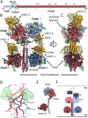
Figure 1.. Architecture of the NiV G homotetramer.
(A) Linear representation of the NiV G ectodomain (as resolved in the cryoEM map), which contains an N-terminal stalk (residue 96 to 147), a neck domain (residue 148 to 165), a linker region (residue 166 to 177) and a C-terminal Head domain (residue 178 to 602). Green arrows indicate N-linked glycosylation sites. Yellow lines refer to cysteine residues. (B-C) Ribbon diagram of the NiV G ectodomain bound to the broadly neutralizing nAH1.3 Fab fragment in two orthogonal orientations. Each of the four NiV G protomers is colored distinctly and resolved N-linked glycans are rendered as green surfaces. The nAH1.3 heavy and light chains are colored gold and yellow, respectively, and only the variable domains were modeled in density. The linkers connecting the neck to the two proximal head domains are shown as dashed lines due to weaker density in the cryoEM reconstruction. (D) Zoomed-in view of the interlaced β-sandwich neck domain showing the four antiparallel inter-protomer disulfide bonds between residues C158 and C162 and the glycan at position N159 protruding from the two chains shown in the foreground. (E) Superimposition of NiV G protomers based on the stalk highlighting that the same polypeptide chain adopts three distinct folds in the homotetrameric assembly. (F) Schematic representation of the NiV G homotetramer showing that only one out of four head domains orients its receptor-binding site (arrow) towards the host cell membrane (light grey) whereas the other three sites point towards the viral membrane (dark grey). EB2: ephrin-B2.
Figure 2.
Figure 2.. Structural basis for nAH1.3-mediated broad neutralization of NiV and HeV
(A) Superimposition of the NiV G head domain (blue surface) bound to nAH1.3 (heavy and light chains colored gold and yellow, respectively) or to ephrin-B2 (purple, PDB 2VSM) showing that they bind to opposite sides of the β-propeller (13). (B) Biolayer interferometry analysis of binding of the nAH1.3 Fab and ephrin-B2 (EB2) to immobilized NiV G ectodomain showing absence of competition irrespective of their order of addition. Each NiV G loaded anti-penta His biosensor probe was sequentially dipped in a solution containing 25 nM nAH1.3 Fab (red) and then 50 nM EB2 + 25 nM nAH1.3 Fab (red) or 50 nM EB2 and then 25 nM of nAH1.3 Fab + 50 nM EB2 (green). Controls with only nAH1.3 Fab (blue) or EB2 (orange) are shown for comparison. (C) Zoomed-in view of the interface between NiV G and nAH1.3 with selected side chains shown as sticks. (D) Ribbon diagram of a NiV G head domain (blue) with the interacting nAH1.3 heavy and light chains CDRs rendered in gold and yellow. (E) Molecular surface representation of the NiV G head showing the nAH1.3 footprint colored by residue conservation between NiV G and HeV G. Conservative sub, conservative substitution. Semi-conservative sub, semi-conservative substitution. (F) Molecular surface representation of the NiV G head showing the nAH1.3 escape mutations identified here (I520T introducing an N-linked glycosylation site at position N518; N186D comes from HeV G escape mutant) and Q450K and R516K (previously described (31)).
Figure 3.
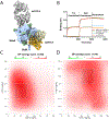
Figure 3.. A HNV G head-directed mAb cocktail with synergistic neutralizing activity
(A) Superimposition of the NiV G head domain (blue surface) bound to nAH1.3 (heavy and light chains colored gold and yellow, respectively) or to the m102.3 Fab (heavy and light chains colored dark and light grey, respectively, PDB 6CMI) showing that they bind to opposite sides of the β-propeller. m102.3 is closely related to the m102.4 mAb. (B) Biolayer interferometry analysis of binding of the nAH1.3 and m102.4 IgGs to the immobilized NiV G ectodomain showing absence of competition irrespective of the order of addition. Each NiV G loaded anti-penta His biosensor probe was sequentially dipped in a solution containing 100 nM nAH1.3 IgG (red) and then 100 nM m102.4 IgG + 100 nM nAH1.3 IgG (red) or 100 nM m102.4 IgG and then 100 nM of nAH1.3 IgG + 100 nM m102.4 IgG (green). Controls with only nAH1.3 IgG (blue) or m102.4 IgG (orange) are shown for comparison. (C-D) Synergy maps for neutralization of replication competent rCedV-NiV-B-GFP (C) and rCedV-HeV-GFP (D) by varying concentrations of the m102.4 and nAH1.3 mAb cocktail analyzed with SynergyFinder (41). ZIP synergy score greater than 10 indicates strong synergistic relationship. The white box indicates the most synergistic region on each plot.
Figure 4.
Figure 4.. The NiV G receptor-binding head domain is immunodominant and accounts for most of the neutralizing activity elicited by vaccination
(A) Study design for vaccination of rhesus macaques, where two rhesus macaques have been immunized three times (4 weeks apart) with 200 μg of an alum-adjuvanted equimolar mixture of the purified NiV-B and NiV-M sG tetramers. Blood was collected on day 42 and day 84 post immunization. (B) Serum neutralizing Ab titers against rCedV-NiV-B-GFP and rCedV-HeV-GFP measured at day 84. The curves for each animal are shown in black and blue. (C-E) Competition ELISAs showing binding of biotinylated nAH1.3 (C), m102.4 (D) and HENV-32 (E) mAbs to the immobilized NiV G ectodomain in the presence of various dilutions of vaccine-elicited rhesus macaque sera (obtained at day 84). The curves for each animal are shown in black and blue whereas binding of the mAb in the absence of sera is shown in green (control). (F-I) Representative EM reconstructions (grey surfaces) of negatively stained complexes formed between purified polyclonal serum Fab fragments and NiV G fitted with atomic models for visualization of antigenic sites targeted. (J) Surface representation of the NiV G tetramer with three head domain-specific Fabs bound (left) and a stalk-directed Fab bound (right) highlighting antigenic sites detected in vaccine elicited polyclonal serum Abs. (K-L) Neutralizing Ab titers against rCedV-NiV-B-GFP (K) and rCedV-HeV-GFP (L) before and after depletion with the NiV G head domain (Head) or the ectodomain tetramer (Ecto).

References

    1. Eaton BT, Broder CC, Middleton D, Wang LF, Hendra and Nipah viruses: different and dangerous. Nat. Rev. Microbiol 4, 23–35 (2006). - PMC - PubMed
    1. Gurley ES, Hegde ST, Hossain K, Sazzad HMS, Hossain MJ, Rahman M, Sharker MAY, Salje H, Islam MS, Epstein JH, Khan SU, Kilpatrick AM, Daszak P, Luby SP, Convergence of Humans, Bats, Trees, and Culture in Nipah Virus Transmission, Bangladesh. Emerg. Infect. Dis 23, 1446–1453 (2017). - PMC - PubMed
    1. Arunkumar G, Chandni R, Mourya DT, Singh SK, Sadanandan R, Sudan P, Bhargava B, People, Nipah Nipah Investigators, Health, Outbreak investigation of Nipah Virus Disease in Kerala, India, 2018. J. Infect. Dis (2018), doi: 10.1093/infdis/jiy612. - DOI - PubMed
    1. Chadha MS, Comer JA, Lowe L, Rota PA, Rollin PE, Bellini WJ, Ksiazek TG, Mishra A, Nipah virus-associated encephalitis outbreak, Siliguri, India. Emerg. Infect. Dis 12, 235–240 (2006). - PMC - PubMed
    1. Ching PK, de los Reyes VC, Sucaldito MN, Tayag E, Columna-Vingno AB, Malbas FF Jr., Bolo GC Jr., Sejvar JJ, Eagles D, Playford G, Dueger E, Kaku Y, Morikawa S, Kuroda M, Marsh GA, McCullough S, Foxwell AR, Outbreak of henipavirus infection, Philippines, 2014. Emerg. Infect. Dis 21, 328–331 (2015). - PMC - PubMed

Publication types

LinkOut - more resources